


- Protocols

- Articles and Issues
- For Authors
- About
- Become a Reviewer


Lodicule Isolation and Morphometric Analysis During Rice Floret Opening

(*contributed equally to this work) Published: Vol 16, Iss 13, Jul 5, 2026 DOI: 10.21769/BioProtoc.5721 Views: 160
Reviewed by: Shweta PanchalPragya BaruaAnonymous reviewer(s)
 PDF
PDF  Ask a question
Original research article
The authors used this protocol in:

Feb 2026
Advertisement


Protocol Collections
Comprehensive collections of detailed, peer-reviewed protocols focusing on specific topics




Related protocols
Abstract
Rice lodicules are specialized floral organs located at the base of the ovary that undergo dynamic morphological changes during the flowering period. Water uptake–driven swelling and subsequent dehydration-induced shrinkage of the lodicules trigger floret opening and closure, respectively. Although lodicules play a central role in floret movement, standardized methods for quantitatively monitoring their temporal morphological changes remain limited. Here, we describe a detailed and reproducible workflow for lodicule sampling, dissection, imaging, and quantitative morphometric analysis. Florets are collected at predefined clock time points during the flowering period, and lodicules are carefully isolated under a stereomicroscope. High-resolution imaging is performed under consistent acquisition settings, followed by precise measurement of lodicule length, width, and thickness using image analysis software. This protocol emphasizes positional consistency in sampling, uniform imaging parameters, and standardized data analysis to enhance reproducibility. This method is suitable for evaluating the effects of genetic background or environmental conditions on lodicule morphology. By providing a standardized analytical framework, this protocol enables accurate and quantitative morphometric analysis of rice lodicules during floret opening.
Key features
• Standardized time-point sampling minimizes variability caused by diurnal fluctuations and handling during lodicule morphometric analysis.
• Enables reproducible isolation and imaging of rice lodicules while preserving native morphology and preventing dehydration-induced artifacts.
• Time-resolved workflow enables analysis of rapid morphological changes associated with floret opening and closure.
• Applicable for comparing genetic and environmental effects on lodicule morphology under controlled experimental conditions.
Keywords: Rice floret
 Lodicule imaging
Lodicule morphometry
Time-course imaging
Morphometric analysis
Lodicule imaging
Lodicule morphometry
Time-course imaging
Morphometric analysis
Graphical overview
Background
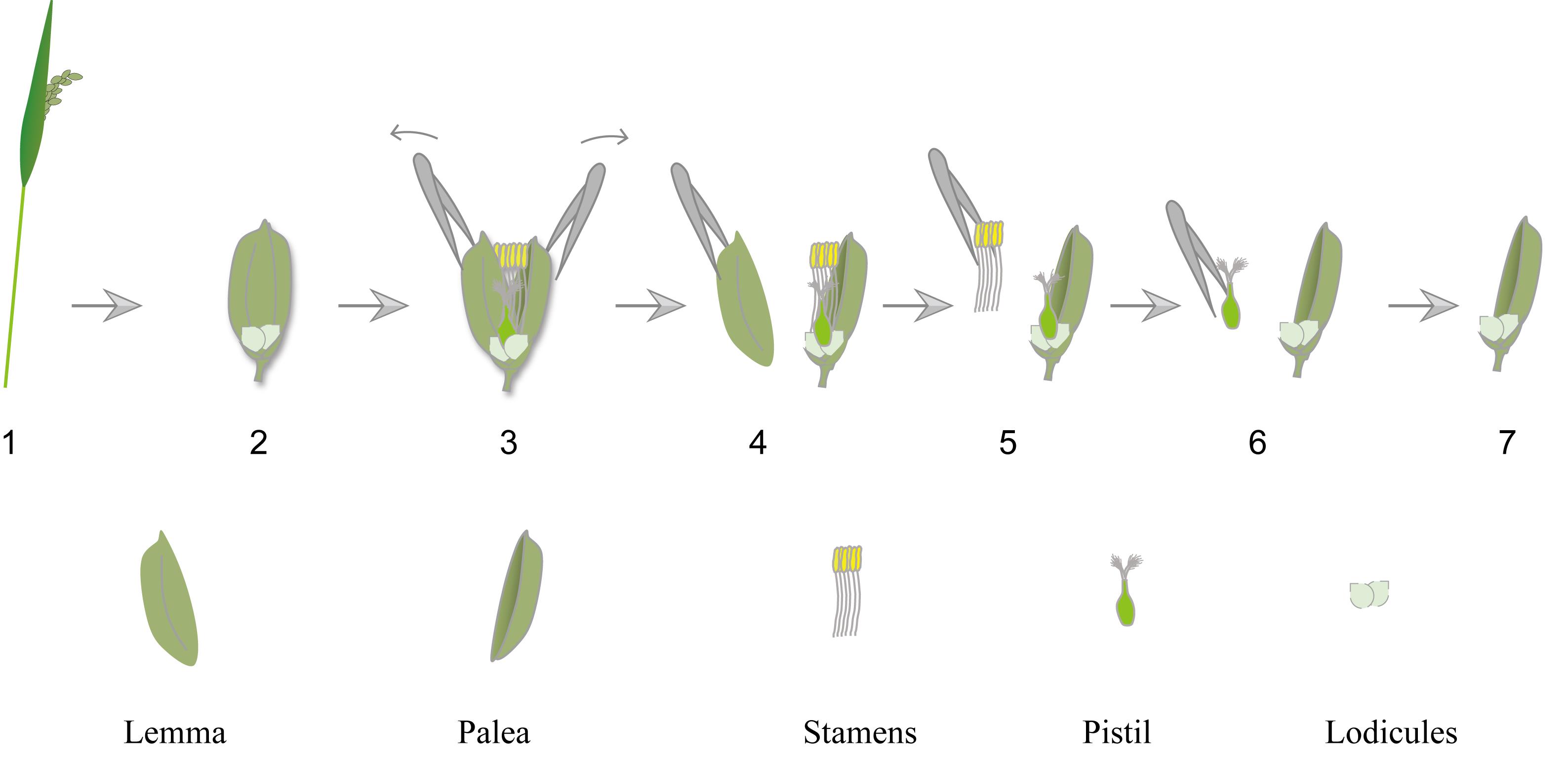
In rice (Oryza sativa), the floret consists of the lemma, palea, stamens, pistil, and two lodicules located at the base of the ovary. Lodicules function analogously to petals [1]. During the flowering period, lodicules absorb water and undergo rapid swelling. The resulting increase in volume generates a mechanical force that separates the lemma and palea, thereby enabling floret opening and pollination. After pollination is completed, lodicules lose water and shrink, which promotes the inward repositioning of the lemma and palea and leads to floret closure [2].
Accurate characterization of lodicule morphology is essential for understanding the mechanisms underlying floral movement, reproductive development, and genotype-dependent variation. Therefore, quantitative analysis of lodicule morphology at defined time points provides valuable phenotypic information. However, lodicule analysis is often influenced by technical variability. Inconsistent sampling intervals, variation of spikelet position in panicle, tissue dehydration during dissection, and differences in imaging parameters can introduce measurement bias. Previous studies have shown that lodicule morphology is highly sensitive to developmental stage, genetic background, and sampling conditions, often resulting in substantial variation in morphometric measurements across experiments [3–5]. In addition, the lack of standardized morphometric criteria limits comparability across studies.
To address these challenges, we describe a workflow for lodicule morphological analysis based on fixed clock-time sampling. This method avoids reliance on subjective developmental staging and instead emphasizes controlled sampling intervals, standardized dissection procedures, and calibrated imaging and measurement settings. By minimizing experimental variability, this protocol enables precise quantification of lodicule length, width, and thickness and facilitates reliable comparison across genotypes or experimental conditions.
Materials and reagents
Biological materials
1. Rice plants (Oryza sativa) grown under controlled conditions and sampled at the flowering stage (0–3 days post-heading)
Reagents
1. Ultrapure water
2. 70% (v/v) ethanol (for sterilization of tools)
3. Soil (Jiang Su Xing Nong Substrate&technology Co., Ltd., catalog number: 161102G0096N)
4. Vermicompost (Wuhan Jiyesheng Chemical Co., Ltd., catalog number: A00970)
Laboratory supplies
1. Fine forceps (RWD, catalog number: F12012-10)
2. Glass slide (MREDA, catalog number: M049985)
3. Dissection needles (MREDA, catalog number: M188991)
4. 1.5 mL microcentrifuge tubes
5. Parafilm (Sigma-Aldrich BS, catalog number: HS120667)
6. Ice and insulated foam box (for temporary storage of samples during transport and handling)
7. Filter paper (Tanon, catalog number: 586-1700)
Equipment
1. Fluorescence stereomicroscope (Leica, catalog number/model: M205 FA)
2. Growth chamber or greenhouse with controlled light and temperature conditions
Software and datasets
1. Leica Application Suite X (LAS X)
2. GraphPad Prism 10
Procedure
A. Plant growth
1. Thoroughly rinse rice seeds (Oryza sativa) with sterile water. Germinate seeds on moist filter paper at 28 °C in the dark for 2–3 days.
2. Transfer germinated seedlings to soil (e.g., paddy soil or soil:vermiculite mixture, 3:1, v/v). Cultivate plants under controlled conditions (28 °C day/28 °C night, 14 h light/10 h dark photoperiod, light intensity of 500 μmol·m-2·s-1, and 60%–70% relative humidity) or under well-documented field conditions.
3. Maintain consistent irrigation and nutrient management throughout vegetative and reproductive stages to ensure uniform plant growth.
4. Record heading date individually for each plant, defined as the day when the first panicle emerges from the flag leaf sheath (0 days post-heading).
Critical: Accurate determination of heading date is essential, as lodicule morphology and floret opening behavior are tightly linked to developmental stage. Inconsistent staging may lead to substantial variation in morphometric measurements.
5. Select plants with the same sowing date and similar developmental stage, plant height, and heading status to minimize biological variation.
6. Inspect panicles daily during the flowering period to determine flowering window and guide precise time-point sampling.
Note: Consistent growth conditions and accurate determination of heading date are critical for reducing variability in lodicule morphology.
B. Time-point sampling
1. Collect florets at a defined developmental stage. Sample within 0–3 days post-heading, when floret opening activity is stable and reproducible.
Caution: Sampling outside this window may introduce variability due to developmental heterogeneity.
2. Because lodicule morphology changes rapidly during floret opening, the time of day is critical. Conduct sampling within a fixed daily time window (e.g., 08:00–11:00) under consistent environmental conditions.
3. Perform time-course sampling using fixed clock intervals, defined as equal time intervals between consecutive sampling points (e.g., every 30 min or every 1 h). A minimum of 4–6 time points is recommended.
Caution: Start with 1-h intervals for general analysis; 30-min intervals are recommended for higher temporal resolution. A minimum of 4–6 time points is suggested. It should be noted that the choice of sampling interval may influence the measured changes in lodicule morphology, particularly given their rapid and transient behavior during floret opening. Therefore, consistent and sufficiently frequent sampling is recommended to ensure comparability across experiments.
4. Prior to sampling, prepare pre-labeled 1.5 mL microcentrifuge tubes for each time point and replicate to avoid delays.
Pause point: Tubes can be prepared and labeled in advance before sampling begins.
5. Collect florets from similar positions within the panicle (e.g., upper or middle spikelets) to minimize positional variation (Figure 1).
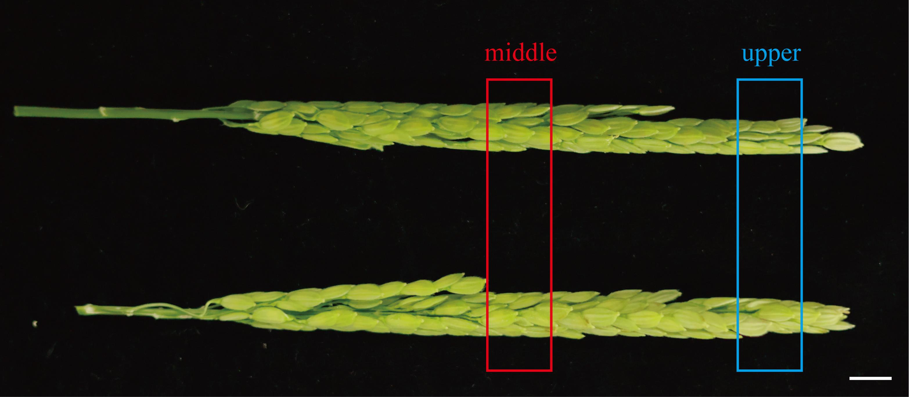
Figure 1. Photograph showing florets from comparable positions within the panicle. The red box indicates florets located in the middle region, while the blue box indicates florets located in the upper region. Scale bar, 1 cm.
6. For each biological replicate, collect 3–5 florets per time point. At least three independent biological replicates are recommended.
7. Immediately place detached florets into collection tubes and keep tubes closed during transport to minimize humidity-induced morphological changes.
Caution: Exposure to ambient air may lead to rapid dehydration and morphological distortion.
8. Transfer samples to the laboratory promptly. The time between collection and dissection should be minimized (preferably within 5 min, and no longer than 10 min).
9. Store samples on ice in an insulated foam box if temporary preservation is required.
Critical: Lodicule morphology is highly sensitive to sampling time, humidity, and handling. Even minor deviations may introduce significant variation in morphometric measurements.
C. Lodicule dissection
1. Place the floret horizontally on a clean glass slide.
2. Hold the floret using fine-tipped forceps and carefully remove the lemma with another forceps (Figure 2A).
Note: Do not remove the palea, as the basal region of the lodicules is partially attached to it (Figure 2B). During subsequent manipulation, the lodicules can be positioned by holding the palea with forceps.
3. Identify the two lodicules located at the base of the ovary. They appear as small, scale-like, and often translucent structures. Carefully remove the stamens and ovary using forceps without damaging the delicate lodicule tissue.
Critical: Mechanical damage during dissection can alter lodicule morphology and compromise downstream measurements.
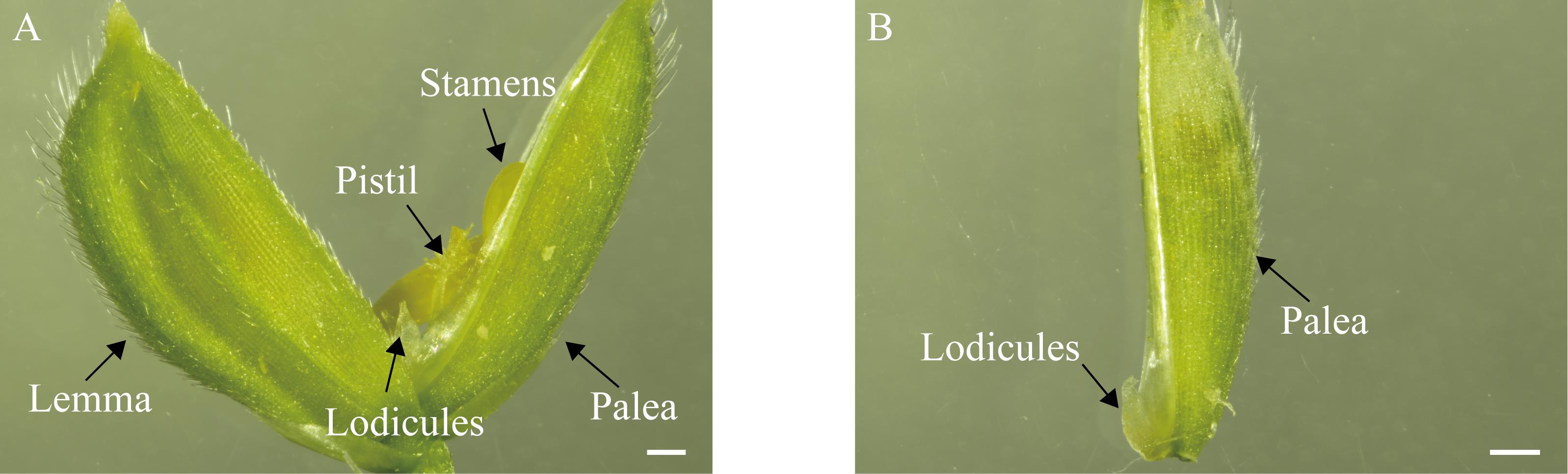
Figure 2. Rice floret structure and dissected sample preparation. (A) Photograph of an intact rice floret, showing all major floral organs, including the lemma, palea, stamens, pistil, and lodicules. (B) Dissected floret after removal of internal floral organs, showing only the palea and lodicules, prepared for imaging analysis. Scale bars, 1 mm.
D. Imaging
1. After lodicule dissection, immediately position the sample on the stage of a stereomicroscope by holding the palea with forceps. Adjust the orientation so that the imaging surface of the lodicule faces the center of the light source. Capture images under consistent illumination conditions.
Critical: Imaging should be performed immediately after dissection to minimize dehydration-induced morphological changes.
2. Use identical microscope magnification (e.g., 10× or 20× objective) for all samples.
3. Maintain constant exposure time, light intensity, white balance, and camera gain settings.
4. Perform calibration using a stage micrometer before the imaging session.
5. Capture images sequentially from both the frontal and lateral views of the lodicules (Figure 3).
E. Morphometric analysis
1. Open images in LAS X software.
2. Set the scale using the micrometer calibration image.
3. Measure three dimensions for each pair of lodicules (Figure 4): length (L), width (W), and thickness (T).
Note: Consistent measurement criteria should be applied across all samples to ensure comparability.
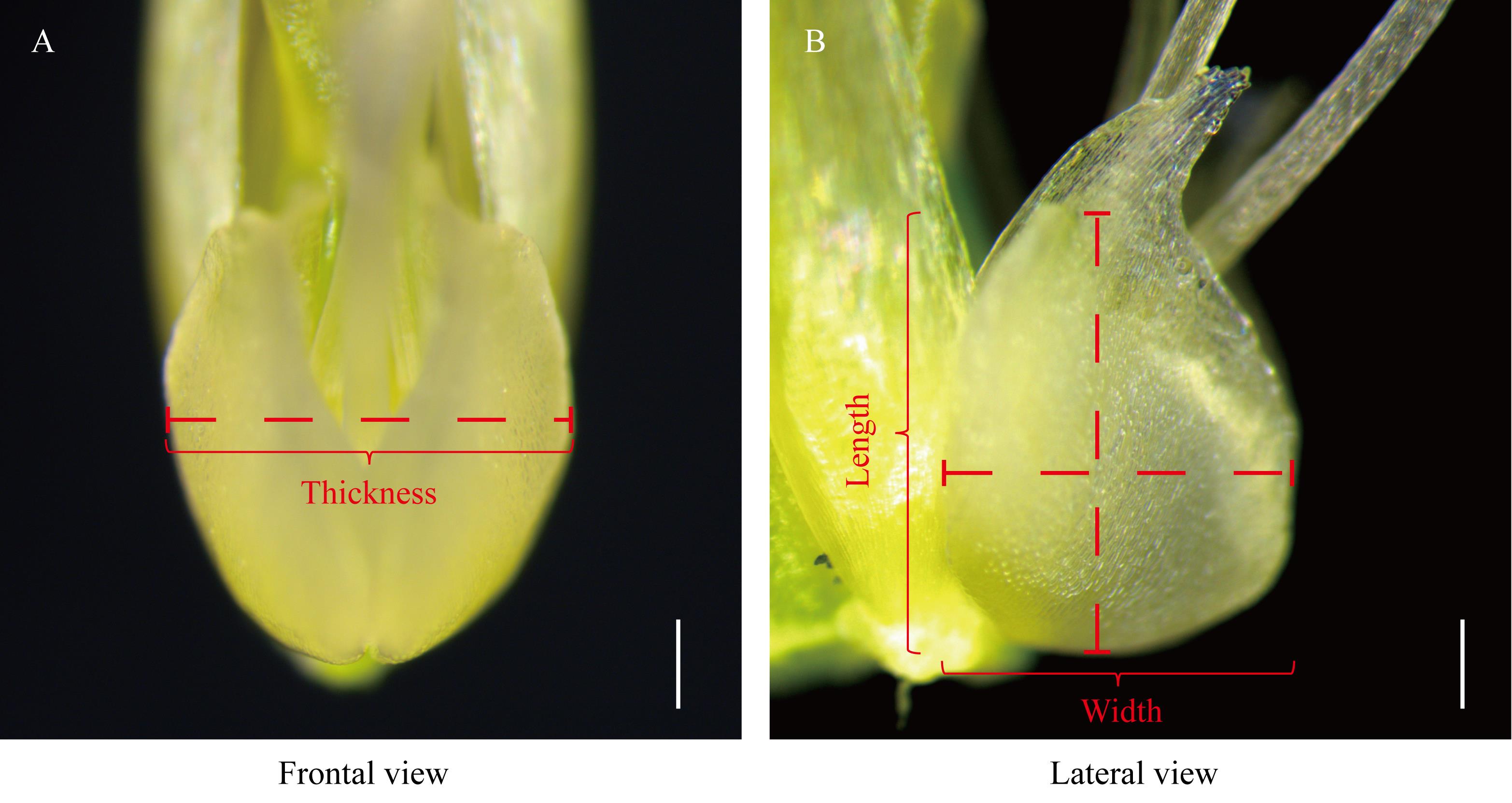
Figure 3. Schematic diagram of lodicule measurement. (A) Frontal view of the lodicule. (B) Lateral view of the lodicule. Length was defined as the maximum vertical distance between the basal and apical surfaces in the lateral view. Width was measured as the maximum horizontal distance in the lateral view. Thickness was defined as the maximum horizontal distance across a pair of lodicules in the frontal view. Scale bars, 200 μm.
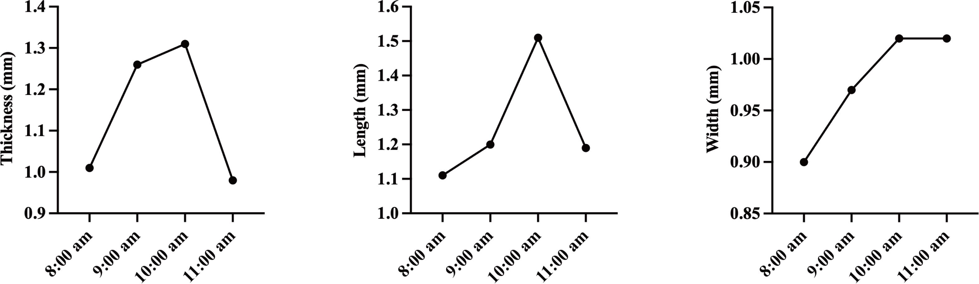
Figure 4. Changes in lodicule morphology during floret opening. Line graphs showing temporal changes in lodicule thickness (T), length (L), and width (W) at different time points during floret opening. One representative dataset was selected for display.
Data analysis
To approximate three-dimensional morphological changes, each pair of lodicules was modeled as an ellipsoid. Lodicule volume (V) was calculated using the ellipsoid volume formula:
Length (L) was defined as the maximum vertical distance between the basal and apical surfaces in the lateral view. Width (W) was defined as the maximum horizontal distance in the lateral view. Thickness (T) was defined as the maximum horizontal distance across a pair of lodicules in the frontal view. An illustrative example is shown in Figure 5.
For quantitative analysis, at least three independent biological replicates should be collected. For each biological replicate, 3–5 florets are sampled per time point, and measurements from individual lodicules are treated as technical replicates.
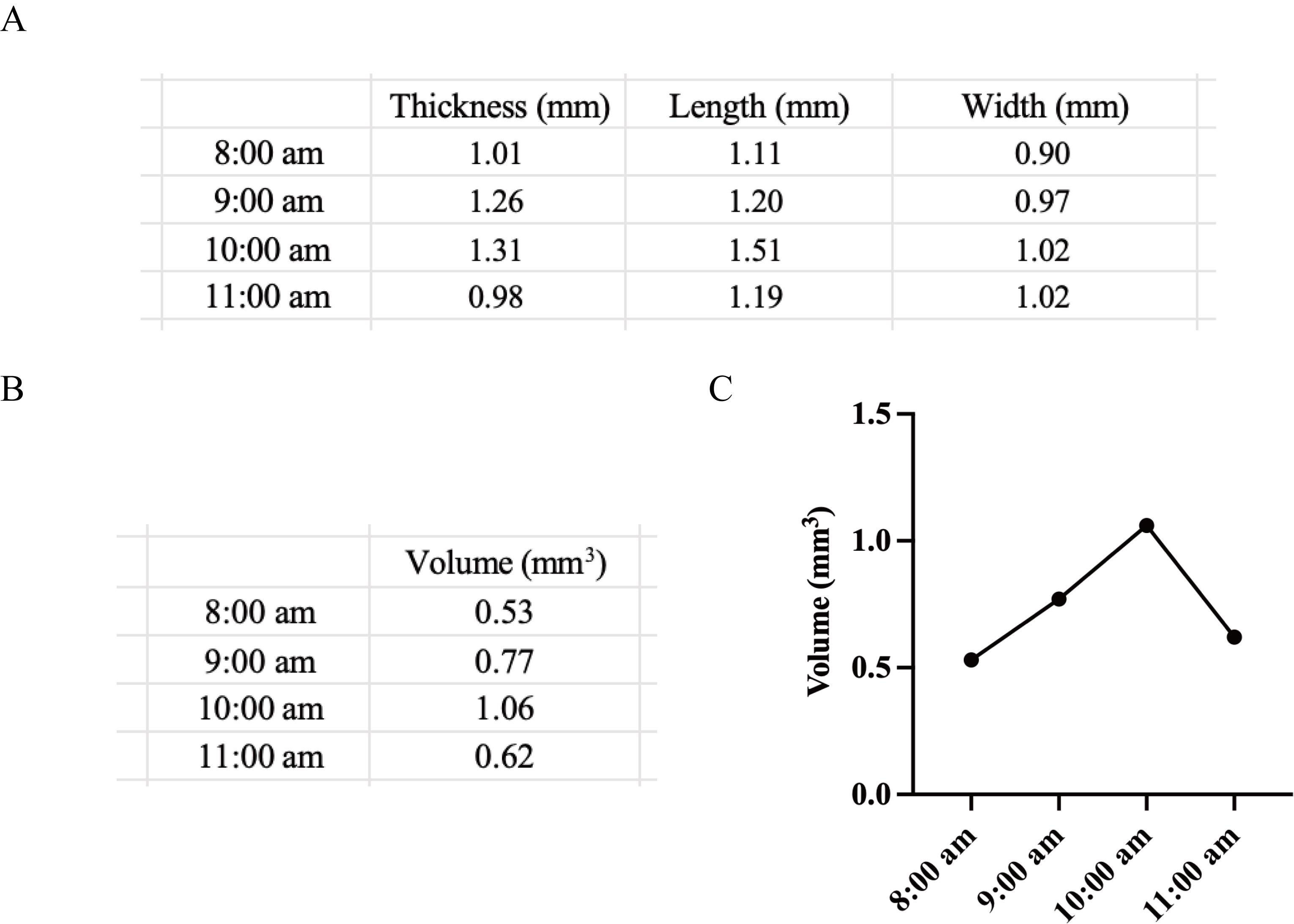
Figure 5. Data analysis of lodicule volume during floret opening. (A) Measurements of lodicule thickness (T), length (L), and width (W) at different time points during floret opening. (B) Lodicule volume calculated at each time point based on an ellipsoid model. (C) Line graph showing dynamic changes in lodicule volume over time. One representative dataset was selected for display.
Validation of protocol
This protocol has been used and validated in the following research article:
Hu et al. [6] The multifunctional protein OsAIM1 regulates floret opening and closure timing via jasmonic acid-mediated lodicule dynamics in rice. Plant physiology. (Figure 4).
General notes and troubleshooting
General notes
1. Developmental stage consistency is critical. Lodicule morphology is highly dynamic and sensitive to developmental progression. Florets collected at slightly different physiological stages may exhibit substantial variation in size and shape. Therefore, sampling should be restricted to florets with uniform external morphology and similar panicle positions to minimize developmental heterogeneity.
2. Mechanical manipulation affects morphology. Lodicules are delicate and easily deformed by forceps pressure or desiccation. Excessive mechanical stress may artificially alter measured length, width, or height. All dissections should be performed gently and consistently across samples.
3. Ellipsoid approximation introduces assumptions. The volume calculation assumes that lodicules approximate an ellipsoid. This geometric simplification provides a standardized comparative metric rather than an absolute anatomical volume. Deviations from ideal ellipsoid geometry should be acknowledged when interpreting quantitative differences.
Troubleshooting
Problem 1: Lodicules appear collapsed or flattened during imaging.
Possible causes: Tissue dehydration or excessive mechanical pressure during manipulation.
Solutions: Minimize exposure time between dissection and imaging. Use fine-tipped forceps with minimal pressure. Ensure that the lodicule is not compressed against the slide.
Problem 2: Inconsistent volumetric measurements between replicates.
Possible causes: Variation in imaging orientation or developmental stage differences among sampled florets.
Solutions: Standardize imaging orientation by aligning the lodicule perpendicular to the optical axis. Restrict sampling to florets from similar panicle positions and with comparable external morphology. Increase the biological replicate number to account for natural variation.
Problem 3: Difficulty separating lodicules from the ovary without damage.
Possible causes: Incomplete removal of surrounding floral tissues or improper removal of the palea.
Solutions: Retain the palea for structural support and use forceps to grasp the palea rather than the lodicules during dissection. Carefully separate the ovary using minimal lateral force.
Acknowledgments
Conceptualization, J.W. and S.Z.; Investigation, Y.H., S.C., H.L., H.H., Z.X., B.H., L.C., L.Z., J.W., K.C., W.L., W.S.; Writing—Original Draft, Y.H.; Writing—Review & Editing, J.W. and S.Z.; Funding acquisition, J.W. and S.Z.; Supervision, J.W., S.Z., L.J., Z.Z., C.L., S.Z., Y.T., X.L., S.L., L.C. Funding support from the Biological Breeding-National Science and Technology Major Project (2024ZD04080), the Foundation of Biological Breeding Zhongshan Lab (ZSBBL-KY2023-07), the Jiangsu Provincial Key Research and Development Program (BE2023362), and the “JBGS” Project of Seed Industry Revitalization in Jiangsu Province [JBGS (2021) 007]. This protocol has been described and validated in the original research paper by Hu et al. [6].
Competing interests
The authors declare no conflicts of interest.
References
- Qin, Y., Yang, J. and Zhao, J. (2005). Calcium changes and the response to methyl jasmonate in rice lodicules during anthesis. Protoplasma. 225: 103–112. https://doi.org/10.1007/s00709-005-0086-6
- Yasuno, N., Ikeda, K., Iida, S., Nagato, Y. and Kyozuka, J. (2009). The expression level of. Plant and Cell Physiology Supplement. 0116–0116
- Xu, P., Wu, T., Ali, A., Zhang, H., Liao, Y., Chen, X., Tian, Y., Wang, W., Fu, X., Li, Y., et al. (2022). Plant Biotechnol J. 20(8): 1441–1443. https://doi.org/10.1111/pbi.13860
- Gou, Y., Heng, Y., Ding, W., Xu, C., Tan, Q., Li, Y., Fang, Y., Li, X., Zhou, D., Zhu, X., et al. (2024). Natural variation in OsMYB8 confers diurnal floret opening time divergence between indica and japonica subspecies. Nat Commun. 15(1): 2262–. https://doi.org/10.1038/s41467-024-46579-z
- Deng, R., Yan, Z., Tang, H. and Zhu, S. (2024). Revealing Physiological Basis for Floret Opening Difference Between Indica and Japonica Rice: Based on Floral Structure, Transcriptome, and Endogenous Floret Opening Regulator. Genes(Basel). 15(11): 1396–. https://doi.org/10.3390/genes15111396
- Hu, Y., Li, H., Hou, H., Cui, S., Xu, Z., Hao, B., Cai, L., Zhu, L., Wang, J., Chang, K., et al. (2026). The multifunctional protein OsAIM1 regulates floret opening and closure timing via jasmonic acid-mediated lodicule dynamics in rice. Plant Physiol. https://doi.org/10.1093/plphys/kiaf690
Article Information
Publication history
Received: Apr 3, 2026
Accepted: May 13, 2026
Available online: May 27, 2026
Published: Jul 5, 2026
Copyright
© 2026 The Author(s); This is an open access article under the CC BY-NC license (https://creativecommons.org/licenses/by-nc/4.0/).
How to cite
Readers should cite both the Bio-protocol article and the original research article where this protocol was used:
- Hu, Y., Cui, S., Li, H., Hou, H., Xu, Z., Hao, B., Cai, L., Zhu, L., Wang, J., Chang, K., Li, W., Shao, W., Zhu, S., Li, C., Zhao, Z., Jiang, L., Tian, Y., Liu, X., Liu, S., Chen, L., Zhou, S. and Wan, J. (2026). Lodicule Isolation and Morphometric Analysis During Rice Floret Opening. Bio-protocol 16(13): e5721. DOI: 10.21769/BioProtoc.5721.
- Hu, Y., Li, H., Hou, H., Cui, S., Xu, Z., Hao, B., Cai, L., Zhu, L., Wang, J., Chang, K., et al. (2026). The multifunctional protein OsAIM1 regulates floret opening and closure timing via jasmonic acid-mediated lodicule dynamics in rice. Plant Physiol. https://doi.org/10.1093/plphys/kiaf690

 Download Citation in RIS Format
Category
Plant Science > Plant developmental biology > Morphogenesis
Plant Science > Plant physiology > Phenotyping
Plant Science > Plant cell biology > Tissue analysis
Do you have any questions about this protocol?
Post your question to gather feedback from the community. We will also invite the authors of this article to respond.
 Post a Question


