


- Protocols

- Articles and Issues
- For Authors
- About
- Become a Reviewer


A Reliable Method for Thawing Primary AML and CMML Mononuclear Cells to Preserve Viability and Function

(*Contributed equally to this work, §Technical contact: kelly.lim@adelaide.edu.au; verity.saunders@sahmri.org) Published: Vol 16, Iss 11, Jun 5, 2026 DOI: 10.21769/BioProtoc.5720 Views: 306
Reviewed by: Olga ZimmermannovaRakesh BamAnonymous reviewer(s)
 PDF
PDF  Ask a question
Original research article
The authors used this protocol in:

Sep 2024
Advertisement


Protocol Collections
Comprehensive collections of detailed, peer-reviewed protocols focusing on specific topics




Abstract
Human mononuclear cells derived from peripheral blood and bone marrow are valuable resources for the study of hematological malignancies, including acute myeloid leukemia (AML) and chronic myelomonocytic leukemia (CMML). Cryopreservation enables long-term storage of patient samples for downstream assays; while thawing protocols have been described, subsequent recovery of viable cells after thawing can be challenging, particularly for fragile blast and monocyte populations. Here, we describe a reliable protocol for thawing cryopreserved AML and CMML mononuclear cells designed to preserve post-thaw viability, recovery, and functional integrity. The method incorporates controlled dilution of cells out of cryoprotectant with anticoagulant-supplemented thaw buffer, DNase I treatment, and gentle resuspension steps. Using this approach, post-thaw viability consistently exceeded 80% with a mean recovery of 55.6% across samples. Recovered cells retained functional capacity, as demonstrated by colony-forming assays, and maintained immunophenotypic characteristics by flow cytometry. This protocol provides a robust and reproducible method for the recovery of cryopreserved AML and CMML mononuclear cells and may be broadly applicable to other fragile or monocyte-rich patient-derived hematopoietic samples.
Key features
• Reliable thawing protocol that preserves viability and recovery of cryopreserved AML and CMML mononuclear cells.
• Controlled dilution with anticoagulant supplementation, DNase I treatment, and gentle resuspension minimizes cell aggregation of fragile blast and monocyte populations.
• Maintains cell functional capacity and immunophenotypic characteristics for downstream assays.
• Applicable to cryopreserved patient-derived mononuclear cells prone to aggregation, low recovery, or poor viability with conventional thawing.
Keywords: AML CMML
Leukemia
Monocyte
Mononuclear cells
Cryopreserved
Thaw
Viability
CMML
Leukemia
Monocyte
Mononuclear cells
Cryopreserved
Thaw
Viability
Graphical overview
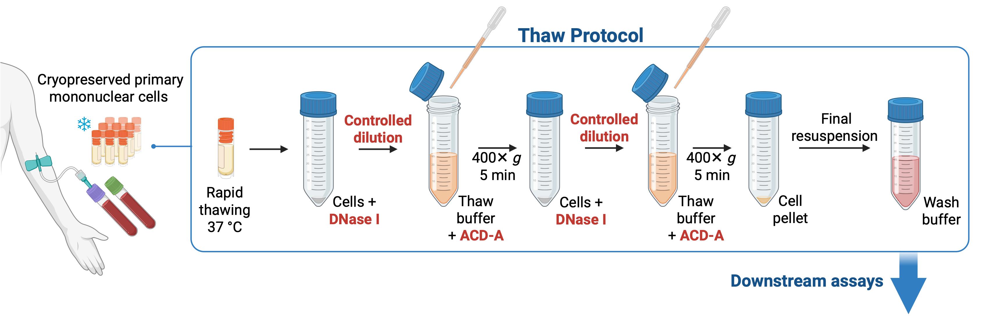
Overview of thaw protocol for cryopreserved primary acute myeloid leukemia (AML) and chronic myelomonocytic leukemia (CMML) mononuclear cells
Background
Human primary mononuclear cells isolated from peripheral blood and bone marrow are valuable in the study of cellular processes and disease mechanisms in hematological malignancies, including acute myeloid leukemia (AML) and chronic myelomonocytic leukemia (CMML). Because access to fresh patient material is limited and experiments cannot always be performed immediately following sample collection, cryopreservation is routinely used to enable long-term storage. However, recovery of viable cells can be challenging, since thawing exposes cells to several concurrent stressors, including dimethyl sulfoxide (DMSO) toxicity at room temperature, osmotic stress leading to membrane damage and extracellular DNA release, mechanical stress from vigorous pipetting, and metabolic stress associated with increased reactive oxygen species.
Certain molecular subtypes of AML, including mutant IDH1 and IDH2, and acute promyelocytic leukemia have been noted to be particularly difficult to recover after cryopreservation. Several protocols for thawing AML cells have been described, most commonly involving rapid thawing in a 37 °C water bath followed by washing steps in serum-containing media, but issues with low viability, poor recovery, and material loss persist [1–4].
To address these challenges, here we describe a reliable step-by-step protocol for thawing cryopreserved mononuclear cells that incorporates controlled dilution of cells out of cryoprotectant to reduce osmotic stress, DNase I treatment, and gentle resuspension in an anticoagulant-supplemented thaw buffer to limit cell aggregation during recovery. These steps are designed to preserve viability, facilitate recovery, reduce sample variability, and maintain the physiological integrity of AML and CMML cells for downstream applications. These include flow cytometry–based characterization of surface markers and cytokine profiling, as well as functional studies such as colony-forming assays, phagocytosis, macrophage differentiation, antigen processing and presentation, oxidative burst, and drug-response assays. We have found our method to be particularly well-suited for CMML samples, which are typically enriched for monocytes that are prone to aggregation during thawing and can be lost during washing steps. Using this method, we consistently achieved post-thaw viability exceeding 80% with a mean recovery of 55.6% across a range of total number of cells per ampoule of cryopreserved sample, with preserved colony-forming capacity and immunophenotypic characteristics.
We routinely apply this protocol for the recovery of cryopreserved AML and CMML mononuclear cells and anticipate it may be useful for other fragile or monocyte-rich primary hematopoietic patient-derived samples.
Materials and reagents
Biological materials
1. Cryopreserved AML and CMML mononuclear cells from peripheral blood or bone marrow, stored at -186 °C (vapor-phase liquid nitrogen tank) in cryoprotectant containing 90% fetal bovine serum and 10% dimethyl sulfoxide
Note: The average number of cells per cryovial used in this protocol was 2.62 × 10⁷ (range 3.0 × 10⁶–1 × 10⁸).
Reagents
1. Hanks’ balanced salt solution, modified with sodium bicarbonate, without calcium chloride and magnesium sulfate (Sigma-Aldrich, catalog number: H9394-500ML)
2. HEPES solution, 1 M (Sigma-Aldrich, catalog number: H0887-100ML)
3. Fetal bovine serum, triple sterile-filtered, pharmaceutical-grade, gamma-irradiated (CellSera, catalog number: AU-FBS/PG), heat-inactivated
4. FenwalTM anticoagulant citrate dextrose solution formula A (ACD-A) (Baxter, catalog number: AHB7898)
5. DNase I grade II 100 mg lyophilized (Roche, catalog number: 10104159001)
6. Trypan Blue stain, 0.4% (Gibco, catalog number: 15250-061)
7. Ethanol, 80% v/v (ChemSupply, catalog number: EL156-20L-P)
8. Glacial acetic acid (Sigma-Aldrich, catalog number: A6283-500ML)
9. Methyl violet (Sigma-Aldrich, catalog number: 69710-25G)
10. Dulbecco’s phosphate-buffered saline (PBS) (Sigma-Aldrich, catalog number: D8537-500ML)
Solutions
1. Wash buffer (see Recipes)
2. Thaw buffer (see Recipes)
3. DNase I (see Recipes)
4. White cell fluid stain (see Recipes)
Recipes
1. Wash buffer
| Reagent | Final concentration | Quantity or volume |
|---|---|---|
| Hanks’ buffered salt solution | 89% | 445 mL |
| Fetal bovine serum | 10% | 50 mL |
| 1 M HEPES | 10 mM | 5 mL |
| Total | 500 mL |
Store at 4 °C and use within 2 months.
2. Thaw buffer
| Reagent | Final concentration | Quantity or volume |
|---|---|---|
| Wash buffer | 97.5% | 39 mL |
| ACD-A | 2.5% | 1 mL |
| Total | 40 mL |
Prepare fresh immediately before using and warm to 37 °C.
3. DNase I
| Reagent | Final concentration | Quantity or volume |
|---|---|---|
| DNase I grade II 100 mg lyophilized | 2.4 mg/mL | 100 mg |
| Sterile distilled water | 99.8% | 41.7 mL |
| Total | 41.8 mL |
Store between -20 and -40 °C and thaw on ice.
4. White cell fluid stain
| Reagent | Final concentration | Quantity or volume |
|---|---|---|
| Glacial acetic acid | 2% | 2 mL |
| Distilled water | 98% | 98 mL |
| Methyl violet | 0.005% | 5 mg |
| Total | 100 mL |
Filter-sterilize and store at ambient temperature.
Laboratory supplies
1. 50 mL conical polypropylene centrifuge tubes (Nunc, catalog number: 339652)
2. Sterile 2.5 mL transfer pipette (Sarstedt, catalog number: 86.1172.001)
3. Mixing cannula (Fairmont Medical, catalog number: MIX1001)
4. Cell strainer, 70 μm (Greiner, catalog number: 542 070)
5. Serological pipette, 5 mL (Corning, Costar Stripette, catalog number: CLS4487-200EA)
6. Serological pipette, 10 mL (Corning Costar Stripette, catalog number: CLS4488-200EA)
7. Pipette tips, 200 μL (Axygen, catalog number: AXTT-200-Y)
Equipment
1. Class II Biosafety cabinet (Nuaire, Cellgard), with suction line for liquid waste removal
2. Benchtop centrifuge (Eppendorf, catalog number: 5810)
3. Light microscope (Olympus, model: BX45)
4. Water bath (Lauda AquaLine, model: AL25)
5. Hemocytometer counting chamber, Neubauer improved
6. 20–200 μL pipette (Gilson, model: Pipetman Classic P200)
7. 2–20 μL pipette (Gilson, model: Pipetman Classic P20)
Procedure
A. Prepare thaw buffer and tubes
1. Prepare 500 mL of wash buffer under aseptic conditions. Wash buffer is used at ambient temperature but can be stably stored at 4 °C for 1–2 months.
2. For each ampoule of cells to be thawed, prepare a sterile 50 mL polypropylene conical centrifuge tube containing 100 μL of DNase I.
Note: DNase I is used in excess and is sufficient for samples containing up to 1 × 10⁸ cells.
3. Prepare 40 mL of thaw buffer per ampoule of cells to be thawed in a sterile 50 mL polypropylene conical centrifuge tube and warm to 37 °C in a water bath.
Critical: ACD-A acidifies the solution; therefore, prepare thaw buffer immediately prior to use by adding 1 mL of ACD-A to 39 mL of wash buffer (final volume 40 mL).
Note: We recommend preparing thaw buffer for no more than four samples concurrently.
B. Thaw samples
1. Thaw an ampoule of mononuclear cells rapidly by immersion in a 37 °C water bath with manual agitation until the contents are almost completely thawed, with only a trace of ice remaining.
2. Spray ampoule with 80% v/v ethanol to disinfect upon removal from the water bath.
3. Using a sterile transfer pipette, immediately transfer thawed cells into the 50 mL tube containing DNase I.
4. Let it sit for 1 min.
5. Using the same sterile transfer pipette, rinse the cryovial by transferring 1 mL of thaw buffer to the empty cryovial and then adding this dropwise to the cells in the 50 mL tube.
6. Add prewarmed thaw buffer to the cells dropwise over 3 min (1 mL every 10 s), gently swirling the tube in a small circular motion for 2 s after each addition, until a final volume of 20 mL is reached.
7. Centrifuge the tube at 400× g for 5 min at ambient temperature to pellet the cells.
8. Discard the supernatant by aspiration using a mixing cannula attached to a suction line.
9. Add another 100 μL of DNase I to the cell pellet with a p200 Pipetman and gently pipette up and down to resuspend the pellet.
10. Repeat step B6 to add another 20 mL of thaw buffer to the cells.
11. Centrifuge the tube at 400× g for 5 min at ambient temperature to pellet the cells.
12. Discard the supernatant by aspiration, leaving approximately 100 μL.
13. Use a p200 Pipetman to gently resuspend the cell pellet in the residual thaw buffer.
14. Gently add 2–5 mL of wash buffer and resuspend the cells.
Notes:
1. If cells have aggregated, pass the cell suspension through a 70 μm nylon cell strainer into a new 50 mL tube.
2. Once cells have been resuspended in wash buffer, they can be kept on ice, allowing for additional samples to be processed up to this point.
15. Determine cell concentration using white cell fluid stain with a 1:10 dilution (10 μL of cells in 90 μL of white cell fluid stain), counted manually on a hemocytometer with a light microscope, according to Formula 1.
Formula 1: (x cells/n squares) × 104 × dilution factor of 10 = cell concentration (cells per mL)
16. Determine cell viability by Trypan Blue exclusion with a 1:10 dilution (10 μL of cells in 40 μL of PBS and 50 μL of 0.4% Trypan Blue solution), counted manually on a hemocytometer with a light microscope, according to Formula 2.
Formula 2: [x viable cells/(x viable cells + y dead cells)] × 100 = cell viability (percentage)
17. Multiply the cell concentration (cells per mL) obtained with Formula 1 by cell viability (percentage) obtained with Formula 2 to determine the overall concentration of viable cells per milliliter.
18. Multiply the overall concentration of viable cells per milliliter by the volume of cell suspension from step B14 to determine the total cell yield from the cryopreserved ampoule. Examples of total cell yield and viability are shown in Figure 1.
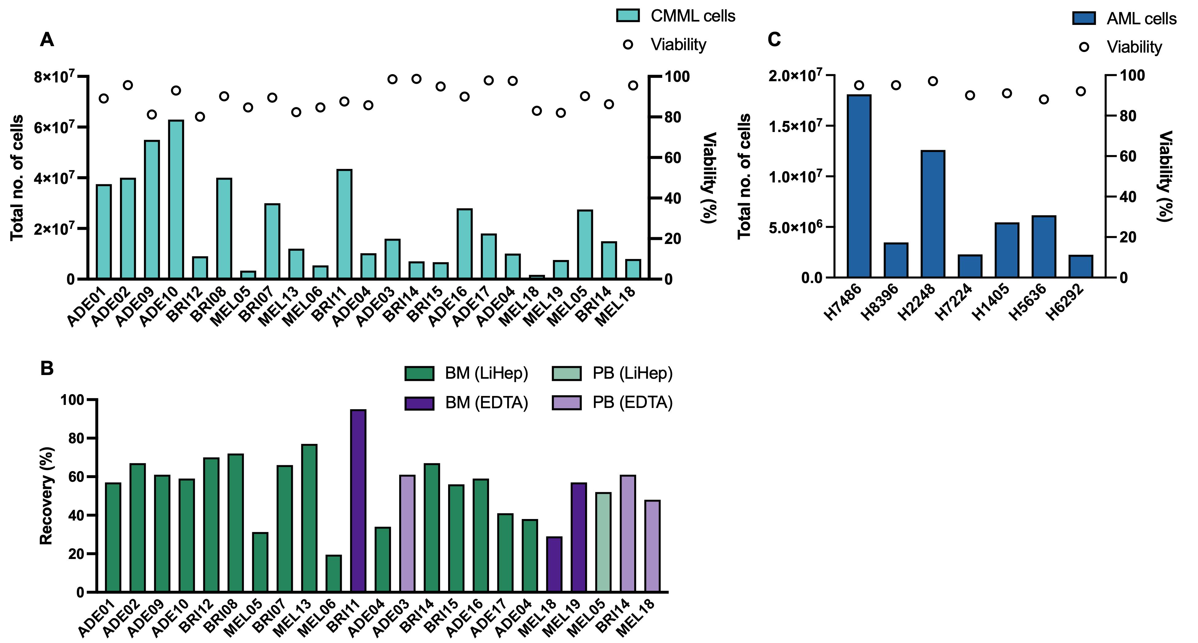
Figure 1. Post-thaw cell numbers, viability, and recovery of cryopreserved primary mononuclear cells from chronic myelomonocytic leukemia (CMML) and acute myeloid leukemia (AML) samples. (A) Total cell yield and viability of CMML mononuclear cells post-thaw. Bars represent the total number of cells recovered (mean 2.15 × 10⁷ ± 3.66 × 10⁶ S.E.M.; range 1.76 × 10⁶–6.3 × 10⁷), while open circles indicate percentage cell viability (mean 89.53% ± 1.26% S.E.M.; range 80.1%–98.8%). (B) Percentage of cell recovery calculated as the number of cells recovered relative to the number of cells cryopreserved (mean 55.6% ± 3.62% S.E.M.; range 19.5%–95%). Green bars indicate samples collected in lithium heparin tubes, and purple bars in EDTA tubes. Darker shades of green and purple indicate samples from bone marrow, and lighter shades from peripheral blood. These data demonstrate that the thaw protocol is applicable to samples collected using different anticoagulants and sample sources. (C) Total cell yield and viability of AML mononuclear cells post-thaw. Bars represent the total number of cells recovered (mean 7.19 × 10⁶ ± 2.26 × 10⁶ S.E.M.; range 2.25 × 10⁶–1.81 × 10⁷), while open circles indicate percentage cell viability (mean 92.57% ± 1.21% S.E.M; range 88.0%–97.0%). The cell recovery percentage is not shown for AML samples because information on the number of cells within the ampoule was unavailable. CMML n = 32, AML n = 7. S.E.M., standard error of mean; BM, bone marrow; PB, peripheral blood; LiHep, lithium heparin; EDTA, ethylenediaminetetraacetic acid.
19. Use cells as required, for example, in colony-forming assays and flow cytometric analysis (Figure 2).
Note: We recommend using the cells immediately rather than resting them in an incubator prior to use.
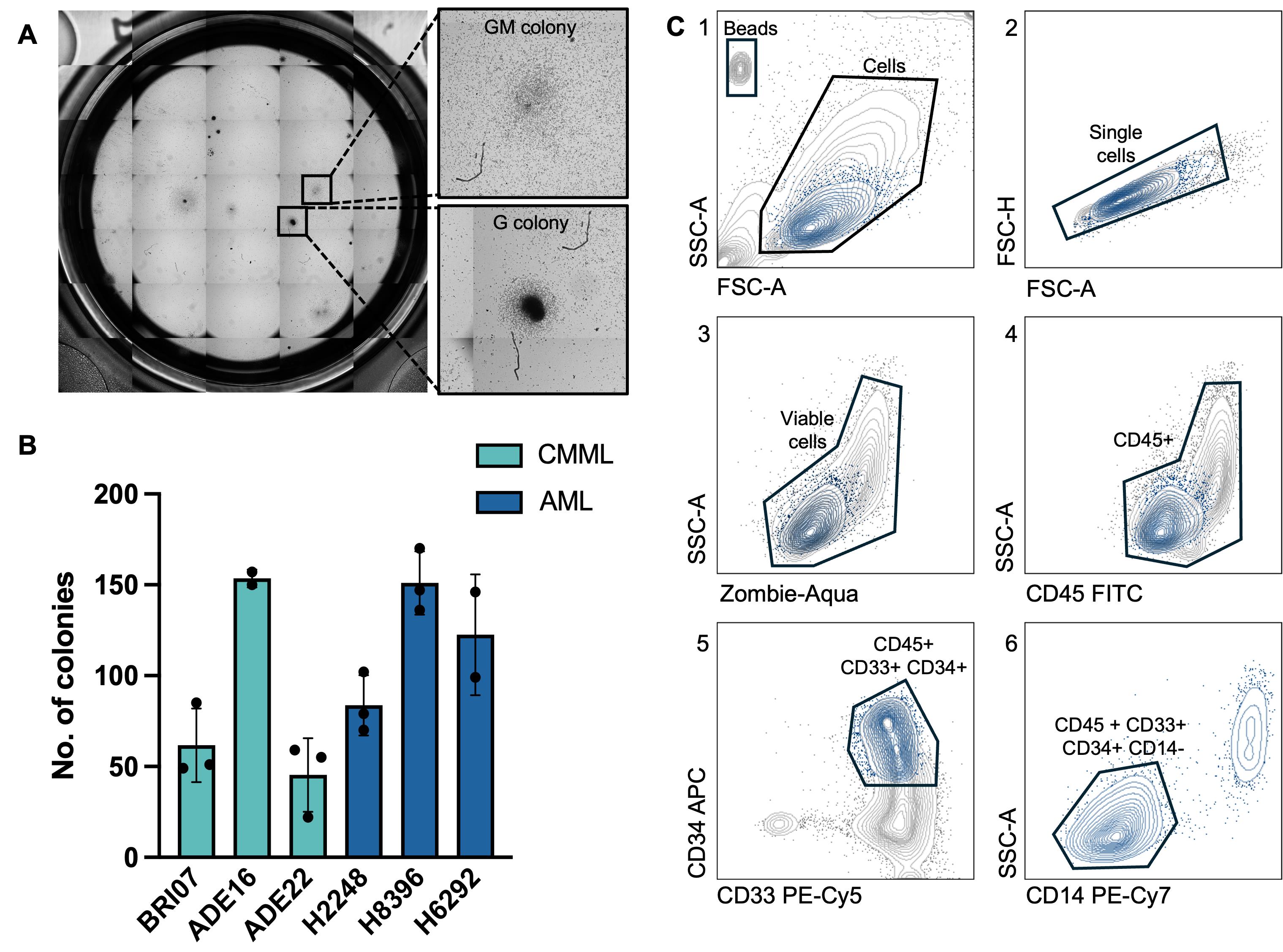
Figure 2. Colony-forming capacity and flow cytometry analysis of chronic myelomonocytic leukemia (CMML) and acute myeloid leukemia (AML) samples demonstrating the preservation of cellular function and immunophenotypic characteristics. (A) Representative image of a methylcellulose-based colony-forming unit (CFU) assay plate. Insets show magnified examples of granulocyte-macrophage (GM) and granulocyte (G) colonies identified. GM-colonies were defined as larger, diffuse, and morphologically heterogeneous, whereas G-colonies were compact clusters of small, uniform cells. (B) Quantification of the number of CFUs formed from 5 × 10⁴ CMML or AML cells seeded per well, demonstrating intrinsic heterogeneity based on underlying molecular profiles. Bars represent the mean total number of colonies enumerated per well with standard deviation, performed in duplicates or triplicates. CMML samples are shown in teal, and AML samples in blue. (C) Representative flow cytometry gating strategy used to identify myeloid blast population in one AML sample. Cells were first gated on forward/side scatter (1), followed by doublet exclusion (2) and viability, with viable cells defined as Zombie-Aqua negative (3). Viable cells were gated for CD45 positivity (4), followed by CD33 and CD34 positivity (5). Final gating identifies CD45+ CD33+ CD34+ CD14- blast population (6), shown in blue throughout the gates. CD, cluster of differentiation.
Validation of protocol
Thaw records and assessment of cellular function by colony-forming assay and immunophenotypic characteristics by flow cytometry analysis are included here and described in the figure legend. Post-thaw cell viability consistently exceeded 80%, independently of the number of cells available per ampoule before and after thawing.
This protocol has also been used and validated in the following publications:
• Lim et al. [5]. CBL mutations in chronic myelomonocytic leukemia often occur in the RING domain with multiple subclones per patient: Implications for targeting. Plos One (Figure 3). DOI: 10.1371/journal.pone.0310641
• Lim et al. [6]. CBL Variants in Chronic Myelomonocytic Leukemia Exhibit a Complex Sub-Clone Architecture with a High Frequency of RING Domain Mutations Sufficient to Induce GM-CSF Hypersensitivity. Blood. DOI: 10.1182/blood-2024-202682
• Lim et al. [7]. Secondary Acute Myeloid Leukemia transformed from chronic myelomonocytic leukemia is strongly resistant to venetoclax but demonstrates sensitivity to anti-GM-CSF lenzilumab immunotherapy. Blood. DOI: 10.1182/blood-2025-1714
Acknowledgments
Conceptualization, Investigation, Writing—Original Draft, K.L., V.S.; Writing—Review & Editing, K.L., V.S., N.R., D.T.; Funding acquisition, Supervision, M.K., D.H., D.T. This work was supported by a Commonwealth Serum Laboratories (2020 Centenary Fellowship), Cancer Council South Australia, Australian Medical Research Future Fund (1201012, 2024427, 2008972), National Health and Medical Research Council (2023/GNT2029809), Blood Cancer United with support from the Mike and Sofia Segal Foundation (LLS 6619-21; LLS 6650-23; CMML Special Initiative) (www.bloodcancerunited.org), Snowdome Foundation (co-funder LLS 6619-21, LLS 6650-23) (www.snowdome.org.au), and the Leukaemia Foundation (co-funder LLS 6619-21, LLS 6650-23) (www.leukaemia.org.au). The graphical abstract was created with Biorender.com. This protocol was described and validated in Lim et al. [5] Plos One (2024), DOI: 10.1371/journal.pone.0310641, Lim et al. [6] Blood (2024), DOI: 10.1182/blood-2024-202682, and Lim et al. [7] Blood (2025), DOI: 10.1182/blood-2025-1714. We thank patients and their families for donating blood for scientific research.
Competing interests
The authors declare no conflicts of interest.
Ethical considerations
Samples were donated for scientific research with written informed consent and used with approval from the Central Adelaide Local Health Network Human Research Ethics Committee (2021/HRE00017; 2020/HREC12986; HREC/15/RAH/496).
References
- Stevens, B. M., O'Brien, C., Jordan, C. T. and Jones, C. L. (2021). Enriching for human acute myeloid leukemia stem cells using reactive oxygen species-based cell sorting. STAR Protoc. 2(1): 100248. https://doi.org/10.1016/j.xpro.2020.100248
- Hassan, N., Yang, J. and Wang, J. Y. (2020). An Improved Protocol for Establishment of AML Patient-Derived Xenograft Models. STAR Protoc. 1(3): 100156. https://doi.org/10.1016/j.xpro.2020.100156
- Wangen, R., Aasebo, E., Trentani, A., Doskeland, S. O., Bruserud, O., Selheim, F. and Hernandez-Valladares, M. (2018). Preservation Method and Phosphate Buffered Saline Washing Affect the Acute Myeloid Leukemia Proteome. Int J Mol Sci. 19(1). https://doi.org/10.3390/ijms19010296
- Degnin, M., Agarwal, A., Tarlock, K., Meshinchi, S., Druker, B. J. and Tognon, C. E. (2017). Novel Method Enabling the Use of Cryopreserved Primary Acute Myeloid Leukemia Cells in Functional Drug Screens. J Pediatr Hematol Oncol. 39(7): e359–e366. https://doi.org/10.1097/MPH.0000000000000946
- Lim, K., Kan, W. L., Nair, P. C., Kutyna, M., Lopez, A. F., Hercus, T., Ross, D. M., Lane, S., Fong, C. Y., Brown, A., et al. (2024). CBL mutations in chronic myelomonocytic leukemia often occur in the RING domain with multiple subclones per patient: Implications for targeting. PLoS One. 19(9): e0310641. https://doi.org/10.1371/journal.pone.0310641
- Lim, K., Kutyna, M., Kan, W., Lopez, A. F., Brown, A., Yong, A. S. M., Lane, S. W., Fong, C. Y., Hughes, T. P., Yeung, D. T., et al. (2024). CBL Variants in Chronic Myelomonocytic Leukemia Exhibit a Complex Sub-Clone Architecture with a High Frequency of RING Domain Mutations Sufficient to Induce GM-CSF Hypersensitivity. Blood. 144(Supplement 1): 3196–3196. https://doi.org/10.1182/blood-2024-202682
- Lim, K., Kutyna, M., Kalro, A., Hiwase, D. and Thomas, D. (2025). Secondary Acute Myeloid Leukemia transformed from chronic myelomonocytic leukemia is strongly resistant to venetoclax but demonstrates sensitivity to anti-GM-CSF lenzilumab immunotherapy. Blood. 146(Supplement 1): 1714–1714. https://doi.org/10.1182/blood-2025-1714
Article Information
Publication history
Received: Mar 17, 2026
Accepted: May 8, 2026
Available online: May 21, 2026
Published: Jun 5, 2026
Copyright
© 2026 The Author(s); This is an open access article under the CC BY-NC license (https://creativecommons.org/licenses/by-nc/4.0/).
How to cite
Lim, K., Saunders, V., Kutyna, M., Robinson, N., Hiwase, D. and Thomas, D. (2026). A Reliable Method for Thawing Primary AML and CMML Mononuclear Cells to Preserve Viability and Function. Bio-protocol 16(11): e5720. DOI: 10.21769/BioProtoc.5720.

 Download Citation in RIS Format
Category
Immunology > Immune cell function
Cell Biology > Cell isolation and culture > Cryopreservation
Do you have any questions about this protocol?
Post your question to gather feedback from the community. We will also invite the authors of this article to respond.
 Post a Question


