


- Protocols

- Articles and Issues
- For Authors
- About
- Become a Reviewer


Isolation and Biophysical Characterization of Extracellular Vesicles Released by Myocytes

Published: Vol 16, Iss 10, May 20, 2026 DOI: 10.21769/BioProtoc.5698 Views: 576
Reviewed by: Anonymous reviewer(s)
 PDF
PDF  Ask a question
Original research article
The authors used this protocol in:

Apr 2026
Advertisement




Protocol Collections
Comprehensive collections of detailed, peer-reviewed protocols focusing on specific topics




Related protocols
Abstract
Extracellular vesicles (EVs) are lipid bilayer–enclosed vesicles released by diverse cell types and found in various body fluids. Because their composition and cargo dynamically respond to physiological and environmental cues, EVs hold promise both as biomarkers and as carriers for therapeutic delivery. Skeletal muscle functions as an endocrine organ, secreting myokines and EVs that modulate a wide range of cellular processes. The murine C2C12 cell line is a widely used in vitro model for investigating muscle biology. Here, we describe a protocol for isolating EVs from differentiated C2C12 myocytes. The isolated EVs are characterized and validated using western blotting, transmission electron microscopy (TEM), and dynamic light scattering (DLS) analysis. This workflow provides a robust platform for studying the molecular composition and functional roles of muscle-derived EVs.
Key features
• Standardized protocol for isolating extracellular vesicles (EVs) from differentiated C2C12 myocytes.
• High-purity EV isolation achieved through sequential ultracentrifugation and size-exclusion chromatography.
• Generates EV samples compatible with diverse downstream applications, including western blotting, transmission electron microscopy (TEM), and dynamic light scattering (DLS) analysis.
• Enables comparative studies of muscle-derived EV composition and functional changes under both control and treatment conditions in C2C12 myocytes.
Keywords: Extracellular vesicles (EVs) Myocytes
In vitro model
Skeletal muscle
Western blotting
Transmission electron microscopy
Dynamic light scattering (DLS)
Myocytes
In vitro model
Skeletal muscle
Western blotting
Transmission electron microscopy
Dynamic light scattering (DLS)
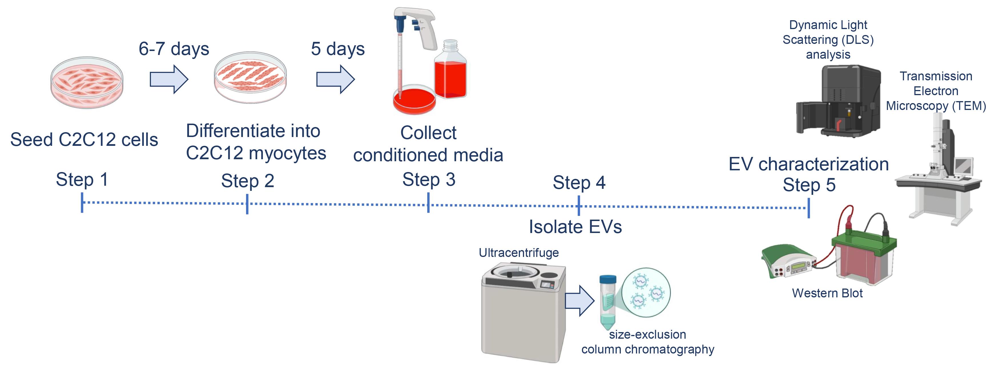
Graphical overview
Background
Extracellular vesicles (EVs) are nanosized, lipid bilayer–enclosed particles (30–150 nm) secreted by various cell types, including immune cells, neurons, adipocytes, and muscle cells [1]. They are present in body fluids such as plasma, urine, cerebrospinal fluid, breast milk, and saliva. EV composition and cargo change dynamically in response to cellular and physiological conditions, making them promising biomarkers and therapeutic tools in disease and regenerative medicine [2,3].
Skeletal muscle accounts for about 40%–50% of total body mass and plays a central role in movement, energy metabolism, and systemic homeostasis [4–8]. It secretes signaling molecules, including myokines and EVs, which regulate lipid and glucose metabolism, myogenesis, lipogenesis, angiogenesis, and inflammation [9,10]. The murine C2C12 skeletal muscle cell line, established by Yaffe and Saxel [11], is a well-characterized in vitro model for studying muscle biology and the muscle secretome [12]. Upon differentiation, C2C12 myoblasts form multinucleated myotubes resembling mature skeletal muscle fibers, providing a robust system to investigate EV biogenesis and function [12].
Isolation of EVs from intact muscle or explant cultures is technically challenging due to mixed cell populations and the lack of markers to determine vesicle origin. To address this, we developed a standardized protocol to isolate EVs from C2C12 myocytes. Conditioned media are collected and subjected to sequential ultracentrifugation followed by size-exclusion chromatography [13,14]. The resulting myocyte-derived EVs can be validated using western blotting, transmission electron microscopy (TEM), and dynamic light scattering (DLS) analysis. This protocol enables the isolation and characterization of muscle-derived EVs with high purity, reducing contamination and avoiding misinterpretation that can arise from isolating EVs directly from heterogeneous muscle tissues.
Materials and reagents
Biological materials
1. C2C12 cells (ATCC, CRL-1772), obtained from Prof. David Virshup (CSCB Department, Duke-NUS Medical School, Singapore)
Reagents
1. PierceTM BCA Protein Assay kit (Thermo Scientific, catalog number: 23225)
2. 250 kDa Plus prestained protein marker (Vazyme, catalog number: MP202)
3. ECLTM Prime western blotting detection reagents (Cytiva, catalog number: RPN2209)
4. Horse serum, exosome-depleted (Gibco, custom-made)
5. Dulbecco’s modified Eagle medium (DMEM) (Cytiva, catalog number: SH30022.01)
6. Fetal bovine serum (FBS), exosome-depleted (Sigma, catalog number: A2720801)
7. 100 U/mL penicillin and 100 μg/mL streptomycin (Cytiva, catalog number: SV30010)
8. Centrifuge tubes, 50 mL (Biobasic Asia Pacific, catalog number: BCT0050)
9. Centrifuge tubes, 15 mL (Biobasic Asia Pacific, catalog number: BCT0015)
10. Corning® 96-well clear flat bottom polystyrene TC-treated microplates, individually wrapped, with lid, sterile (Corning, catalog number: 3596)
11. T175 cell culture flask (SPL Life Sciences, catalog number: 74175)
12. T75 Cellstar® cell culture flask (Greiner, catalog number: 658175)
13. SmartSECTM single column (System Biosciences, catalog number: SSEC200A-1)
14. LabSelect 1.5 mL microcentrifuge tube (LabSelect, catalog number: MCT-001-150)
15. Rabbit monoclonal anti-Alix antibody (Cell Signaling Technology, catalog number: 92880; 1:1,000)
16. Mouse monoclonal anti-Tsg101 antibody (BD Biosciences, catalog number: 612696; 1:500)
17. Mouse monoclonal anti-GM130 antibody (Proteintech, catalog number: 66662; 1:10,000)
18. Goat anti-rabbit IgG, HRP-linked antibody (Cell Signaling Technology, catalog number: 7074; 1:1,000)
19. Goat anti-mouse IgG, HRP-linked antibody (Cell Signaling Technology, catalog number: 7076; 1:1,000)
20. 1× Dulbecco’s phosphate-buffered saline (D-PBS), without Ca and Mg (1ST Base, catalog number: BUF-2046)
21. Trypsin-EDTA solution, with phenol red (Biobasic Asia Pacific, catalog number: UCC8105)
22. Bovine serum albumin (BSA) (Sigma-Aldrich, catalog number: A3059)
23. 20× tris buffered saline (TBS) buffer (1st Base, catalog number: BUF-3030)
24. Methanol, for analysis EMSURE® ACS, ISO, Reag. Ph Eur (Supelco, catalog number: 1.06009)
25. 2× RIPA buffer I (pH 7.4) (BioBasic Asia Pacific, catalog number: RB4475)
26. Protease inhibitor cocktail (200×) (Cell Signaling Technology, catalog number: 7012)
27. Polyoxyethylene-20 (Tween 20) (Biobasic Asia Pacific, catalog number: TB0560)
28. Amersham Hybond P 0.45 PVDF blotting membrane (Cytiva, catalog number: 10600023)
29. Formvar/carbon-supported copper grids (Merck, catalog number: 930288)
30. Phosphotungstic acid, crystal, reagent, highest purity (Electron Microscopy Sciences, catalog number: 19500)
31. VitrobotTM filter paper, grade 595, 50 mm (Electron Microscopy Sciences, catalog number: 71166-65)
32. Millipore® Stericup® quick-release vacuum filtration system (Millipore, catalog number: S2GPU05RE)
33. Cuvette, 4.2 mL, 45 × 12 mm (H × W), PS, transparent, 4 optical sides (Sarstedt, catalog number: 67.754)
34. 10× tris glycine-sodium dodecyl sulfate (TG-SDS) buffer (1st Base, catalog number: BUF-2030)
35. Bromophenol blue (Sigma-Aldrich, catalog number: B0126)
36. Ammonium persulfate (APS) (Bio-Rad, catalog number: 1610700)
37. 30% acrylamide/bis solution 37.5:1 (Bio-Rad, catalog number: 1610158)
38. 1.0 M tris buffer, pH 6.8 (1st Base, catalog number: BUF-1415)
39. 1.5 M tris buffer, pH 8.8 (1st Base, catalog number: BUF-1419)
40. 10% SDS solution (BioBasic Asia Pacific, catalog number: SD8118)
41. TEMED (Bio-Rad, catalog number: 161-0800)
42. Glycerol (Biobasic Asia Pacific, catalog number: GB0232)
43. Sodium dodecyl sulfate (SDS) (Wako, catalog number: 194-13985)
44. Trypan blue stain, 0.4% (Gibco, catalog number: 15250-061)
45. Bright-LineTM hemacytometer (Merck, catalog number: Z359629)
Solutions
1. 10% resolving gel (see Recipes)
2. 4% stacking gel (see Recipes)
3. Growth media (see Recipes)
4. Differentiation media (see Recipes)
5. Electrophoresis running buffer (see Recipes)
6. Electrophoresis transfer buffer (see Recipes)
7. Electrophoresis wash buffer (see Recipes)
8. Sample buffer (see Recipes)
9. BCA working solution (see Recipes)
10. BCA standard curve preparation (see Recipes)
Recipes
1. 10% resolving gel
| Component | Concentration | Volume |
|---|---|---|
| Water | n/a | 4.05 mL |
| Tris buffer, pH 8.8 | 1.5 M | 2.5 mL |
| 30% acrylamide/bis solution | 30% | 3.3 mL |
| SDS Solution | 10% | 100 μL |
| APS | 10% | 50 μL |
| TEMED | 5 μL |
2. 4% stacking gel
| Component | Concentration | Volume |
|---|---|---|
| Water | n/a | 3.625 mL |
| Tris buffer, pH 6.8 | 1.0 M | 0.625 mL |
| 30% acrylamide/bis solution | 30% | 0.650 mL |
| SDS | 10% | 50 μL |
| APS | 10% | 50 μL |
| TEMED | 5 μL |
3. Growth media
| Component | Concentration | Volume |
|---|---|---|
| DMEM | n/a | 445 mL |
| Exosome-depleted FBS | 10% | 50 mL |
| Penicillin/Streptomycin | 1% | 5 mL |
Note: Preparation of exosome-depleted FBS from standard serum requires ultracentrifugation at 100,000–120,000× g for 16–18 h at 4 °C.
4. Differentiation media
| Component | Concentration | Volume |
|---|---|---|
| DMEM | n/a | 485 mL |
| Exosome-depleted horse serum | 2% | 10 mL |
| Penicillin/Streptomycin | 1% | 5 mL |
Note: Preparation of exosome-depleted horse serum (HS) from standard serum requires ultracentrifugation at 100,000–120,000× g for 16–18 h at 4 °C.
5. Electrophoresis running buffer (2 L)
| Component | Volume |
|---|---|
| MilliQ water | 1.8 L |
| 10× TG-SDS buffer | 200 mL |
6. Electrophoresis transfer buffer (2 L)
| Component | Volume |
|---|---|
| MilliQ water | 1.4 L |
| 10× TG-SDS buffer | 200 mL |
| Methanol | 400 mL |
7. TBST wash buffer (1 L)
| Component | Volume |
|---|---|
| MilliQ water | 1.4 L |
| 20× TBS buffer | 500 mL |
| Tween 20 | 1 mL |
8. Sample buffer
| Component | Concentration | Volume |
|---|---|---|
| Tris buffer, pH 6.8 | 1.0 M | 6.25 mL |
| Glycerol | n/a | 25 mL |
| SDS | n/a | 2 g |
| Bromophenol blue | 0.25% | 4 mL |
| MilliQ water | n/a | 14.75 mL |
Aliquot into a 1.5 mL tube and store at -20 °C until use.
9. BCA working solution
| Reagent | Amount |
|---|---|
| Part A | 25 mL |
| Part B | 500 μL |
Note: Part A and Part B are included in the PierceTM BCA Protein Assay kit.
10. BCA standard curve preparation
| Volume of RIPA (μL) | Volume and source of BSA (μL) | Final BSA concentration (μg/mL) | |
|---|---|---|---|
| A | 0 | 60 of stock | 2,000 |
| B | 30 | 30 of stock | 1,000 |
| C | 30 | 30 of vial B dilution | 500 |
| D | 30 | 30 of vial C dilution | 250 |
| E | 30 | 30 of vial D dilution | 125 |
| F | 48 | 12 of vial E dilution | 25 |
| G | 48 | 0 | Blank |
Equipment
1. OptimaTM L-100 XP ultracentrifuge (Beckman Coulter, model: OPTIMA L-100)
2. SW 28 Ti swinging-bucket rotor with buckets (Beckman Coulter, model: SW 28)
3. Zetasizer Nano ZS (Malvern Panalytical, UK; discontinued; current replacement model: Zetasizer Ultra)
4. Allegra X-30 benchtop centrifuge (Beckman Coulter, model: Allegra X-30)
5. iBrightTM CL1500 imaging system (Invitrogen, model: CL1500)
6. HT7800 RuliTEM (Hitachi, Japan)
7. Mini-PROTEAN Tetra Cell (Bio-Rad, catalog number: 165-8033)
8. FormaTM Steri-CycleTM i160 dual CO2 incubator (Thermo Scientific, catalog number: 50163013)
9. Labculture® Class II Type A2 biological safety cabinet (ESCO, catalog number: LA2-4A1)
10. Sub Aqua Pro unstirred water bath (Grant, catalog number: SAP12)
11. Bright-LineTM hemacytometer (Merck, catalog number: Z359629)
12. Nikon Eclipse TS100 inverted microscope (Nikon, model: TS100)
13. Tecan Infinite M200 microplate reader (Tecan, model: M200)
Software and datasets
1. ZS explorer software (Malvern Panalytical, v4.0.0)
2. I-controlTM software (Tecan)
3. iBright Analysis Software (ThermoFisher Scientific, v5.3.0)
Procedure
A. Thawing and seeding C2C12 cells
1. Prewarm growth media (see Recipe 3) to 37 °C.
2. Prepare a 15 mL conical tube containing fresh growth media.
3. Rapidly thaw 1 mL aliquot of C2C12 cells in a 37 °C water bath.
4. Transfer the thawed cells into the conical tube containing prewarmed growth medium.
5. Mix gently by inverting the tube.
6. Centrifuge at 200× g for 5 min at room temperature to pellet the cells.
7. Remove the supernatant and resuspend the cell pellet in 5 mL of fresh growth medium.
8. Count the cells using the trypan blue exclusion method and seed 2.25 × 10⁵ cells into each T75 flask.
9. Incubate at 37 °C in a humidified incubator with 5% CO2 for 3 days.
Note: No media change is required during this period.
B. Passaging and scaling up C2C12 cells
1. On day 3, passage the cells when 70%–80% confluent.
2. Remove culture media and wash cells with 5 mL of sterile PBS.
3. Remove PBS and add 3 mL of trypsin-EDTA.
4. Incubate at 37 °C for 2 min and monitor under a microscope to make sure all cells detach.
5. Add 7 mL of prewarmed growth medium to neutralize trypsin and resuspend cells.
6. Transfer the cell suspension to a 15 mL conical tube.
7. Centrifuge at 200× g for 5 min to pellet the cells.
8. Remove supernatant and resuspend the cell pellet in 5 mL of growth medium.
9. Mix 50 μL of cell suspension with 50 μL of trypan blue.
10. Count viable cells using a hemacytometer and calculate the total cell number.
Note: Ensure that cell viability is high before proceeding.
11. Seed 1.75 × 10⁶ cells into each of two T175 flasks, resuspending in 30 mL of growth medium per flask.
12. Incubate at 37 °C humidified incubator with 5% CO2 for 3–4 days.
Note: No media change is required during this period.
13. Observe under a microscope to confirm confluency.
C. Differentiation of C2C12 myoblasts into myocytes and collection of conditioned media
1. When cells reach 80%–90% confluency (days 3–4), remove growth media and replace with differentiation media (see Recipe 4).
2. Incubate in a 37 °C humidified incubator with 5% CO2 for 3 days.
3. Replace with fresh differentiation medium and continue incubation for another 3 days.
4. Monitor morphology under a microscope. Myotube formation typically begins from day 5 (Figure 1).
5. Incubate in a 37 °C humidified incubator with 5% CO2 for 3 days.
6. On day 8, collect conditioned medium for EV isolation.
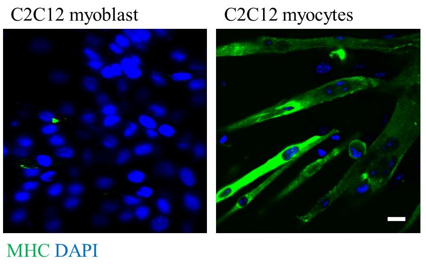
Figure 1. C2C12 myoblast (left panel) and C2C12 myocytes (right panel). Scale bar: 20 μm.
D. Isolation of EVs from C2C12 myocytes
1. Filter the conditioned medium through a 0.22-μm filter to remove large particles.
2. Performed sequential centrifugation steps as follows:
a. 200× g for 10 min at 4 °C to remove residual cells.
b. 2,000× g for 20 min at 4 °C to remove debris and apoptotic bodies.
c. 10,000× g for 2 h at 4 °C to remove large vesicles and remaining debris.
3. Wash the pellet once with PBS and ultracentrifuge at 100,000× g for 70 min at 4 °C to collect small EVs.
4. Resuspend the EV pellet in 500 μL of PBS.
5. Load the resuspended EVs onto a SmartSECTM single column.
6. Incubate the column at room temperature for 20–30 min while spinning gently to allow EV binding.
7. Loosen the cap slightly before removing the bottom closure.
8. Place the column into a 1.5-mL centrifuge tube and centrifuge at 500× g for 30 s to elute purified EVs.
9. The purified EVs are now ready for quantification and downstream characterization.
E. EV characterization by western blot
1. Determine protein concentration using PierceTM BCA Protein Assay kit:
a. Prepare the BSA standard curve as described in Recipe 10.
b. Resuspend the purified EV pellet in 100 μL of 1× RIPA lysis buffer supplemented with protease inhibitors.
c. Vortex briefly and incubate on ice for 30 min with intermittent mixing.
d. Centrifuge at 14,000× g for 30 min at 4 °C and transfer the supernatant to a new microcentrifuge tube.
e. Pipette 12.5 μL of each BSA standard and diluted EV sample in duplicate into a 96-well plate.
f. Add 100 μL of BCA working solution (see Recipe 9) to each well (1:8 sample to working reagent ratio for microplate procedure).
g. Incubate the plate at 37 °C for 30 min.
h. Measure absorbance at 562 nm using a microplate reader.
i. Calculate protein concentration of EV samples based on the BSA standard curve (Figure 2).
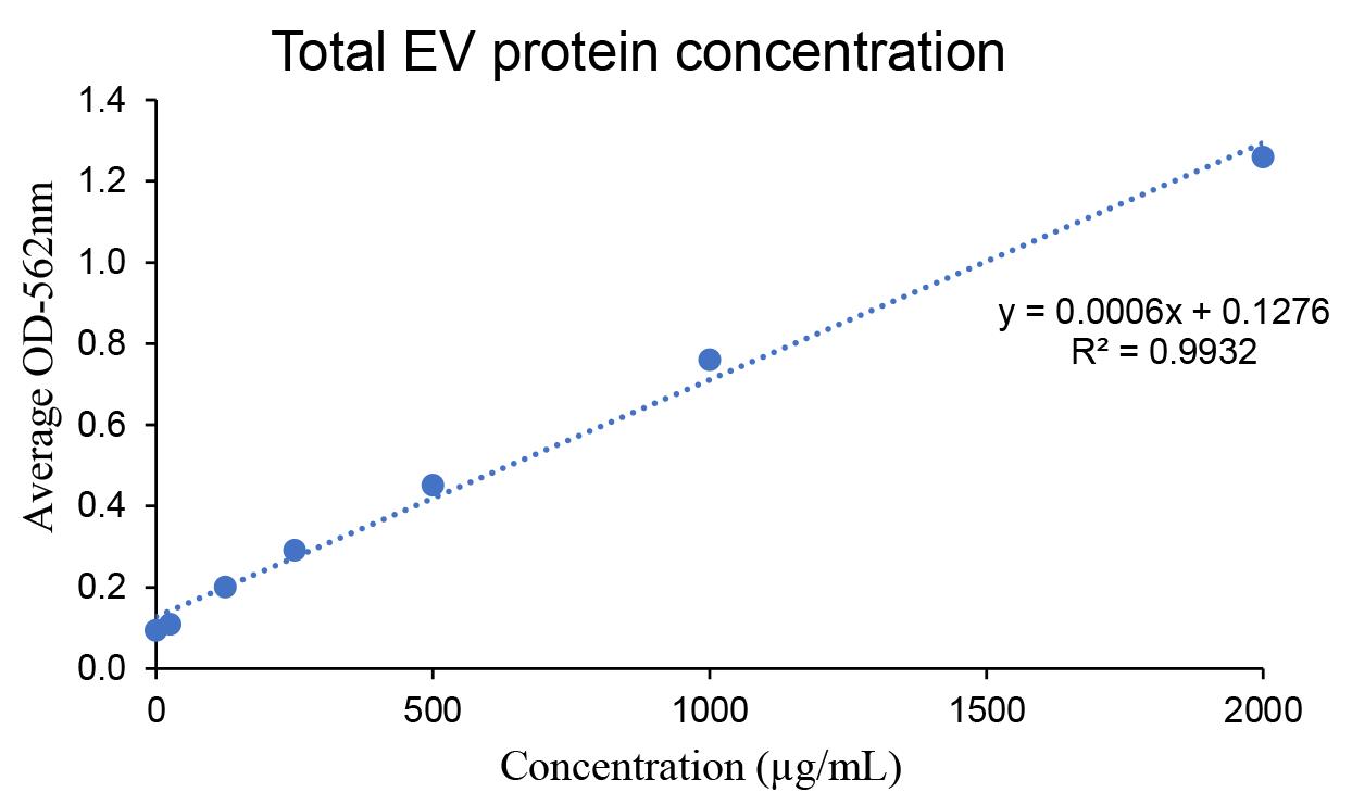
Figure 2. BCA standard curve for protein quantification. The standard curve was prepared using serial dilutions of BSA (see Recipe 10). Protein concentrations of C2C12 myocyte-derived extracellular vesicles (EVs) were calculated based on the linear equation derived from the standard curve. Values represent the mean of two technical replicates.
2. Mix EV lysates with sample buffer (see Recipe 8) and heat at 75 °C for 5 min to denature proteins.
3. Cast the SDS-PAGE gel with a 10% resolving gel (see Recipe 1) at the bottom and a 4% stacking gel (see Recipe 2) on top.
3. Load 10 μg of protein onto the SDS-PAGE gel and perform electrophoresis.
4. Run the gel at 110 V for 1.5 h at room temperature with electrophoresis running buffer (see Recipe 5).
5. Transfer proteins to a PVDF membrane at 100 V and 4 °C for 1 h with electrophoresis transfer buffer (see Recipe 6).
6. Wash the membrane two times with TBST wash buffer (see Recipe 7).
6. Block the membrane in 5% BSA in TBST wash buffer for 1 h at room temperature.
7. Incubate the membrane overnight at 4 °C with primary antibodies against EV markers (e.g., ALIX, TSG101) and non-EV markers (e.g., GM130).
8. Wash the membrane three times with TBST wash buffer (5 min each), then incubate with HRP-conjugate secondary antibodies for 1 h at room temperature.
9. Wash the membrane three times with TBST wash buffer (5 min each).
10. Incubate the membrane with ECLTM Prime western blotting detection reagents for 1–2 min.
11. Acquire western blot images using the iBrightTM CL1500 imaging system (Figure 3).
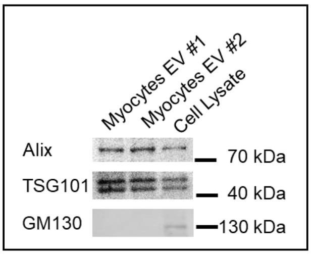
Figure 3. Isolated extracellular vesicle (EV) proteins are characterized by western blot analysis. Alix and TSG101 were used as positive EV markers, and GM130 was used as a negative control to confirm the purity of EVs.
F. Size determination of EVs by dynamic light scattering (DLS) analysis
1. Turn on the Zetasizer Nano ZS machine and launch the ZS explorer software.
2. Resuspend the purified EV pellet in 100 μL of PBS and transfer to a cuvette. Avoid air bubbles during pipetting.
3. Prepare a blank control by filling another cuvette with 100 μL of PBS and place it in the holder.
4. Close the chamber lid and allow the system to stabilize for 3 min.
5. In the software, create a new measurement file.
6. On the Measure page:
a. Enter a descriptive name for the sample.
b. Select the DTS0012 cuvette.
c. Set material type to polystrene latex.
d. Set dispersant type to PBS.
e. Choose measurement type as “Size.”
7. Click Start to begin the blank measurement [this establishes the dispersant scattering mean count rate (kcps)].
8. Replace the PBS blank with the EV sample cuvette.
9. Choose measurement type: Size and Cumulative particle concentration.
10. Click Start to measure the EV sample.
11. Once the run is complete, go to the Analyze page to view the size distribution and record particle size and cumulative particle concentration (Figure 4).
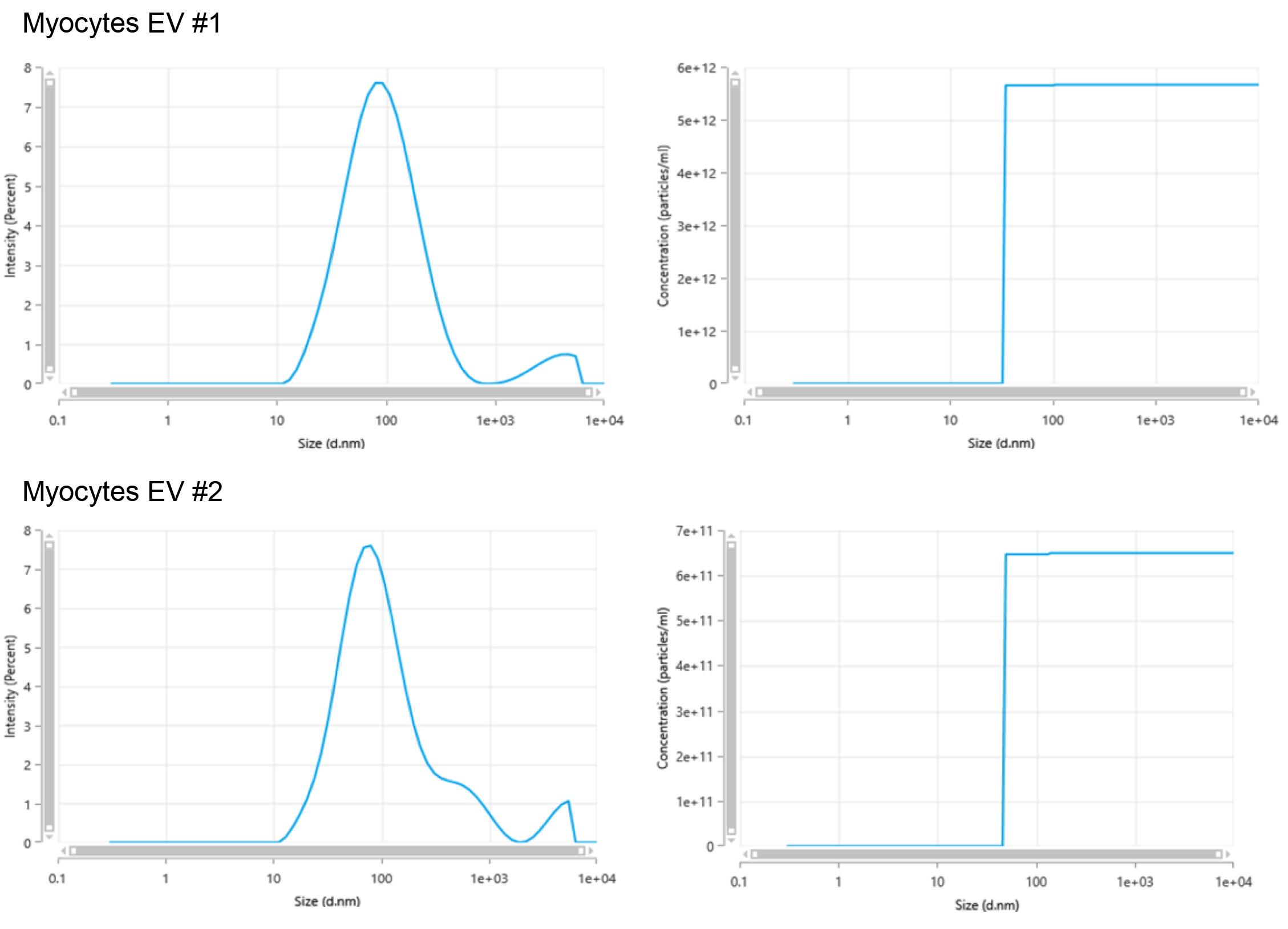
Figure 4. Dynamic light scattering (DLS) analysis characterization of C2C12 myocyte-derived extracellular vesicles (EVs). EVs exhibit a size peak at 79.88 nm in diameter. The cumulative particle concentrations were quantified at 5.673 × 10¹² and 6.497 × 10¹¹ particles/mL.
H. Visualization of EVs with transmission electron microscopy (TEM)
1. Place 10 μL of purified EV suspension onto a formvar-carbon-coated copper grid and incubate for 2 min at room temperature to allow adsorption.
2. Gently remove excess liquid using filter paper.
3. Stain the grid with 3% (v/v) phosphotungstic acid (PTA), pH 7.0 (adjusted with 1 M sodium hydroxide), prepared in distilled water, for 1 min at room temperature.
4. Remove excess stain using filter paper.
5. Air-dry the grid for 15 min at room temperature.
6. Observe the samples using a Hitachi HT7800 Ruli Transmission Electron Microscope operated at 80 kV.
7. Acquire representative images from three randomly selected fields of view at a magnification of 40,000× (Figure 5).
Note: Phosphotungstic acid provides negative staining that enhances the contrast of EV membranes, enabling visualization of their morphology and size.
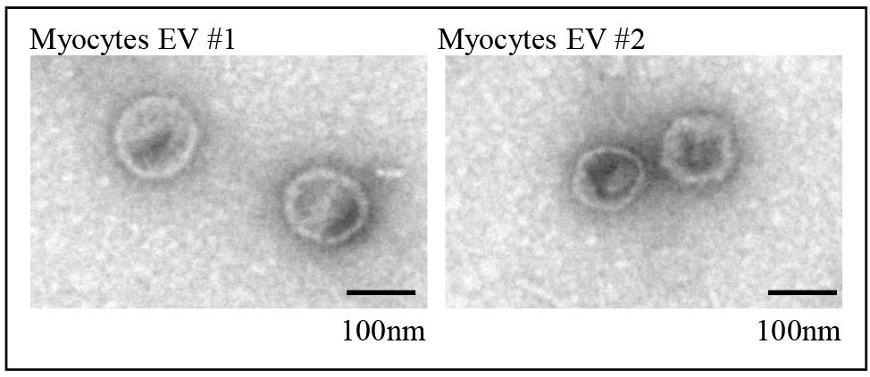
Figure 5. Transmission electron microscopy (TEM) images show the morphology of extracellular vesicles (EVs) isolated from C2C12 myocytes. The figures show isolated EVs from C2C12 myocytes #1 and #2 in the size range of 90–100 nm. Magnification: 40,000×; scale bar: 100 nm.
Data analysis
1. BCA analysis: The total protein concentrations of extracellular vesicles isolated from C2C12 myocytes were 134.43 μg/mL and 83.83 μg/mL, as summarized in Table 1.
Table 1. BCA analysis
| Sample | OD1 | OD2 | Average OD | Concentration (μg/mL) |
|---|---|---|---|---|
| Std A | 1.297 | 1.221 | 1.259 | 2,000 |
| Std B | 0.761 | 0.760 | 0.7606 | 1,000 |
| Std C | 0.446 | 0.458 | 0.452 | 500 |
| Std D | 0.292 | 0.292 | 0.292 | 250 |
| Std E | 0.202 | 0.198 | 0.200 | 125 |
| Std F | 0.110 | 0.109 | 0.109 | 25 |
| Std G | 0.097 | 0.092 | 0.094 | 0 |
| Myocyte-derived EV #1 | 0.2086 | 0.2078 | 0.2082 | 134.43 |
| Myocyte-derived EV #2 | 0.1845 | 0.1713 | 0.1779 | 83.83 |
2. Western blot: EV-specific markers (Alix and TSG101) were detected on myocyte-derived EV #1 and #2, while the non-EV-specific marker (GM130) was absent, as shown in Figure 3.
3. DLS analysis: The size distribution of both myocyte-derived EV #1 and #2 showed a peak diameter of 79.88 nm. The cumulative particle concentrations were at 5.673 × 10¹² particles/mL for myocyte EV #1 and 6.497 × 10¹¹ particles/mL for myocyte EV #2.
4. TEM analysis: Both myocytes EV #1 and #2 exhibited similar morphology, with vesicle diameter around 100 nm.
Validation of protocol
The EVs isolated from C2C12 myocytes meet the Minimal Information for Studies of Extracellular Vesicles (MISEV) guidelines. Specifically, the EV population was validated by demonstrating (A) the presence of EV-positive markers (ALIX and TSG101), (B) appropriate size distribution and cumulative particle concentration, and (C) EV morphology.
This protocol has been used and validated in the following research article:
1. Characterization and use of C2C12 myocytes-derived EVs (Figures 2 and 3): Goh et al. (2026). Sarcopenia promotes tumorigenesis by disrupting NOTCH-SDC2-regulated biogenesis of muscle-derived extracellular vesicles. Nature Communications. https://doi.org/10.1038/s41467-026-72410-y [15].
2. Successful differentiation of C2C12 progenitor cells into myocytes (Figure 3A, F): Choy et al. (2025). Exercise suppresses DEAF1 to normalize mTORC1 activity and reverse muscle aging, Proc. Natl. Acad. Sci. U.S.A. 122(48): e2508893122, https://doi.org/10.1073/pnas.2508893122 [6].
General notes and troubleshooting
General notes
1. This protocol can be adapted for C2C12 myoblasts or myocytes subjected to drug treatments or genetic modification.
2. It is crucial to perform column chromatography in order to obtain a highly purified EV population.
Troubleshooting
| Problem | Possible cause | Solution |
|---|---|---|
| 1. Low EV protein concentration | 1. The EV pellet was not fully resuspended. 2. An insufficient volume of conditioned media was used for EV isolation. | 1. Incubate the EV pellet in PBS at 37 °C for 30 min to ensure complete resuspension. 2. Increase EV yield by collecting and pooling extra batches of conditioned media. |
| 2. Multiple peaks across a broad size distribution were detected in DLS analysis | 1. Contamination with microvesicles, apoptotic bodies, or debris. (Note: Some microvesicles fall within the 100–200 nm range, which can partially overlap with the EV of interest). | Perform differential centrifugation, pre-filter the supernatant, and follow with column chromatography to purify the EV population. |
Acknowledgments
Conceptualization, K.Y.G.; Investigation, K.Y.G., W.X.L.; Writing—Original Draft, K.Y.G.; Writing—Review & Editing, H.-W.T.; Funding acquisition, K.Y.G, H.-W.T.; Supervision, H.-W.T. This work was supported by Singapore’s Ministry of Education AcRF Award (2022-MOET1-0004 and FY2025-MOET1-0004 to H.-W.T.), Diana Koh Innovative Cancer Research Award (Duke-NUS-DKICRA/2024/0001 to H.-W.T.), National Academy of Medicine grant (MOH-001189-00 to H.-W.T.), and National Medical Research Council (NMRC) (MOH-001208-00 and MOH-001885-00 to H.-W.T.; MOH-001831-00 to K.Y.G.).
This protocol was used in [6,15].
Graphical overview was partially created using BioRender.
Competing interests
The authors declare no conflict of interest.
Ethical considerations
All animal procedures described in this protocol were conducted in accordance with institutional ethical guidelines and were approved by the Institutional Animal Care and Use Committee (IACUC) (IACUC Protocol No. 2025/SHS/1974).
References
- Welsh, J. A., Goberdhan, D. C. I., O'Driscoll, L., Buzas, E. I., Blenkiron, C., Bussolati, B., Cai, H., Di Vizio, D., Driedonks, T. A. P., Erdbrugger, U., et al. (2024). Minimal information for studies of extracellular vesicles (MISEV2023): From basic to advanced approaches. J Extracell Vesicles. 13(2): e12404. https://doi.org/10.1002/jev2.12404
- Boukouris, S. and Mathivanan, S. (2015). Exosomes in bodily fluids are a highly stable resource of disease biomarkers. Proteomics Clin Appl. 9(3–4): 358–367. https://doi.org/10.1002/prca.201400114
- Muthu, S., Bapat, A., Jain, R., Jeyaraman, N. and Jeyaraman, M. (2021). Exosomal therapy-a new frontier in regenerative medicine. Stem Cell Investig 8: 7. https://doi.org/10.21037/sci-2020-037
- Han, X., Goh, K. Y., Lee, W. X., Choy, S. M. and Tang, H. W. (2022). The Importance of mTORC1-Autophagy Axis for Skeletal Muscle Diseases. Int J Mol Sci. 24(1): 297. https://doi.org/10.3390/ijms24010297
- Gopal Krishnan, P. D., Lee, W. X., Goh, K. Y., Choy, S. M., Turqueza, L. R. R., Lim, Z. H. and Tang, H. W. (2025). Transcriptional regulation of autophagy in skeletal muscle stem cells. Dis Model Mech. 18(2): e052007. https://doi.org/10.1242/dmm.052007
- Choy, S. M., Goh, K. Y., Lee, W. X., Jiang, W., Gou, Q., Gopal Krishnan, P. D., Chee Ong, S., Chua, K., Harmston, N. and Tang, H. W. (2025). Exercise suppresses DEAF1 to normalize mTORC1 activity and reverse muscle aging. Proc Natl Acad Sci USA. 122(48): e2508893122. https://doi.org/10.1073/pnas.2508893122
- Goh, K. Y., Lee, W. X., Choy, S. M., Priyadarshini, G. K., Chua, K., Tan, Q. H., Low, S. Y., Chin, H. S., Wong, C. S., Huang, S. Y., et al. (2024). FOXO-regulated DEAF1 controls muscle regeneration through autophagy. Autophagy. 20(12): 2632–2654. https://doi.org/10.1080/15548627.2024.2374693
- Lee, W. X., Goh, K. Y., Choy, S. M. and Tang, H.-W. (2025). DEAF1 - a transcriptional brake on muscle autophagy. Autophagy. 1–2. https://doi.org/10.1080/15548627.2025.2610451
- Iglesias, P. (2025). Muscle in Endocrinology: From Skeletal Muscle Hormone Regulation to Myokine Secretion and Its Implications in Endocrine–Metabolic Diseases. J Clin Med. 14(13): 4490. https://doi.org/10.3390/jcm14134490
- Severinsen, M. C. K. and Pedersen, B. K. (2020). Muscle-Organ Crosstalk: The Emerging Roles of Myokines. Endocr Rev. 41(4): 594–609. https://doi.org/10.1210/endrev/bnaa016
- Yaffe, D. and Saxel, O. (1977). Serial passaging and differentiation of myogenic cells isolated from dystrophic mouse muscle. Nature. 270(5639): 725–727. https://doi.org/10.1038/270725a0
- Lautaoja-Kivipelto, J. H., Karvinen, S., Korhonen, T. M., O'Connell, T. M., Tiirola, M., Hulmi, J. J. and Pekkala, S. (2024). Interaction of the C2C12 myotube contractions and glucose availability on transcriptome and extracellular vesicle microRNAs. Am J Physiol Cell Physiol. 326(2): C348–C361. https://doi.org/10.1152/ajpcell.00401.2023
- Corso, G., Mager, I., Lee, Y., Gorgens, A., Bultema, J., Giebel, B., Wood, M. J. A., Nordin, J. Z. and Andaloussi, S. E. (2017). Reproducible and scalable purification of extracellular vesicles using combined bind-elute and size exclusion chromatography. Sci Rep. 7(1): 11561. https://doi.org/10.1038/s41598-017-10646-x
- Guescini, M., Maggio, S., Ceccaroli, P., Battistelli, M., Annibalini, G., Piccoli, G., Sestili, P. and Stocchi, V. (2017). Extracellular Vesicles Released by Oxidatively Injured or Intact C2C12 Myotubes Promote Distinct Responses Converging toward Myogenesis. Int J Mol Sci. 18(11): 2488. https://doi.org/10.3390/ijms18112488
- Goh, K. Y., Lee, W. X., Gou, Q., Choy, S. M., Ong, S.C., Gopal Krishnan. P. D., Wang, H. X., Turqueza, L. R. R., Tan, Q. H., Chua, K., et al. (2026). Sarcopenia promotes tumorigenesis by disrupting NOTCH-SDC2-regulated biogenesis of muscle-derived extracellular vesicles. Nat Commun. https://doi.org/10.1038/s41467-026-72410-y
Article Information
Publication history
Received: Nov 27, 2025
Accepted: Feb 4, 2026
Available online: Apr 30, 2026
Published: May 20, 2026
Copyright
© 2026 The Author(s); This is an open access article under the CC BY-NC license (https://creativecommons.org/licenses/by-nc/4.0/).
How to cite
Goh, K. Y., Lee, W. X. and Tang, H. W. (2026). Isolation and Biophysical Characterization of Extracellular Vesicles Released by Myocytes. Bio-protocol 16(10): e5698. DOI: 10.21769/BioProtoc.5698.

 Download Citation in RIS Format
Category
Cell Biology > Organelle isolation > Extracellular vesicle
Biophysics > Scattering spectroscopy
Cell Biology > Cell imaging > Electron microscopy
Do you have any questions about this protocol?
Post your question to gather feedback from the community. We will also invite the authors of this article to respond.
 Post a Question


