


- Protocols

- Articles and Issues
- For Authors
- About
- Become a Reviewer


Generation of Functional Patient-Specific Thymus Organoids From Human Pluripotent Stem Cells (hPSCs) Using Air–Liquid Interface Culture

Published: Vol 16, Iss 9, May 5, 2026 DOI: 10.21769/BioProtoc.5684 Views: 1145
Reviewed by: Anonymous reviewer(s)
 PDF
PDF  Ask a question
Original research article
The authors used this protocol in:
Apr 2023
Advertisement



Protocol Collections
Comprehensive collections of detailed, peer-reviewed protocols focusing on specific topics




Related protocols
Abstract
The thymus is critical for the establishment of a functional and self-tolerant adaptive immune system, but it involutes with age, resulting in reduced naive T-cell output. Generation of a functional human thymus from human pluripotent stem cells (hPSCs) is an attractive regenerative medicine strategy. Direct differentiation of thymic epithelial progenitors (TEPs) from hPSCs has been demonstrated in vitro, but functional thymic epithelial cells (TECs) develop only after transplantation of TEPs in vivo. Functional human reaggregated thymic organoid cultures (RTOCs) and artificial thymic organoids (ATOs) cultured at the air–liquid interface support T-cell development in vitro and in vivo and permit the interrogation of human thymic function and T-cell development. However, these approaches require access to primary human tissues or murine bone marrow stromal cells, are allogeneic, and do not support negative selection. Recently, we reported the directed differentiation of induced PSCs (iPSCs) to functional thymic epithelial progenitors (TEPs) that support murine T-cell development after transplantation in nude mice. Here, we combined hPSC-derived TEPs, hematopoietic progenitor cells (HPCs), and mesenchymal cells, differentiated from the same hPSC line, and generated functional isogenic stem cell–derived thymic organoids (sTOs). Our revised protocol improves our TEP differentiation process and allows the generation of functional isogenic, patient-specific thymic organoids in vitro.
Key features
• This protocol offers a reproducible approach to generate functional multicellular stem cell–derived thymic organoids (sTOs).
• sTOs support human thymic epithelial cell development and maturation in vitro in a patient-specific manner.
• sTOs support human T-cell development from stem cell–derived hematopoietic progenitor cells.
• sTOs may facilitate positive and negative thymic selection of developing T cells in vitro.
Keywords: Stem cell–derived thymic organoids Thymic epithelial cells
Human pluripotent stem cells
Human T-cell development
Positive and negative selection
Cell therapy
Thymic epithelial cells
Human pluripotent stem cells
Human T-cell development
Positive and negative selection
Cell therapy
Graphical overview
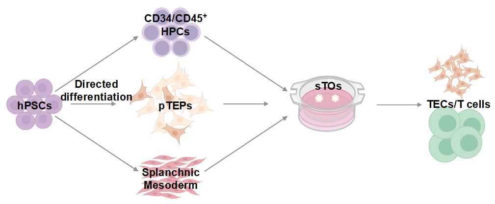
Generation of functional thymic organoids from hPSCs. Human pluripotent stem cells (hPSCs), hematopoietic progenitor cells (HPCs), patient-specific thymic epithelial progenitors (pTEPs), stem cell–derived thymic organoids (sTOs), thymic epithelial cells (TECs).
Background
The thymus is required for the development of a functional adaptive immune system, facilitating the generation of self-tolerant T cells that can respond to foreign antigens. Thymic epithelial cells (TECs) are divided into cortical and medullary (c/m) TECs, based on their location and function, and can be identified by the expression of KRTs 8 and 5, respectively. c- and mTECs originate from common bipotent progenitors, which first express KRT8 only and then co-express both keratins prior to bifurcating into c- and mTEC lineages [4]. Age-related involution of the thymus results in decreased thymic function and naive T-cell output and increased autoimmunity and disease risk [5,6]. Thymic dysfunction or absence can result from intrinsic or extrinsic factors, namely DiGeorge syndrome (DGS), FOXN1 deficiency, infection, or radiation therapy [7,8]. Complete athymia can be treated with an allogeneic thymus transplant; however, such treatment is associated with various complications and relies on limited donor tissue [8,9]. Thus, an experimental model system to interrogate the mechanisms of thymic insufficiency and function is necessary and could serve to further the development of cell-based treatments for thymic defects.
Thymic organoids cultured at the air–liquid interface allow the interrogation of thymic function and T-cell development [10–15]. Functional human reaggregated thymic organoid cultures (RTOCs) made with expanded primary (1°) TECs and thymic mesenchyme (TM) combined with allogeneic cord blood-derived hematopoietic stem cells (HSCs) support T-cell development in vitro and in vivo [16]. However, RTOCs require access to 1° tissue, utilize allogeneic components, and do not support negative selection.
TECs express Notch ligands, such as DLL4, which are critical for T-cell commitment and differentiation. In vitro, transgenic OP9 or MS5 murine bone marrow stromal cells expressing human DLL1 or DLL4 support positive selection of developing murine and human T-cell progenitors [17]. Combining primary HSCs [18] or human pluripotent stem cell (hPSC)-derived embryonic mesodermal progenitors (EMPs) [19] with MS5-DLL1/DLL4 cells, the Crooks group elegantly demonstrated T-cell development in artificial thymic organoids (ATOs) in vitro. Though OP9-DLL1 and ATO cultures eliminate the need for primary tissue, the lack of thymic stromal cells and class II major histocompatibility complex/human leukocyte antigen (MHC/HLA) expression on supporting stromal cells results in a skew toward CD8 T-cell development. Thus, we developed a thymic organoid model derived from isogenic hPSC-derived cell compartments that supports patient-specific TEC and T-cell development in vitro. This platform eliminates the need for primary human and/or mouse tissues and transgenic murine bone marrow stromal cell lines. Furthermore, our approach permits the study of aspects of thymic epithelial cell development, and potentially negative thymic selection, in the human context and in a patient-specific manner.
Materials and reagents
Biological materials
1. Embryonic stem cell (ESC) line (human MEL-1 INSGFP/W) [20]
2. ESC line (Human MEL-1 FOXN1GFP/W) [21]
3. Induced pluripotent stem cell (iPSC) line (human CB2) [1]
4. iPSC line (human CB3) [1]
5. iPSC line (human CUHM008) [1]
6. iPSC line (human CUHM009) [1]
Reagents
1. 2-phospho-L-ascorbic acid trisodium salt (Sigma-Aldrich, catalog number: 49752-10G)
2. 2-Mercaptoethanol (Thermo Fisher Scientific, catalog number: 21985023)
3. A83-A8301 (MedChem Express, catalog number: HY-10432)
4. Activin A (R&D Systems, catalog number: 338-AC-01M)
5. AccutaseTM (STEMCELL Technologies, catalog number: 07920)
6. Advanced DMEM/F12 (Thermo Fisher Scientific, catalog number: 12634010)
7. BMP-4 (R&D Systems, catalog number: 314-BPE-050)
8. C59 (Cellagen Technologies, catalog number: C76412S)
9. CellAdhereTM dilution buffer (STEMCELL Technologies, catalog number: 07183)
10. CHIR99021 (STEMCELL Technologies, catalog number: 72054)
11. Collagenase D (Millipore Sigma, catalog number: 11088858001)
12. Collagenase/dispase (Millipore Sigma, catalog number: 10269638001)
13. CryoStor® cell cryopreservation media (Millipore Sigma, catalog number: C2874)
14. Dulbecco’s phosphate buffered saline (D-PBS) (Thermo Fisher Scientific, catalog number: 14190144)
15. DMEM, high glucose (Thermo Fisher Scientific, catalog number: 11965092)
16. DNase I (Roche, catalog number: 10104159001)
17. Ethylenediaminetetraacetic acid, (EDTA), 0.5 M solution, molecular biology grade, ultrapure (Thermo Fisher Scientific, catalog number: J15694.AP)
18. EGF (R&D Systems, catalog number: 236-EG-01M)
19. Fetal bovine serum (FBS) (Thermo Fisher Scientific, catalog number: A5670801)
20. FGF2 (R&D Systems, catalog number: 233-FB-500)
21. FGF8b (PeproTech/Thermo Fisher Scientific, catalog number: 100-25-250UG)
22. FLT-3L (PeproTech/Thermo Fisher Scientific, catalog number: 300-19-250UG)
23. GlutaMAX (Gibco, catalog number: 35050-061)
24. Ham’s F-12 Nutrient Mix (Thermo Fisher Scientific, catalog number: 11765054)
25. Heparin (Sigma-Aldrich, catalog number: H3149-250KU)
26. HEPES (Thermo Fisher Scientific, catalog number: 15630080)
27. Human serum (Gemini, catalog number: 100-512)
28. Human TruStain FcXTM (BioLegend, catalog number: 422302)
29. Hybridoma mix (Thermo Fisher Scientific, catalog number: 12045084)
30. Hydrocortisone (Sigma-Aldrich, catalog number: H0888-1G)
31. IGF-II (PeproTech/Thermo Fisher Scientific, catalog number: AF-100-12-250UG)
32. IL-7 (PeproTech, catalog number: AF-200-07-250UG)
33. IMDM/F12 (Thermo Fisher Scientific, catalog number: 12440053)
34. ITSE AF (Invitria, catalog number: 777ITS091)
35. Insulin-transferrin-selenium (ITS) (Gibco, catalog number: 41400-045)
36. Knockout DMEM/F12 (Thermo Fisher Scientific, catalog number: 12660012)
37. LDN193189 (Stemcell Technologies, catalog number: 72149)
38. LiberaseTM (Roche, catalog number: 5401127001)
39. Lipid mixture 1 (Sigma-Aldrich, catalog number: L0288-100 mL)
40. LY364947 (R&D Systems, catalog number: 2718)
41. Matrigel (Corning, catalog number: 354277)
42. Methyl cellulose (Sigma-Aldrich, catalog number: M7027-100G)
43. mTeSR Plus (STEMCELL Technologies, catalog number: 05826)
44. N21-MAX media supplement (R&D Systems, catalog number: AR008)
45. Non-essential amino acids (Gibco, catalog number: 11140-050)
46. Paraformaldehyde 16% aqueous solution EM grade (Electron Microscopy Sciences, catalog number: 15710)
47. Penicillin-streptomycin (Thermo Fisher Scientific, catalog number: 15140122)
48. Phosphate buffered saline (PBS) (Thermo Fisher Scientific, catalog number: 10010023)
49. PIK90 (Cayman Chemical Company, catalog number: 10010749)
50. Polyvinyl alcohol (Sigma-Aldrich, catalog number: P1763-250G)
51. Propidium iodide (Thermo Fisher Scientific, catalog number: P1304MP)
52. ReLeSRTM (STEMCELL Technologies, catalog number: 100-0483)
53. Rock inhibitor Y-27632 (R&D Systems, catalog number: 1254-50)
54. SAG (R&D Systems, catalog number: 4366/10)
55. SANT1 (Tocris, catalog number: 1974)
56. SB431542 (STEMCELL Technologies, catalog number: 100-1051)
57. SCF (PeproTech/Thermo Fisher Scientific, catalog number: 300-07-250UG)
58. Sucrose, molecular biology grade (Thermo Fisher Scientific, catalog number: J65148.36)
59. TheraPEAKTM X-VIVOTM 10 (Lonza Bioscience, catalog number: BP04-743Q)
60. TrypLE (Gibco, catalog number: 12-604-021)
61. Trolox (Millipore Sigma, catalog number: 648471)
62. TTNPB (R&D Systems, catalog number: 0761)
63. VEGF (PeproTech/Thermo Fisher Scientific, catalog number: AF-100-20-250UG)
64. Vitronectin XFTM (STEMCELL Technologies, catalog number: 07180)
65. Wnt-3a (R&D Systems and Bio-Techne: 5036-WN-010)
66. Antibodies (flow cytometry)
a. CD3 (BioLegend, catalog numbers: 317308 and 344818), 1:100
b. CD4 (BioLegend, catalog number: 300520), 1:100
c. CD5 (BioLegend, catalog number: 364010), 1:180
d. CD7 (BioLegend, catalog numbers: 343104 and 343114), 1:60
e. CD8 (BioLegend, catalog numbers: 300916 and 300910), 1:100
f. CD25 (BioLegend, catalog number: 302614), 1:20
g. CD34 (BioLegend, catalog number: 343610), 1:40
h. CD45 (BioLegend, catalog numbers: 304036 and 304017), 1:180
i. CD104 (BioLegend, catalog number: 327806 and 327808), 1:50
j. CD127 (BioLegend, catalog number: 351344), 1:50
k. CD205 (BioLegend, catalog number: 342210), 1:50
l. EPCAM (eBioscience, catalog number: 56-9326-42), 1:50
m. PD-1 (R&D Systems, catalog number: 1615114), 1:50
n. HLA-DR (BioLegend, catalog number: 307636), 1:50
67. Primers (HPLC-purified from IDT):
ACTB F: CATGTACGTTGCTATCCAGGC
ACTB R: CTCCTTAATGTCACGCACGAT
Pro-Insulin F: GCAGCCTTTGTGAACCAACAC
Pro-Insulin R: CCCCGCACACTAGGTAGAGA
Islet Antigen 2 F: CGGGACACATGATTCTGGCAT
Islet Antigen 2 R: CTGCTTGGTAGGCACAGAGG
GAD1 F: GCGGACCCCAATACCACTAAC
GAD1 R: CACAAGGCGACTCTTCTCTTC
MBP F: GGCCGGACCCAAGATGAAAA
MBP R: CCCCAGCTAAATCTGCTCAGG
TG F: AGACACCTCCTACCTCCCTCA
TG R: TCCTTGGACATCGCTTTGGC
68. qPCR iTAQ probes:
AIRE (Bio-Rad, qHsaCIP0029272)
DLL4 (Bio-Rad, qHsaCEP0051500)
FOXN1 (ThermoFisher, Hs00919266_m1)
HLA-DRA (Bio-Rad, qHsaCEP0040019)
KRT5 (Bio-Rad, qHsaCEP0055058)
KRT8 (Bio-Rad, qHsaCEP0041467)
OAZ1 (ThermoFisher, Hs00427923_m1)
Solutions
1. Digestion buffer (see Recipes)
2. Fluorescence-activated cell sorting (FACS) buffer (see Recipes)
3. mTeSR Plus complete medium (see Recipes)
4. Quench media (see Recipes)
5. SMD base media (see Recipes)
6. SMD-1 (see Recipes)
7. SMD-2 (see Recipes)
8. SMD-3 (see Recipes)
9. Thymic epithelial progenitor cell differentiation medium (TCD)-1 (see Recipes)
10. TCD-2 (see Recipes)
11. TCD-3 (see Recipes)
12. TCD-4 (see Recipes)
13. TCD-5 (see Recipes)
14. Hematopoietic progenitor cell differentiation (HCD) base media (see Recipes)
15. HCD-1 (see Recipes)
16. HCD-2 (see Recipes)
17. HCD-3 (see Recipes)
18. HCD-4 (see Recipes)
19. HCD-5 (see Recipes)
Recipes
1. Digestion buffer
| Reagent | Final concentration | Volume for 15 mL |
|---|---|---|
| DMEM (high glucose) | N/A | 15 mL |
| FBS | 2% (v/v) | 300 μL |
| DNase I (100 mg/mL) | 100 μg/mL | 15 μL |
| LiberaseTM (5 mg/mL) | 96 μg/mL | 288 μL |
| Collagenase D | 1.25 mg/mL | 13.2 mg |
| Collagenase/Dispase | 0.88 mg/mL | 18.8 mg |
Prepare fresh.
2. FACS buffer
| Reagent | Final concentration | Volume for 500 mL |
|---|---|---|
| PBS | 1× | 493 mL |
| FBS | 2% (v/v) | 10 mL |
| EDTA | 2 mM | 2 mL |
If not used immediately, store at 2–8 °C for up to 2 weeks.
3. mTeSR Plus complete medium
| Reagent | Final concentration | Volume for 500 mL |
|---|---|---|
| mTeSR Plus basal medium | 80% (v/v) | 400 mL |
| mTeSR Plus 5× supplement | 20% (v/v) | 100 mL |
If not used immediately, store mTeSR Plus complete medium at 2–8 °C for up to 2 weeks.
4. Quench media
| Reagent | Final concentration | Volume for 50 mL |
|---|---|---|
| DMEM (high glucose) | N/A | 40 mL |
| FBS | 10% (v/v) | 10 mL |
If not used immediately, store at 2–8 °C for up to 2 weeks.
5. SMD base media
| Reagent | Final concentration | Volume for 100 mL |
|---|---|---|
| Advanced DMEM/F12 | N/A | 472.5 mL |
| N21-MAX Media supplement (50×) | 1× | 10 mL |
| GlutaMAX (100×) | 1× | 5 mL |
| HEPES (1 M) | 15 mM | 7.5 mL |
| Penicillin-streptomycin (100×) | 1× | 5 mL |
If not used immediately, store at 2–8 °C for up to 2 weeks.
6. SMD-1
| Reagent | Final concentration | Volume for 100 mL |
|---|---|---|
| SMD base media | N/A | 100 mL |
| Activin A (50 μg/mL) | 30 ng/mL | 60 μL |
| BMP-4 (50 μg/mL) | 40 ng/mL | 80 μL |
| CHIR99021 (4 mM) | 6 μM | 150 μL |
| FGF2 (50 μg/mL) | 20 ng/mL | 40 μL |
| PIK90 (100 mM) | 100 nM | 100 μL |
If not used immediately, store at 2–8 °C for up to 2 weeks.
7. SMD-2
| Reagent | Final concentration | Volume for 100 mL |
|---|---|---|
| SMD base media | N/A | 100 mL |
| A83-A8301 (10 mM) | 1 μM | 10 μL |
| BMP-4 (50 μg/mL) | 30 ng/mL | 60 μL |
| C59 (10 mM) | 1 μM | 10 μL |
If not used immediately, store at 2–8 °C for up to 2 weeks.
8. SMD-3
| Reagent | Final concentration | Volume for 100 mL |
|---|---|---|
| SMD base media | N/A | 100 mL |
| A83-A8301 (10 mM) | 1 μM | 10 μL |
| BMP-4 (50 μg/mL) | 30 ng/mL | 60 μL |
| C59 (10 mM) | 1 μM | 10 μL |
| FGF2 (50 μg/mL) | 20 ng/mL | 40 μL |
| TTNPB (30 μM) | 6 nM | 20 μL |
If not used immediately, store at 2–8 °C for up to 2 weeks.
9. TCD-1
| Reagent | Final concentration | Volume for 100 mL |
|---|---|---|
| TheraPEAKTM X-VIVOTM 10 | N/A | 100 mL |
| Activin A (50 μg/mL) | 100 ng/mL | 200 μL |
| Wnt-3a (25 μg/mL) | 50 ng/mL | 200 μL |
| ITS (100×) | 0.02% (v/v) | 20 μL |
If not used immediately, store at 2–8 °C for up to 2 weeks.
10. TCD-2
| Reagent | Final concentration | Volume for 100 mL |
|---|---|---|
| TheraPEAKTM X-VIVOTM 10 | N/A | 100 mL |
| Activin A (50 μg/mL) | 100 ng/mL | 200 μL |
| ITS (100×) | 0.05%(v/v) | 50 μL |
If not used immediately, store at 2–8 °C for up to 2 weeks.
11. TCD-3
| Reagent | Final concentration | Volume for 100 mL |
|---|---|---|
| TheraPEAKTM X-VIVOTM 10 | N/A | 100 mL |
| Activin A | 100 ng/mL | 200 μL |
| TTNPB (30 μM) | 6 nM | 20 μL |
| ITS (100×) | 0.05% (v/v) | 50 μL |
If not used immediately, store at 2–8 °C for up to 2 weeks.
12. TCD-4
| Reagent | Final concentration | Volume for 100 mL |
|---|---|---|
| TheraPEAKTM X-VIVOTM 10 | N/A | 100 mL |
| BMP-4 (50 μg/mL) | 20 ng/mL | 40 μL |
| LY364947 (10 mM) | 5 μM | 50 μL |
| TTNPB (30 μM) | 6 nM | 20 μL |
| SAG (100 μg/mL) | 100 ng/mL | 100 μL |
If not used immediately, store at 2–8 °C for up to 2 weeks.
13. TCD-5
| Reagent | Final concentration | Volume for 100 mL |
|---|---|---|
| TheraPEAKTM X-VIVOTM 10 | N/A | 100 mL |
| Activin A (50 μg/mL) | 20 ng/mL | 40 μL |
| EGF (200 μg/mL) | 20 ng/mL | 10 μL |
| FGF-8b (100 μg/mL) | 50 ng/mL | 50 μL |
| LDN193189 (1.25 mM) | 500 nM | 40 μL |
| SANT-1 (2.5 mM) | 250 nM | 20 μL |
| TTNPB (30 μM) | 6 nM | 20 μL |
| Wnt-3a (25 μg/mL) | 50 ng/mL | 200 μL |
| 2-phospho-L-ascorbic acid (50 mg/mL) | 50 μg/mL | 1:1,000 of working volume |
| Heparin (10 mg/mL) | 10 μg/mL | 1:1,000 of working volume |
| Hydrocortisone (50 μg/mL) | 0.5 μg/mL | 1:100 of working volume |
| ITS (100×) | 0.05% (v/v) | 1:2,000 of working volume |
| Non-essential amino acids (100×) | 1% (v/v) | 1:100 of working volume |
| Trolox (100 mM) | 0.1 mM | 1:1,000 of working volume |
2-phospho-L-ascorbic acid trisodium salt, heparin, and Trolox should be added to the medium immediately before use.
If not used immediately, store TCD-5 at 2–8 °C for up to 2 weeks.
14. Hematopoietic cell differentiation (HCD) base media
| Reagent | Final concentration | Volume for 500 mL |
| IMDM | N/A | 232 mL |
| Ham’s F12 Nutrient Mix | N/A | 232 mL |
| Hybridoma Mix | 4% (v/v) | 20 mL |
| GlutaMAX (100×) | 1× | 5 mL |
| Human serum | 0.1% (v/v) | 500 μL |
| ITSE AF | 0.1% (v/v) | 500 μL |
| Polyvinyl alcohol (100 mg/mL) | 1 mg/mL | 5 mL |
| Lipid Mix 1 | 1% (v/v) | 5 mL |
| 2-Mercaptoethanol (55 mM) | 22 μM | 200 μL |
| 2-phospho-L-ascorbic acid (50 mg/mL) | 50 μg/mL | 1:1,000 of working volume |
2-phospho-L-ascorbic acid trisodium salt should be added to the medium immediately before use.
If not used immediately, store HCD base media at 2–8 °C for up to 2 weeks.
15. HCD-1
| Reagent | Final concentration | Volume for 100 mL |
|---|---|---|
| HCD base media | N/A | 100 mL |
| Activin A (50 μg/mL) | 10 ng/mL | 20 μL |
| BMP-4 (50 μg/mL) | 20 ng/mL | 40 μL |
| CHIR99021 (4 mM) | 0.5 μM | 12.5 μL |
| FGF-2 (50 μg/mL) | 10 ng/mL | 20 μL |
| Rock inhibitor Y-27632 (10 mM) | 10 μM | 100 μL |
| SCF (100 μg/mL) | 20 ng/mL | 20 μL |
| VEGF (100 μg/mL) | 20 ng/mL | 20 μL |
If not used immediately, store at 2–8 °C for up to 2 weeks.
16. HCD-2
| Reagent | Final concentration | Volume for 100 mL |
|---|---|---|
| HCD base media | N/A | 100 mL |
| Activin A (50 μg/mL) | 10 ng/mL | 20 μL |
| BMP-4 (50 μg/mL) | 20 ng/mL | 40 μL |
| CHIR99021 (4 mM) | 0.5 μM | 12.5 μL |
| FGF-2 (50 μg/mL) | 10 ng/mL | 20 μL |
| SCF (100 μg/mL) | 20 ng/mL | 20 μL |
| VEGF (100 μg/mL) | 20 ng/mL | 20 μL |
If not used immediately, store at 2–8 °C for up to 2 weeks.
17. HCD-3
| Reagent | Final concentration | Volume for 100 mL |
|---|---|---|
| HCD base media | N/A | 100 mL |
| BMP-4 (50 μg/mL) | 20 ng/mL | 40 μL |
| CHIR99021 (4 mM) | 3 μM | 75 μL |
| FGF-2 (50 μg/mL) | 10 ng/mL | 20 μL |
| SB431542 (10 mM) | 3 μM | 30 μL |
| SCF (100 μg/mL) | 20 ng/mL | 20 μL |
| VEGF (100 μg/mL) | 20 ng/mL | 20 μL |
If not used immediately, store at 2–8 °C for up to 2 weeks.
18. HCD-4
| Reagent | Final concentration | Volume for 100 mL |
|---|---|---|
| HCD base media | N/A | 100 mL |
| BMP-4 (50 μg/mL) | 20 ng/mL | 40 μL |
| FGF-2 (50 μg/mL) | 10 ng/mL | 20 μL |
| IGF-II (50 μg/mL) | 20 ng/mL | 40 μL |
| SCF (100 μg/mL) | 50 ng/mL | 50 μL |
| VEGF (100 μg/mL) | 50 ng/mL | 50 μL |
If not used immediately, store at 2–8 °C for up to 2 weeks.
19. HCD-5
| Reagent | Final concentration | Volume for 100 mL |
|---|---|---|
| HCD base media | N/A | 100 mL |
| FGF-2 (50 μg/mL) | 10 ng/mL | 20 μL |
| FLT-3L (50 μg/mL) | 10 ng/mL | 20 μL |
| IL-7 (50 μg/mL) | 20 ng/mL | 20 μL |
| SCF (100 μg/mL) | 100 ng/mL | 100 μL |
| VEGF (100 μg/mL) | 50 ng/mL | 50 μL |
If not used immediately, store at 2–8 °C for up to 2 weeks.
Laboratory supplies
1. 70 μm reversible cell strainer (STEMCELL Technologies, catalog number: 27260)
2. 100 μm cell strainer (Millipore Sigma, catalog number: CLS431752)
3. Falcon® 5 mL round-bottom polystyrene test tube, with cell strainer snap cap (Corning, catalog number: 352235)
4. Standard tissue culture-treated 6-well multi-well plate (Corning, catalog number: 3516)
5. Millicell® standing cell culture inserts (Millipore Sigma, catalog number: PICM03050)
6. 6-well ultra-low adherent plate for suspension culture (Millipore Sigma, catalog number: CLS3471)
Equipment
1. Swing bucket centrifuge
2. Cytek Aurora 5-laser cytometer.
3. CountessTM 3 FL Automated Cell Counter (Thermo Fisher Scientific, catalog number: AMQAF2000)
Software and datasets
1. Prism v10.4.1 (GraphPad, 12/05/2024)
2. FlowJo v10.10 (FlowJo LLC, 11/16/2023)
Procedure
A. Stem cell culture and maintenance
1. Maintain human pluripotent stem cell (hPSC) lines at 37 °C in a humidified 5% CO2 incubator in mTeSR Plus complete medium (Recipe 3) on human embryonic stem cell qualified (hESC) Matrigel-coated 6-well tissue culture plates (Figure 1).
Note: Culture iPSCs in 6-well tissue culture-treated plates coated with 0.5–1 mg/mL Corning hESC-qualified Matrigel. Dilute Matrigel in cold KO DMEM/F12 (1 mL/6-well) according to the lot-specific dilution provided by the manufacturer and incubate for 30–60 min at 37 °C in a humidified 5% CO2 incubator.
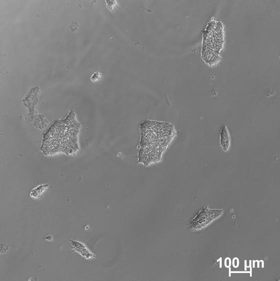
Figure 1. Example of quality human pluripotent stem cell (hPSC) cultures
2. Passage cells when ~80% confluent.
Note: To prevent spontaneous differentiation, stem cells should be passaged when colony edges start to merge.
3. Maintenance cells should be passaged as aggregates with ReLeSRTM, according to the manufacturer’s instructions.
4. Briefly aspirate medium from hPSCs.
5. Add 1.5 mL of 1× D-PBS to clean/rinse the cell monolayer and aspirate.
6. Add 1 mL of ReLeSRTM and aspirate to completely remove ReLeSRTM within 1 min, so that colonies are only exposed to the residual liquid.
7. Incubate at 37 °C for 6–8 min.
Note: We recommend that users optimize the dissociation time whenever a new cell line is used.
8. After 6–8 min, remove the plate from the incubator and add 1 mL of room-temperature mTeSR Plus medium per well.
9. Hold the plate with one hand and use the other to firmly tap the side of the plate for approximately 30–60 s.
10. Plate the cell aggregate mixture at the desired density onto coated wells containing mTeSR Plus medium.
Note: If the colonies are at an optimal density, the culture can be split every 4–7 days using 1:10 to 1:50 splits (i.e., cell aggregates from one well can be plated in 10–50 wells).
11. Place the plate in the incubator and move the plate in several quick, short, back-and-forth and side-to-side motions to evenly distribute the cell aggregates. Do not disturb the plate for 24 h.
Note: Uneven distribution of aggregates may result in increased spontaneous differentiation of the hPSCs.
12. Perform daily medium changes and visually assess the cultures to monitor growth until the next passaging time.
Note: High-quality hPSC cultures with minimal spontaneous differentiation must be maintained.
B. Thymic epithelial progenitor cell differentiation
1. Prepare a Matrigel-coated 24-well plate for TCD cells ≥ 1 h prior to single cell dissociation of hPSCs.
2. Day (-1): One day prior to TCD induction, aspirate medium from hPSCs.
3. Add 1.5 mL of 1× D-PBS to clean/rinse the cell monolayer and aspirate.
4. Add 1 mL of TrypLE and incubate at 37 °C for 7 min.
Note: We recommend that users optimize the dissociation time whenever a new cell line is used.
5. After 7 min, remove the plate from the incubator and gently tap the side of the plate to dislodge the cells from the plate.
6. Check that the cells have been dissociated under the microscope.
7. Add 1.5 mL of mTeSR Plus to neutralize the TrypLE.
8. Gently wash the cells off the plate using a p1000 pipette and transfer to a 50 mL conical tube containing 23 mL of D-PBS.
Note: Ensure that cells are fully dissociated into single cells, but do not over pipette, or excessive cell death will occur.
9. Mix the cells well to obtain a homogenous cell suspension.
10. Transfer 100 μL of the cell suspension to a 1.5 mL microcentrifuge tube containing 900 μL of D-PBS + 1 μL of propidium iodide for cell counting.
Notes:
1. Propidium iodide can be replaced by your viability marker of choice.
2. The cell counter used was the MOXI GO II, but alternatives can be used.
3. Discard if cell death is >10%, as cells are not fit for differentiation.
11. Centrifuge the cell suspension in a swinging bucket centrifuge at 300× g for 3 min at room temperature. In the meantime, obtain a cell count using your preferred method.
12. Aspirate supernatant and resuspend the cells in mTeSR Plus medium, supplemented with 10 μM Rock inhibitor Y-27632 (RI), to obtain a concentration of 1 × 10⁶ cells/100 μL.
Calculation example: [final] = 1 × 10⁶ cells/100 μL
25 mL within a conical tube with a concentration of 1.25 × 10⁶ cells/mL
1.25 × 10⁶ cells/mL × 25 mL (total volume in conical tube) = 31.25 × 10⁶ cells
31.25 × 10⁶ cells × 100 μL = 3,125 μL (3.125 mL)
Resuspend 31.25 × 10⁶ cells in 3.125 mL to obtain a [final] of 1 × 10⁶ cells/100 μL
13. Prepare a Matrigel-coated plate by removing the Matrigel solution and adding 0.5 mL of mTeSR Plus medium, supplemented with 10 μM RI, to each well.
Note: We recommend preparing the plate with medium ahead of time.
14. Plate 600,000 hPSCs per 24-well (at 1× 10⁶/100 μL, you will need to add 60 μL/well):
600,000 hPSC per 24-well = 3.15 × 10⁵ hPSC/cm2
15. Place the plate in the incubator and gently rock side to side to evenly distribute the cells across the well.
16. (Day 0) Twenty-four hours after plating hPSCs, remove the plate from the incubator and inspect the cells under the microscope (Figure 2).
Note: Cells should be 95%–100% confluent and completely cover the bottom of the well. If not, discard the plate.
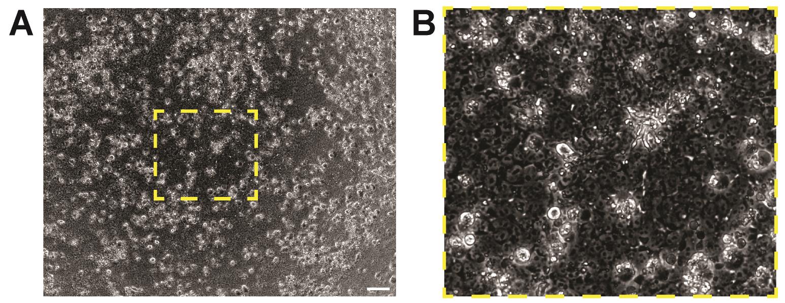
Figure 2. Day 0 thymic cell differentiation culture. (A) Cells form a complete monolayer, covering the entirety of the well (4× magnification, scale = 200 μm). (B) Enlarged image of the inset in (A).
17. Aspirate the medium and wash once with 0.5 mL of knockout DMEM/F12 (KO-DMEM/F12) to remove trace amounts of hPSC medium.
18. Induce definitive endoderm (DE) differentiation by adding 350 μL of “day 0” medium (TCD-1) (see Recipes; Table 1).
Table 1. Media, volumes, and associated times for hPSC–TEP differentiation
| Day | 0 | 1 | 2 | 3 | 4 | 5–8 | 9+ |
|---|---|---|---|---|---|---|---|
| Media | TCD-1 | TCD-2 | TCD-2 | TCD-2 | TCD-3 | TCD-4 | TCD-5 |
| Volume (μL) | 300 | 350 | 450 | 600 | 750 | 750 | 750 |
19. (Day 1) Twenty-two to twenty-four hours after DE induction, inspect cells under the microscope (cell morphology will have begun to change) (Figure 3).
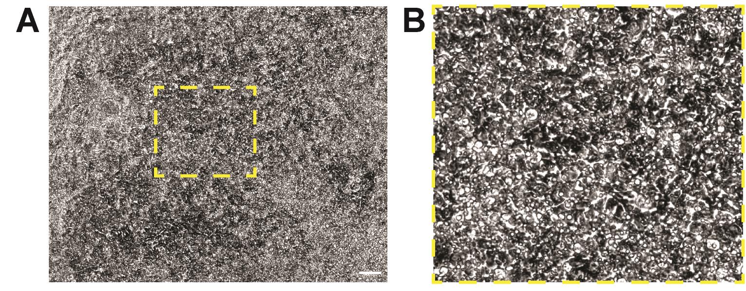
Figure 3. Thymic cell differentiation cultures. (A) Thymic cell differentiation cultures 24 h after definitive endoderm (DE) induction (4× magnification, scale = 200 μm). (B) Enlarged image of the inset in (A).
20. Aspirate day-0 medium, wash cells with 0.5 mL of KO-DMEM/F12, and add 450 μL of day-1 medium (TCD-2) (see Recipes; Table 1).
Note: Timing of medium changes is imperative. Medium should be changed every 24 h for optimal outcomes.
21. Perform daily media changes according to the schedule outlined in Table 1.
22. You may observe a high amount of cell death on days 1–4. This is normal and to be expected.
23. Around day 4 of differentiation, cells should start forming a thick layer and/or ridges throughout the well (Figure 4).
Note: Ridges should be very prominent by differentiation day 9.
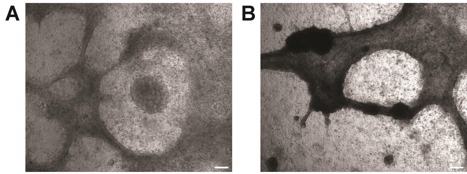
Figure 4. Ridge formation in thymic cell differentiation (TCD) cultures. Ridges in TCD cultures at (A) day 4 and (B) day 9 (4× magnification, scale = 200 μm).
C. Splanchnic mesoderm differentiation
1. To initiate splanchnic mesoderm differentiation, follow steps B1–17.
2. (Day 0) Induce splanchnic mesoderm differentiation by adding 300 μL of day 0 medium (SMD-1) (see Recipes; Table 2).
Table 2. Media, volumes, and associated times for hPSC to splanchnic mesoderm differentiation
| Day | 0 | 1 | 2 | 3 | 4 |
|---|---|---|---|---|---|
| Media | SMD-1 | SMD-2 | SMD-3 | SMD-3 | SMD-3 |
| Volume (μL) | 300 | 350 | 450 | 600 | 750 |
3. (Day 1) Twenty-four hours after splanchnic mesoderm induction, aspirate day 0 medium, wash cells with 0.5 mL of KO-DMEM/F12, and add 350 μL of day-1 medium (SMD-2) (see Recipes; Table 2).
Note: Timing of medium changes is imperative. Medium should be changed every 24 h for optimal outcomes.
4. Perform daily media changes according to the schedule outlined in Table 2.
5. (Day 5) Twenty-four hours after the addition of day-4 medium, aspirate all medium.
6. Wash cells once with 1 mL of D-PBS and aspirate.
7. Add 0.75 mL of room-temperature Accutase to each well and incubate at 37 °C for 15–25 min (monitor until you see cells start to detach from the well bottom).
8. Gently tap the side of the plate until you see the cells start to detach from the bottom of the well and form small clumps.
9. Add 1 mL of quench media (Recipe 4) and gently triturate with a p1000 pipette until cells dissociate to single cells.
10. Pass the cell suspension through a 100 μm filter into a 50 mL conical tube containing at least 10 mL of D-PBS.
11. Mix cells well to obtain a homogenous cell suspension.
12. Transfer 100 μL of the cell suspension to a 1.5 mL microcentrifuge tube containing 900 μL of D-PBS + 1 μL of propidium iodide for cell counting.
Notes:
1. Propidium iodide can be replaced by your viability marker of choice.
2. The cell counter used was the MOXI GO II; alternative cell counters can be used.
3. Discard if cell death is >10%, as cells are not fit for use.
13. Centrifuge the cell suspension in a swinging bucket centrifuge at 300× g for 3 min at RT. In the meantime, obtain a cell count using your preferred method.
14. Aspirate and resuspend the cells in CryoStore freezing medium at a concentration of 1 × 10⁶ cells/100 μL.
15. Transfer cells to cryovials in 1 × 10⁶–2 × 10⁶ cell aliquots.
16. Freeze down and store until needed.
Note: If cells are to be used immediately, skip steps C14–16.
D. Hematopoietic cell differentiation
1. To initiate hematopoietic cell differentiation, follow steps B1–11.
2. Aspirate and resuspend the cells in HCD-1 media (see Recipes), at a concentration of 1 × 10⁶ cells/100 μL.
3. Transfer 5.5 × 10⁶ cells (550 μL) into a single well of a 6-well suspension plate containing 5 mL of HCD-1 medium.
Final concentration = 1 × 10⁶ cells/mL; 5.5 mL total volume.
4. Incubate the cells for 24 h at 37 °C in a humidified 5% CO2 incubator on an orbital shaker shaking at 100 rpm.
a. Cells will self-aggregate into spheroids/clusters.
b. Clusters should be homogenous in size and shape (~200 μm).
5. Perform medium changes as outlined in Table 3, changing the media on the underlined days.
Table 3. Hematopoietic progenitor cell (HPC) differentiation days and corresponding media
| Day | 0 | 1 | 2 | 3 | 4 | 5 | 6 | 7 | 8+ |
|---|---|---|---|---|---|---|---|---|---|
| Media | HCD-1 | HCD-2 | HCD-3 | HCD-3 | HCD-4 | HCD-4 | HCD-4 | HCD-4 | HCD-5 |
6. To change medium in suspension plates:
a. Remove the plate lid to access the well.
b. Tilt the plate ~45° on the horizontal axis toward you.
c. Without aspirating the cell clusters, remove most of the spent medium (leaving ~2 mL in the well).
d. Set the plate back down and add 5.5 mL of fresh medium.
7. Gradually start increasing the medium volume by 1–2 mL per day as you notice the media yellowing. In some cases, it is best to split a single 6-well into two wells to allow better nutrient distribution.
8. (Day 8) At least 1 h prior to use, prepare Vitronectin solution (10 μg/mL; 40 μL in 960 μL of CellAdhere dilution buffer).
a. Place Millicell® standing cell culture inserts in the wells of an empty 6-well plate.
b. Coat the Millicell® standing cell culture inserts with 1 mL of prepared Vitronectin solution and gently rock the plate back and forth to evenly distribute the Vitronectin solution evenly across the surface.
c. Incubate at room temperature for at least 1 h before use.
9. After Vitronectin has set, transfer the cell clusters from the 6-well plate to a 50 mL conical tube and allow the clusters to settle by gravity for 3–5 min.
Note: 18–24 Millicell® standing cell culture inserts can be plated from a single 6-well of cell clusters.
10. Aspirate and resuspend in HCD-5 media. Resuspend in ~10 μL/Millicell® standing cell culture insert being plated.
11. Completely remove the Vitronectin solution from the Millicell® standing cell culture inserts and transfer 10 μL (~20 clusters) of cluster suspension to Vitronectin-coated Millicell® standing cell culture inserts, using a pipette tip to distribute the clusters evenly on the Millicell® standing cell culture inserts.
Note: Use a wide-bore 20 μL pipette tip or cut the tip of a standard 20 μL tip to accommodate the diameter of the cell clusters.
12. Carefully add 1.5 mL of HCD-5 media underneath each Millicell® standing cell culture insert, taking care to not to flood the top of the insert.
13. Change media every other day with HCD-5 media.
a. Tilt the plate ~45° toward you to pool the media on the side of the well, taking caution not to flood the top of the insert with media.
b. Aspirate the media from around the cell culture insert, leaving approximately 0.5 mL of media under the insert.
c. Add 1 mL of room-temperature HCD-5 between the cell culture insert and the edge of the well, allowing the media to spread under the cell culture insert without being in direct contact with the stem cell–derived thymic organoids (sTOs).
14. On day 12, aspirate media from underneath the cell culture inserts.
15. Using a p1000 pipette and 1 mL of HCD-5 media, mechanically release/wash the cells from the membrane of the Millicell® standing cell culture insert and pass the cell suspension through a 70 μm reversible strainer on top of a 50 mL conical tube.
Note: Pre-wet the filter to prevent cells from sticking.
16. Wash the membrane with another 0.5 mL of HCD-5 media and transfer to the same 50 mL conical tube.
17. Wash the 70 μm filter with 10 mL of DMEM, or D-PBS, supplemented with 10% (w/v) FBS.
18. Flip the reversible 70 μm strainer over onto a new 50 mL conical tube.
19. With 10 mL of TrypLE + 1:1,000 DNase I, wash the hematopoietic progenitor cell clusters from the filter into the 50 mL conical tube.
20. Incubate at 37 °C for 5 min.
21. Using a p1000 pipette, gently triturate to break the clusters apart.
22. Incubate at 37 °C for another 3 min.
23. Gently triturate (cells should be in a single-cell suspension at this point) and add 1 volume (10 mL) of DMEM supplemented with 10% (w/v) FBS.
24. Pass the cell suspension through a pre-wetted 100 μm filter affixed on top of the original 50 mL conical tube.
25. Take an aliquot for cell counting (see step B10).
26. Centrifuge the cell suspension in a swinging bucket centrifuge at 300× g for 3 min at RT.
27. Aspirate and resuspend in HCD-5 media (see Recipes) at a concentration of 1 × 10⁶ cells/100 μL.
28. Alternatively, cells can be frozen down for later use (see steps C14–16).
E. Generation of stem cell–derived thymic organoids
1. Differentiate hPSCs to the three cell lineages as described in sections B–D.
2. Dissociate splanchnic mesoderm and hematopoietic progenitor cells to single cells as described above (steps C6–11, D14–27). If frozen stocks of splanchnic mesoderm or hematopoietic progenitors are to be used:
a. Remove frozen stocks from cryo-storage.
b. Thaw in a 37 °C water bath until only a small ice crystal remains.
c. Add 1 mL of prewarmed DMEM + 10% (w/v) FBS to the vial and transfer thawed cells to a 15 mL conical tube containing 10 mL of prewarmed DMEM + 10% (w/v) FBS.
d. Gently mix by inverting 2–3 times and take an aliquot for cell counting (see step B10).
e. Centrifuge the cell suspension in a swinging bucket centrifuge at 300× g for 3 min at RT.
f. Resuspend cells (both lineages) in HCD-5 media at a concentration of 1 × 10⁶ cells/100 μL.
3. At day 21–30 of TCD, remove about half of the media from however many TCD wells are needed.
Note: One TCD well can be used to generate three sTOs.
4. Using a p1000 pipette, mechanically dissociate the TCD cultures into small cell clumps and transfer to a 1.5 mL microcentrifuge tube.
Note: You may combine three TCD wells in one 1.5 mL microcentrifuge tube for a total of nine sTOs per 1.5 mL microcentrifuge tube.
5. Triturate until TCD cultures are well broken up.
6. For each TCD well added to the 1.5 mL microcentrifuge tube, add 30 μL each of splanchnic mesoderm and hematopoietic progenitor cell suspensions (300,000 cells). The desired mesenchyme:HPC:TEP ratio is 1:1:20.
Note: 1 × 24-well TCD culture yields ~6 × 10⁶ TEPs, on average.
7. Mix well with a p1000 pipette.
8. Centrifuge the cell suspension in a swinging bucket centrifuge at 300× g for 3 min at RT.
9. Carefully remove as much of the supernatant as possible, without disturbing the cell pellet.
10. Resuspend the cell pellet in 5 μL of HCD-5 media/sTO.
Note: Subtract ~20 from the volume added to account for the volume of the cell pellet, i.e., 9 sTOs in a 1.5 mL microcentrifuge tube: 9 sTOs × 5 μL/sTO = 45 μL of resuspension volume.
45 μL − 20 μL = 25 μL of HCD-5 media added to cell pellet.
11. Measure the total volume with a p200 pipette and divide by the number of sTOs wanted, to ensure that each sTO has the same volume. Adjust the volume as needed.
12. Using a p20 pipette with wide-bore tips, draw up 5 μL of cell slurry and gently release the cell slurry as a drop at the end of the tip, gently touching the drop to the top of the cell insert membrane to release it. The cell slurry will form a small sphere on the surface of a Millicell® standing cell culture insert (Figure 5).
Notes:
1. Larger volume/sTO could be used if the volume of the cell pellet is larger than anticipated.
2. Three sTOs can be placed on a single insert. sTOs should be spaced ~1 cm apart and ~1 cm from the edge of the insert.
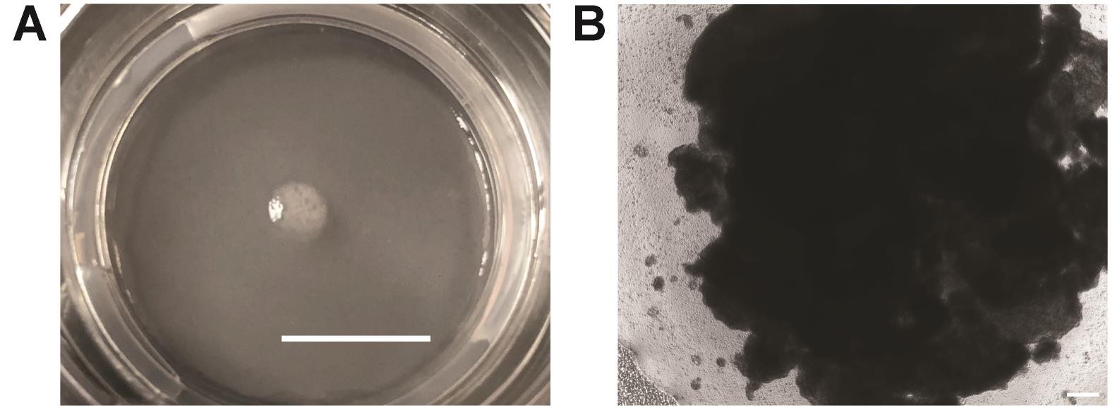
Figure 5. Stem cell–derived thymic organoid (sTO) generation and plating. (A) sTO immediately after plating (scale = 10mm). (B) sTO 24 h after plating (4× magnification, scale = 200 μm).
13. Carefully add 1.5 mL of HCD-5 media underneath each Millicell® standing cell culture insert, taking care not to flood the top of the insert.
14. Incubate at 37 °C in a humidified 5% CO2 incubator.
15. Change media every other day (see step D13).
16. sTOs can be cultured for up to 6 weeks, with the best lymphocyte production observed 3–5 weeks after sTO formation.
F. Collecting sTOs for analysis
1. Aspirate as much media as possible from underneath the cell culture insert.
2. With a p1000 pipette, add 250 μL of HCD-5 media to the top of the cell culture insert membrane, mechanically scrape/detach the sTOs from the membrane, and transfer them to a 1.5 mL microcentrifuge tube.
3. Wash the membrane with 0.5 mL of HCD-5 media, collecting as many free-floating cells from the top of the membrane as possible, and transfer them to the 1.5 mL tube.
4. Centrifuge the cell suspension in a swinging bucket centrifuge at 300× g for 3 min at room temperature.
5. Aspirate and resuspend in 750 μL of digestion buffer (see Recipes).
Note: To prepare sTOs for qPCR analysis, resuspend the cell pellet in cell lysis buffer (IN PLACE of digestion buffer) for your preferred RNA isolation method. qPCR analysis could then be performed to assess the expression levels of key thymic markers (see Materials and reagents).
6. Incubate at 37 °C on a heat block or in a water bath for 6 min.
7. After 6 min, triturate with a p1000 pipette ~10 times.
8. Repeat steps F6–7 until sTOs are mostly dissociated (typically requires ~4 rounds). On the third round, use a p200 pipette to triturate.
9. Once a mostly single-cell suspension has been obtained, quench the reaction by adding 750 μL of quench media (see Recipes).
10. Centrifuge the cell suspension in a swinging bucket centrifuge at 300× g for 3 min at room temperature.
11. Aspirate the supernatant, resuspend in 500 μL of FACS buffer (see Recipes), and pass the cell suspension through a 35 μm filter into a 5 mL round-bottom polystyrene FACS tube.
12. Perform FC receptor block using Human TruStain FcXTM (5 μL/sample) and incubate for 10 min at room temperature.
13. After FC block, add the antibody cocktail with desired markers to each sample and incubate for 30 min on ice in the dark.
14. Wash cells with 2 mL of FACS buffer.
15. Centrifuge the cell suspension in a swinging bucket centrifuge at 300× g for 3 min at RT.
16. Aspirate and resuspend in 0.2 mL of FACS buffer with 2.5 ng/mL DAPI.
17. Analyze on a cytometer; see Figure 6 for representative flow cytometry and qPCR analysis.
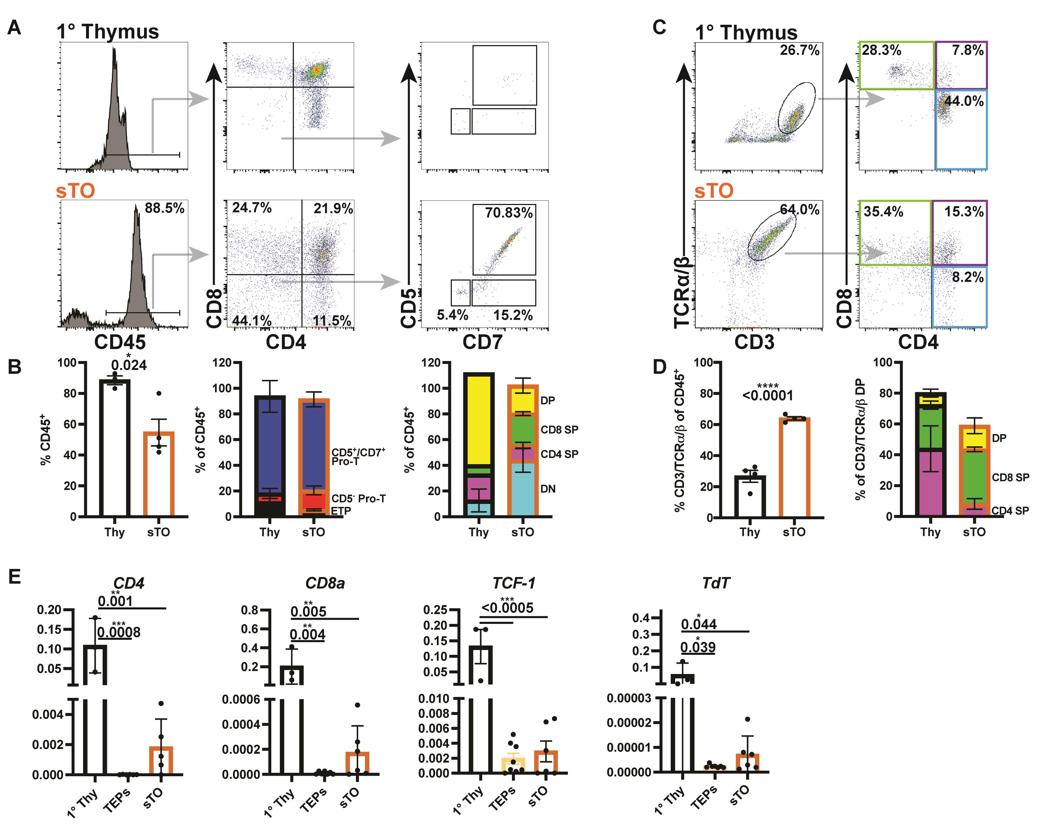
Figure 6. Stem cell–derived thymic organoids (sTOs) support T-cell development. (A–D) FC plots (A and C) and quantification (B and D) of thymocytes in primary thymus and week-4 sTOs [thymi, n = 6; sTOs, n = 4, two human pluripotent stem cell (hPSC) lines]. (E) qPCR analysis of CD4, CD8, and TdT in 1 thymus, thymic epithelial progenitors (TEPs), and sTOs (thymi, n = 3; TEPs, n = 6–7, four hPSC lines; sTOs, n = 5–6, 4 hPSC lines). Data are shown as mean ± SEM. P values were determined by t-test (B, D) or one-way ANOVA with Tukey’s multiple comparison test (E).
Validation of protocol
This protocol or parts of it has been used and validated in the following research article(s):
• Ramos et al. [22]. Generation of functional thymic organoids from human pluripotent stem cells. Stem Cell Reports.
Acknowledgments
Authors’ contribution
Conceptualization, S.A.R., H.A.R.; Investigation, S.A.R., H.A.R.; Writing—Original Draft, S.A.R.; Writing—Review & Editing, S.A.R., H.A.R.; Funding acquisition, H.A.R.; Supervision, H.A.R.
The Russ lab is or was supported by NIH/NIDDK grants R01DK12044, R01DK132387, and UG3DK142184; NIDDK/HIRN RRID: SCR_014393. Additional support was provided by Breakthrough T1D (formerly JDRF) 2-SRA-2023-1313-S-B, 3-SRA-2023-1367-S-B, and 3-SRA-2026-1817-S-B. We also acknowledge our previous work published in Stem Cell Reports (2023), DOI: 10.1016/j.stemcr.2023.02.013 [22] on which the current protocol is based.
Competing interests
H.A.R. scientific co-founder of Tolerance Bio and consults for Tolerance Bio, Sernova Biotherapeutics, Axxon Advisors and Guidepoint Global. S.A.R. consults for Tolerance Bio.
References
- Ramos, S. A., Morton, J. J., Yadav, P., Reed, B., Alizadeh, S. I., Shilleh, A. H., Perrenoud, L., Jaggers, J., Kappler, J., Jimeno, A., et al. (2022). Generation of functional human thymic cells from induced pluripotent stem cells. J Allergy Clin Immunol. 149(2): 767–781.e6. https://doi.org/10.1016/j.jaci.2021.07.021
- Han, L., Chaturvedi, P., Kishimoto, K., Koike, H., Nasr, T., Iwasawa, K., Giesbrecht, K., Witcher, P. C., Eicher, A., Haines, L., et al. (2020). Single cell transcriptomics identifies a signaling network coordinating endoderm and mesoderm diversification during foregut organogenesis. Nat Commun. 11(1): 4158. https://doi.org/10.1038/s41467-020-17968-x
- Motazedian, A., Bruveris, F. F., Kumar, S. V., Schiesser, J. V., Chen, T., Ng, E. S., Chidgey, A. P., Wells, C. A., Elefanty, A. G., Stanley, E. G., et al. (2020). Multipotent RAG1+ progenitors emerge directly from haemogenic endothelium in human pluripotent stem cell-derived haematopoietic organoids. Nat Cell Biol. 22(1): 60–73. https://doi.org/10.1038/s41556-019-0445-8
- Kadouri, N., Nevo, S., Goldfarb, Y. and Abramson, J. (2019). Thymic epithelial cell heterogeneity: TEC by TEC. Nat Rev Immunol. 20(4): 239–253. https://doi.org/10.1038/s41577-019-0238-0
- Coder, B. D., Wang, H., Ruan, L. and Su, D. M. (2015). Thymic Involution Perturbs Negative Selection Leading to Autoreactive T Cells That Induce Chronic Inflammation. J Immunol. 194(12): 5825–5837. https://doi.org/10.4049/jimmunol.1500082
- Palmer, D. B. (2013). The Effect of Age on Thymic Function. Front Immunol. 4: e00316. https://doi.org/10.3389/fimmu.2013.00316
- Duah, M., Li, L., Shen, J., Lan, Q., Pan, B. and Xu, K. (2021). Thymus Degeneration and Regeneration. Front Immunol. 12: e706244. https://doi.org/10.3389/fimmu.2021.706244
- Kreins, A. Y., Bonfanti, P. and Davies, E. G. (2021). Current and Future Therapeutic Approaches for Thymic Stromal Cell Defects. Front Immunol. 12: e655354. https://doi.org/10.3389/fimmu.2021.655354
- Janda, A., Sedlacek, P., Hönig, M., Friedrich, W., Champagne, M., Matsumoto, T., Fischer, A., Neven, B., Contet, A., Bensoussan, D., et al. (2010). Multicenter survey on the outcome of transplantation of hematopoietic cells in patients with the complete form of DiGeorge anomaly. Blood. 116(13): 2229–2236. https://doi.org/10.1182/blood-2010-03-275966
- Auerbach, R. (1960). Morphogenetic interactions in the development of the mouse thymus gland. Dev Biol. 2(3): 271–284. https://doi.org/10.1016/0012-1606(60)90009-9
- Ceredig, R., Medveczky, J. and Skulimowski, A. (1989). Mouse fetal thymus lobes cultured in IL-2 generate CD3+, TCR-gamma delta-expressing CD4-/CD8+ and CD4-/CD8- cells. J Immunol. 142(10): 3353–3360. https://doi.org/10.4049/jimmunol.142.10.3353
- Han, J. and Zúñiga-Pflücker, J. C. (2021). High-Oxygen Submersion Fetal Thymus Organ Cultures Enable FOXN1-Dependent and -Independent Support of T Lymphopoiesis. Front Immunol. 12: e652665. https://doi.org/10.3389/fimmu.2021.652665
- Mandel, T. E. and Kennedy, M. M. (1978). The differentiation of murine thymocytes in vivo and in vitro. Immunology. 35: 317–331.
- Watanabe, Y. and Katsura, Y. (1993). Development of T cell receptor αβ‐bearing T cells in the submersion organ culture of murine fetal thymus at high oxygen concentration. Eur J Immunol. 23(1): 200–205. https://doi.org/10.1002/eji.1830230131
- Watson, J. D., Morrissey, P. J., Namen, A. E., Conlon, P. J. and Widmer, M. B. (1989). Effect of IL-7 on the growth of fetal thymocytes in culture. J Immunol. 143(4): 1215–1222. https://doi.org/10.4049/jimmunol.143.4.1215
- Chung, B., Montel-Hagen, A., Ge, S., Blumberg, G., Kim, K., Klein, S., Zhu, Y., Parekh, C., Balamurugan, A., Yang, O. O., et al. (2014). Engineering the Human Thymic Microenvironment to Support Thymopoiesis In Vivo. Stem Cells. 32(9): 2386–2396. https://doi.org/10.1002/stem.1731
- Schmitt, T. M. and Zúñiga-Pflücker, J. C. (2002). Induction of T Cell Development from Hematopoietic Progenitor Cells by Delta-like-1 In Vitro. Immunity. 17(6): 749–756. https://doi.org/10.1016/s1074-7613(02)00474-0
- Seet, C. S., He, C., Bethune, M. T., Li, S., Chick, B., Gschweng, E. H., Zhu, Y., Kim, K., Kohn, D. B., Baltimore, D., et al. (2017). Generation of mature T cells from human hematopoietic stem and progenitor cells in artificial thymic organoids. Nat Methods. 14(5): 521–530. https://doi.org/10.1038/nmeth.4237
- Montel-Hagen, A., Seet, C. S., Li, S., Chick, B., Zhu, Y., Chang, P., Tsai, S., Sun, V., Lopez, S., Chen, H. C., et al. (2019). Organoid-Induced Differentiation of Conventional T Cells from Human Pluripotent Stem Cells. Cell Stem Cell. 24(3): 376–389.e8. https://doi.org/10.1016/j.stem.2018.12.011
- Micallef, S. J., Li, X., Schiesser, J. V., Hirst, C. E., Yu, Q. C., Lim, S. M., Nostro, M. C., Elliott, D. A., Sarangi, F., Harrison, L. C., et al. (2011). INS GFP/w human embryonic stem cells facilitate isolation of in vitro derived insulin-producing cells. Diabetologia. 55(3): 694–706. https://doi.org/10.1007/s00125-011-2379-y
- Soh, C. L., Giudice, A., Jenny, R. A., Elliott, D. A., Hatzistavrou, T., Micallef, S. J., Kianizad, K., Seach, N., Zúñiga-Pflücker, J. C., Chidgey, A. P., et al. (2014). FOXN1GFP/w Reporter hESCs Enable Identification of Integrin-β4, HLA-DR, and EpCAM as Markers of Human PSC-Derived FOXN1+ Thymic Epithelial Progenitors. Stem Cell Rep. 2(6): 925–937. https://doi.org/10.1016/j.stemcr.2014.04.009
- Ramos, S. A., Armitage, L. H., Morton, J. J., Alzofon, N., Handler, D., Kelly, G., Homann, D., Jimeno, A. and Russ, H. A. (2023). Generation of functional thymic organoids from human pluripotent stem cells. Stem Cell Rep. 18(4): 829–840. https://doi.org/10.1016/j.stemcr.2023.02.013
Article Information
Publication history
Received: Aug 26, 2025
Accepted: Mar 25, 2026
Available online: Apr 27, 2026
Published: May 5, 2026
Copyright
© 2026 The Author(s); This is an open access article under the CC BY-NC license (https://creativecommons.org/licenses/by-nc/4.0/).
How to cite
Ramos, S. A. and Russ, H. A. (2026). Generation of Functional Patient-Specific Thymus Organoids From Human Pluripotent Stem Cells (hPSCs) Using Air–Liquid Interface Culture. Bio-protocol 16(9): e5684. DOI: 10.21769/BioProtoc.5684.

 Download Citation in RIS Format
Category
Stem Cell > Organoid culture
Stem Cell > Pluripotent stem cell > Cell differentiation
Immunology > Immune cell differentiation > T cell
Do you have any questions about this protocol?
Post your question to gather feedback from the community. We will also invite the authors of this article to respond.
 Post a Question


