


- Protocols

- Articles and Issues
- For Authors
- About
- Become a Reviewer


Reconstitution of Active Plant H+-ATPase AHA2 in Giant Unilamellar Vesicles

Published: Vol 16, Iss 9, May 5, 2026 DOI: 10.21769/BioProtoc.5675 Views: 493
Reviewed by: Willy R Carrasquel-UrsulaezAnonymous reviewer(s)
 PDF
PDF  Ask a question
Original research article
The authors used this protocol in:

Mar 2025
Advertisement



Protocol Collections
Comprehensive collections of detailed, peer-reviewed protocols focusing on specific topics




Abstract
Membrane transporters mediate the selective movement of ions and molecules across biological membranes and are essential for cellular homeostasis. However, their functional characterization in living cells is often complicated by the complexity of the native membrane environment. Reconstitution into model membrane systems provides a powerful alternative by enabling precise control over lipid composition and experimental conditions. Giant unilamellar vesicles (GUVs) are particularly well suited for transporter studies, as their cell-sized dimensions allow direct microscopic observation and fluorescence-based measurements of protein activity. Here, we describe a two-step reconstitution protocol in which transport proteins are first incorporated into large unilamellar vesicles and then used to generate protein-containing giant unilamellar vesicles (proteo-GUVs) via the poly(vinyl alcohol) swelling method. This two-step approach enhances protein incorporation efficiency and preserves transporter functionality. The method is exemplified using the P3-type ATPase Arabidopsis thaliana plasma membrane H+-ATPase isoform 2 (AHA2). We further describe a fluorescence-based assay to assess proton transport activity in proteo-GUVs. Our approach provides a versatile and controlled platform for biochemical, biophysical, and single-molecule analysis of membrane transporters.
Key features
• Describes a SNAP-based labeling method.
• Provides a reconstitution approach based on detergent removal using size-exclusion chromatography and bio-beads.
• Introduces a gentle dehydration–rehydration strategy using gel-assisted swelling for the generation of proteo-GUVs.
• Measures proton pump activity in GUVs under a confocal microscope using the pH-sensitive dye pyranine.
Keywords: GUV proteo-GUV
Gel-assisted swelling
LUV
PVA
Proteoliposome
proteo-GUV
Gel-assisted swelling
LUV
PVA
Proteoliposome
Graphical overview
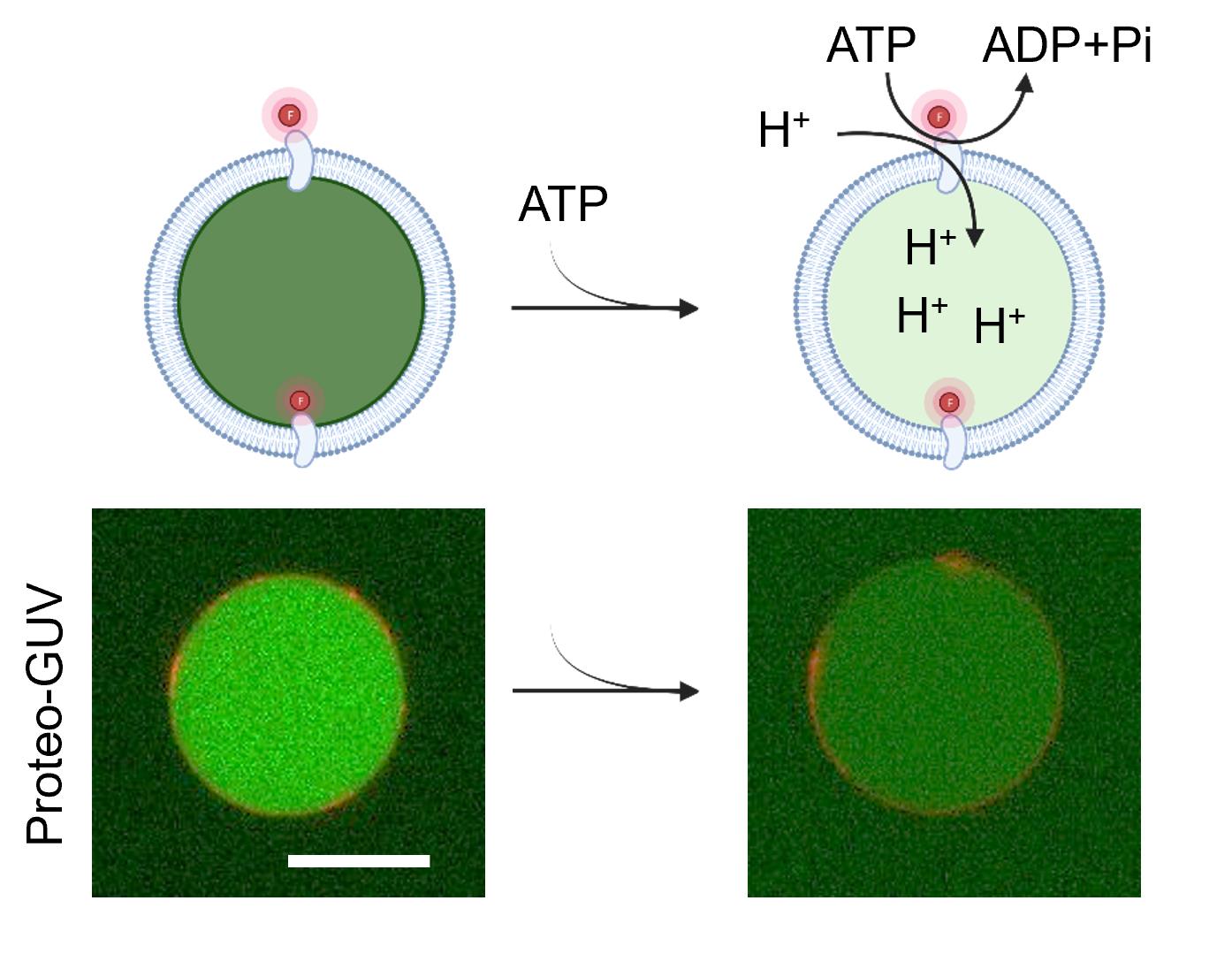
Functional SNAP-AHA2 in giant unilamellar vesicle (GUV)
Background
Membrane transporters are essential for cellular function by mediating the transport of ions and molecules across biological membranes. Studying them in cells is challenging due to the complexity of the cellular environment. Reconstitution into model membrane systems, such as liposomes, enables investigations under well-defined chemical conditions and supports detailed biochemical, biophysical, and single-molecule analyses [1]. This approach is particularly useful for studying P-type ATPases, exemplified here by the P3-ATPase Arabidopsis thaliana H+-ATPase isoform 2 (AHA2), which uses ATP hydrolysis to generate a proton gradient that drives processes such as nutrient uptake, pH regulation, stomatal opening, and cell growth [2,3]. Giant unilamellar vesicles (GUVs) extend these capabilities by offering cell-sized membranes amenable to direct microscopic observation and quantitative fluorescence analysis [4–6]. GUVs with defined lipid compositions allow systematic assessment of membrane protein activity using fluorophores [7]. Various strategies exist for generating membrane protein–containing GUVs (proteo-GUVs), including electroformation, swelling methods, charge-mediated fusion, and direct reconstitution [8–14]. Here, we employ the poly(vinyl alcohol) (PVA) swelling method to generate proteo-GUVs from proteoliposomes (protein–containing large unilamellar vesicles). The method is simple and compatible with both neutral and charged lipids, works under physiological ionic strength conditions, and preserves membrane protein integrity and activity.
Materials and reagents
Biological materials
1. Purified membrane protein
The C-terminally truncated Arabidopsis thaliana plasma membrane H+-ATPase isoform 2 (AHA2), containing an N-terminal hexahistidine tag for purification and a SNAP tag for fluorescent labeling (hereafter referred to as SNAP–AHA2), was used in this protocol. SNAP–AHA2 was heterologously expressed in Saccharomyces cerevisiae strain RS-72 (MATa, ade1-100 his4-519 leu2-3,112; the endogenous proton pump PMA1 gene is under the control of the GAL1 promoter) [15] and purified via the His-tag, resulting in protein concentrations of 5–10 mg/mL. Purified SNAP–AHA2 was stored in storage buffer composed of 50 mM MOPS-KOH (pH 7.0), 20% (w/v) glycerol, 50 mM KCl, 1 mM ethylenediaminetetraacetic acid, 1 mM dithiothreitol, and 0.04% (w/v) n-Dodecyl-β-D-maltoside (DDM). Aliquots were frozen in liquid nitrogen and stored at -80 °C [16].
2. Large unilamellar vesicles (LUVs)
LUVs were composed of lecithin (Sigma-Aldrich, catalog number: P5638-500G) and prepared according to previously published protocols [17,18]. Briefly, 15 mg of lecithin was dissolved in 1 mL of buffer (10 mM MOPS-KOH, pH 7.0, 50 mM K2SO4), yielding a lipid concentration of 15 mg/mL. The lipid solution was extruded 15 times through 200-nm polycarbonate membranes to obtain LUVs with a uniform size distribution.
Note: Lecithin is a heterogeneous mixture of phospholipids. For experiments requiring precise control over lipid composition, synthetic phospholipids can be used as an alternative. However, membrane proteins may display specific lipid requirements with respect to phospholipid headgroup identity and fatty acid chain length and/or degree of saturation. Consequently, empirical optimization of lipid composition may be necessary to achieve optimal protein stability and activity.
Reagents
1. Adenosine 5′-triphosphate disodium salt (ATP) (Roth, catalog number: HN35.3)
2. Bio-beads SM-2 resin (Bio-Rad Laboratories Inc., catalog number: 1523920)
3. Deionized water
4. Detergent/Soap
5. DMSO (Sigma-Aldrich, catalog number: 276855)
6. Dithiothreitol (Millipore, catalog number: 1.11474)
7. Ethanol (96%) (Sigma-Aldrich, catalog number: 64-17-5)
8. Ethylenediaminetetraacetic acid (EDTA) (Sigma, catalog number: E6758)
9. Glucose (Duchefa Biochemie, catalog number: G0802.5000)
10. Glycerol (VWR, catalog number: 24388.295)
11. HEPES (Carl Roth, catalog number: 6763.3)
12. Lecithin (Sigma-Aldrich, catalog number: 429415)
13. Magnesium sulfate heptahydrate (MgSO4·7H2O) (Merck, catalog number 10034-99-8)
14. Methanol (PROLABO® CHEMICALS, VWR, catalog number: 20834.291)
15. MOPS (Sigma-Aldrich, catalog number: M1254)
16. Mowiol®28-99, Poly(vinyl)alcohol) (PVA), MW 145,000 (Sigma-Aldrich, catalog number: 10849-250G)
17. n-Dodecyl-β-D-maltoside (DDM) (Glycon, catalog number: D97002)
18. Octyl β-D-glucopyranoside (OG) (Glycon, catalog number: D97002)
19. Potassium chloride (KCl) (Merck, catalog number: 7447-40-7)
20. Potassium hydroxide (KOH), 1 M (Sigma-Aldrich, catalog number: 1310-58-3)
21. Potassium sulfate (K2SO4) (Sigma-Aldrich, catalog number: P9458-250G)
22. Pyranine (8-Hydroxypyrene-1,3,6-trisulfonic acid trisodium salt) (Sigma-Aldrich, catalog number: H1529)
23. Sephadex G-50 fine (Sigma-Aldrich, catalog number: S5897)
24. SNAP-Surface® Alexa Fluor® 647 (New England Biolabs, catalog number: S9136S) dissolved in DMSO and stored at -20 °C
25. Sodium chloride (NaCl) (Carl Roth, catalog number: 7647-14-5)
26. Sucrose (Duchefa Biochemie, catalog number: S0809.5000)
27. Valinomycin (Thermo Fischer, catalog number: J62312.MC)
Solutions
1. ATP (0.5 M) (see Recipes)
2. DDM (20%) (see Recipes)
3. Ethanol (70%) (see Recipes)
4. HEPES-KOH pH 7.4 (0.5 M) (see Recipes)
5. KCl (1 M) (see Recipes)
6. K2SO4 (0.5 M) (see Recipes)
7. KOH (1 M) (see Recipes)
8. MgSO4 (1 M) (see Recipes)
9. Microscopic buffer (300 mM) (see Recipes)
10. MOPS-KOH pH 7 (0.5 M) (see Recipes)
11. NaCl (1 M) (see Recipes)
12. OG (1 M) (see Recipes)
13. PVA (5%) (see Recipes)
14. Pyranine (0.1 M) (see Recipes)
15. Reconstitution buffer (see Recipes)
16. Sephadex G-50 fine gel (see Recipes)
17. SNAP labeling buffer (see Recipes)
18. Swelling buffer (300 mM) (see Recipes)
19. Valinomycin (125 μM) (see Recipes)
Recipes
Note: All solutions are stored at 4 °C if not stated otherwise.
1. ATP (0.5 M)
| Reagent | Final concentration | Quantity or volume |
|---|---|---|
| ATP | 0.5 M | 2.25 g |
| ddH2O | n/a | Fill up to 10 mL |
| Total | n/a | 10 mL |
Before adjusting the solution to its final volume with ddH2O, check the pH and adjust it to 7 using 1 M KOH. Store in aliquots at -20 °C.
2. DDM (20%, w/v)
| Reagent | Final concentration | Quantity or volume |
|---|---|---|
| DDM | 20% | 2 g |
| ddH2O | n/a | 10 mL |
Aliquot into 1 mL portions and store at -20 °C.
3. Ethanol (70%, v/v)
| Reagent | Final concentration | Quantity or volume |
|---|---|---|
| Ethanol, 96% | 70% | 729.17 mL |
| ddH2O | n/a | 270.83 mL |
| Total | n/a | 1,000 mL |
Store at room temperature.
4. HEPES-KOH pH 7.4 (0.5 M)
| Reagent | Final concentration | Quantity or volume |
|---|---|---|
| HEPES | 0.5 M | 23.83 g |
| ddH2O | n/a | Fill up to 200 mL |
| Total | n/a | 200 mL |
Before adjusting the solution to its final volume with ddH2O, check the pH and adjust it to 7.4 using 1 M KOH.
5. KCl (1 M)
| Reagent | Final concentration | Quantity or volume |
|---|---|---|
| KCl | 1 M | 3.728 g |
| ddH2O | n/a | Fill up to 50 mL |
| Total | n/a | 50 mL |
Store at room temperature.
6. K2SO4 (0.5 M)
| Reagent | Final concentration | Quantity or volume |
|---|---|---|
| K2SO4 | 0.5 M | 17.43 g |
| ddH2O | n/a | Fill up to 200 mL |
| Total | n/a | 200 mL |
Store at room temperature.
7. KOH (1 M)
| Reagent | Final concentration | Quantity or volume |
|---|---|---|
| KOH | 1 M | 5.611 g |
| ddH2O | n/a | Fill up to 100 mL |
| Total | n/a | 100 mL |
8. MgSO4 (1 M)
| Reagent | Final concentration | Quantity or volume |
|---|---|---|
| MgSO4·7H2O | 1 M | 1.233 g |
| ddH2O | n/a | Fill up to 5 mL |
| Total | n/a | 5 mL |
Store at room temperature.
9. Microscopic buffer (300 mM)
| Reagent | Final concentration | Quantity or volume |
|---|---|---|
| MOPS-KOH pH 7, 0.5 M | 10 mM | 1 mL |
| K2SO4, 0.5 M | 50 mM | 5 mL |
| MgSO4, 1M | 4 mM | 0.2 mL |
| Valinomycin, 125 μM | 62.5 nM | 25 μL |
| Glucose | 132 mM | 1.189 g |
| ddH2O | n/a | Fill up to 50 mL |
| Total | n/a | 50 mL |
Filter-sterilize using a 0.2 μm Acrodisc® syringe filter.
10. MOPS-KOH pH 7 (0.5 M)
| Reagent | Final concentration | Quantity or volume |
|---|---|---|
| MOPS | 0.5 M | 20.93 g |
| ddH2O | n/a | Fill up to 200 mL |
| Total | n/a | 200 mL |
Before adjusting the solution to its final volume with ddH2O, check the pH and adjust it to 7 using 1 M KOH.
11. NaCl (1 M)
| Reagent | Final concentration | Quantity or volume |
|---|---|---|
| NaCl | 1 M | 2.922 g |
| ddH2O | n/a | Fill up to 50 mL |
| Total | n/a | 50 mL |
Store at room temperature.
12. OG (1 M)
| Reagent | Final concentration | Quantity or volume |
|---|---|---|
| OG | 1 M | 2.924 g |
| ddH2O | n/a | Fill up to 10 mL |
| Total | n/a | 10 mL |
Aliquot into 1 mL portions and store at -20 °C.
13. PVA (5%, w/v)
| Reagent | Final concentration | Quantity or volume |
|---|---|---|
| PVA | 5% | 2 g |
| ddH2O | n/a | Fill up to 40 mL |
| Total | n/a | 40 mL |
Dissolve 2 g of PVA in 40 mL of deionized water while stirring in a fume hood at ~90 °C for approximately 5 h. Cover the glass flask with aluminum foil to prevent water evaporation. Transfer the 5% PVA solution into a Falcon tube, seal with Parafilm, and store at 60 °C.
14. Pyranine (0.1 M)
| Reagent | Final concentration | Quantity or volume |
|---|---|---|
| Pyranine | 0.1 M | 0.5 g |
| ddH2O | n/a | 9.535 mL |
Aliquot into 1 mL portions and store at -20 °C.
15. Reconstitution buffer
| Reagent | Final concentration | Quantity or volume |
|---|---|---|
| MOPS-KOH pH 7, 0.5 M | 10 mM | 2 mL |
| K2SO4, 0.5 M | 50 mM | 10 mL |
| ddH2O | n/a | Fill up to 100 mL |
| Total | n/a | 100 mL |
Filter-sterilize using a 0.2 μm Acrodisc® syringe filter.
16. Sephadex G-50 fine gel
| Reagent | Final concentration | Quantity or volume |
|---|---|---|
| Sephadex G-50 fine | 4% | 2 g |
| Reconstitution buffer | n/a | Fill up to 50 mL |
| Total | n/a | 50 mL |
The gel has to swell overnight at room temperature.
17. SNAP labeling buffer
| Reagent | Final concentration | Quantity or volume |
|---|---|---|
| HEPES-KOH, pH 7.4, 0.5 M | 50 mM | 1.5 mL |
| KCl, 1 M | 100 mM | 1.5 mL |
| Dithiothreitol, 1 M | 1 mM | 1.5 μL |
| DDM, 20% | 0.04% | 30 μL |
| ddH2O | n/a | Fill up to 15 mL |
| Total | n/a | 15 mL |
Seal the falcon tube with parafilm.
18. Swelling buffer (300 mM)
| Reagent | Final concentration | Quantity or volume |
|---|---|---|
| MOPS-KOH pH 7, 0.5 M | 10 mM | 1 mL |
| K2SO4, 0.5 M | 50 mM | 5 mL |
| MgSO4, 1M | 4 mM | 0.2 mL |
| Pyranine, 0.1 M | 100 μM | 50 μL |
| Sucrose | 132 mM | 2.259 g |
| ddH2O | n/a | Fill up to 50 mL |
| Total | n/a | 50 mL |
Filter-sterilize using a 0.2 μm Acrodisc® syringe filter.
19. Valinomycin (125 μM)
| Reagent | Final concentration | Quantity or volume |
|---|---|---|
| Valinomycin | 125 μM | 1.39 mg |
| Ethanol, 96% | n/a | 10 mL |
Aliquot in 100 μL batches at -20 °C.
Laboratory supplies
1. 3 mL disposable syringes (Henry Schein, catalog number: 9003017)
2. Ice
3. Ice bucket (e.g., Magic Touch 2TM ice bucket with lid; Sigma-Aldrich, catalog number: BAM168072002)
4. Microcentrifuge tubes of 1.5 mL capacity (SARSTEDT AG & Co. KG, catalog number: 72.690.001)
5. Microcentrifuge tubes of 2 mL capacity (SARSTEDT AG & Co. KG, catalog number: 72.691)
6. Microscope glass slides (26 × 76 mm, #1.5) (Thermo Fisher Scientific, Life Technologies Corporation Eugene)
7. O-ring (28 × 1 mm) (Dichtelemente arcus GmbH, catalog number: CR-70)
8. Parafilm (Sigma-Aldrich, catalog number: P7793-1EA)
9. Polyethersulfone membrane with a pore size of 0.2 μm (Filtropur, SARSTEDT AG & Co. KG, catalog number: 83.1826.001)
10. Wipes (Precision Wipes, KIMTECH Science, Kimberly-Clark® Professional, catalog number: 7552)
11. Aluminum foil
12. Coverslips (26 × 76 × 0.16–0.19 mm, #1.5) (Epredia, catalog number: BC02600760AC40MNZO)
13. Detergents for cleaning glass slides
14. Disposable glass Pasteur pipettes (150 mm) (VWR, catalog number: 612-1701)
15. Falcon tubes, 15 and 50 mL (Thermo Fisher Scientific, catalog numbers: 10773501 and 10788561)
16. Glass beads, 3 mm (Merck, catalog number: 104015)
17. Glass pipettes (e.g., graduated pipettes BLAUBRAND® Type 3 Class AS, 10 mL, graduation: 10 mL; Carl Roth, catalog number: HXT8.1)
18. High vacuum grease (Dow Corning, catalog number: 0315)
Equipment
1. Analytical balance (e.g., Sartorius Entris-I II, 220 g/0.1 mg; Buch Holm, catalog number: 4669128)
2. Magnets
3. pH-meter (pH-Meter 761 Calimatic)
4. Pipette tips 20, 200, and 1,000 μL (SARSTEDT AG & Co. KG, catalog numbers: 70.3021, 70.760.002, and 70.3050.020)
5. Pipettes P20, P200, P1000 (GILSON®, catalog numbers: FD10001, FD10005, and FD10006)
6. Refrigerator (5 °C)
7. Rotavapor® R-100 Evaporator with I-100 Controller and V-100 vacuum pump (Flawil, Switzerland)
8. Scissors
9. Tabletop centrifuge (Eppendorf, model: 5810 R, rotor A-4-62)
10. Vortexer (Vortex Genie 2 TM, BENDER & HOBEIN AG)
11. Confocal laser scanning microscope
Note: For this protocol, a Leica TCS SP8 equipped with 63×/1.20, NA water objective was used. Images were acquired using a 400 Hz unidirectional scanner, a pixel size of 246.27 × 246.27 μm, a pinhole of 100 μm (1 AU) with Leica HyD detectors.
12. End-over-end rotator (INTELLI-MIXER, neoLab®, catalog number: 7-0045)
13. Flow cabinet to work with organic solvents
14. Freezer (-20 °C)
15. Glass desiccator (Boro 3.3 with a socket in the lid, 20 cm, including stopcock; BRAND GmbH, catalog number: 65238)
16. Heating block (Rotilabo®-Block-Heater H 250; CARL ROTH, catalog number: Y264.1)
17. Ice machine
18. Magnetic stirrer (e.g., IKAMAG®, DREHZAHL ELECTRONIC, IKA)
Software and datasets
1. ImageJ (Wayne, Rasband, S., U. S. National Institutes of Health, Bethesda, Maryland, USA, Version 2.16.0/1.54p Java 21.0.7)
2. Leica LAS X software (LAS AF, Leitz, Wetzlar, Germany)
3. Microsoft® Excel® for Microsoft 365 MSO (Version 2511)
Procedure
The overall strategy relies on a two-step approach in which proteoliposomes (proteins containing large unilamellar vesicles, LUVs) are first generated and subsequently used as starting material for the production of protein-containing giant unilamellar vesicles (proteo-GUVs). A key advantage of this approach is that protein incorporation and functionality can be verified at the proteoliposome stage, prior to GUV formation, thereby ensuring the integrity of the protein before proceeding to the final vesicle preparation. This two-step strategy is implemented through four main experimental steps: (A) Labeling of SNAP-AHA2, (B) reconstitution of SNAP-AHA2 into preformed LUVs, (C) formation of GUVs from the proteoliposomes using PVA-assisted swelling, and (D) microscopic observation and analysis of proteo-GUVs.
A. Labeling of SNAP-AHA2
A final concentration of 10 μM label (corresponding to 0.2 nmol) and 5 μM SNAP-AHA2 (corresponding to 0.1 nmol or 12 μg) is used per labeling reaction. Calculate the required volume of the SNAP-AHA2 stock solution, using the molecular weight of SNAP-AHA2 (114,000 g/mol).
Note: If alternative labeling systems are used, such as CLIP-tag or ACP-tag, the corresponding reactive substrate must be selected to match the labeling chemistry of the chosen tag.
1. Prepare LUV solution (15 mg/mL total lipid) as described previously [17,18].
2. Into a 1.5 mL microcentrifuge tube, combine 1.46 μL of 15 g/L LUVs (final concentration 0.5 g/L), 0.43 μL of 1 mM SNAP-Surface® Alexa Fluor® 647 (final concentration 10 μM), and SNAP labeling buffer to bring the total volume to 43.87 μL (adjust to account for the volume of SNAP-AHA2 fusion protein to be added later). Keep the tube on ice during preparation.
Note: To maintain protein stability and prevent loss of activity, detergents and LUVs should be added to ensure the presence of detergent–lipid micelles. Mix well by carefully pipetting up and down (do not vortex).
3. Next, add the appropriate amount of SNAP-AHA2 fusion protein to reach a final concentration of 5 μM in the reaction.
4. Again, mix well by inverting the tube and finger flick. Make sure no solution is on the surface of the microcentrifuge tubes. Wrap the tube in aluminum foil to protect from light and incubate in the refrigerator (5 °C) for 30 min.
5. After 30 min, labeling is complete. Alexa647-labeled SNAP-AHA2 solution can now be used directly for downstream experiments.
6. (Optional) To confirm labeling, samples can be analyzed by SDS-PAGE using 10% polyacrylamide gels and visualized on a ChemiDoc XRS Imaging System using the Image LabTM software and pre-programmed option for Coomassie-stained gels and Alexa647 fluorophores.
B. Reconstitution of SNAP-AHA2 into LUVs
Note: AHA2-containing LUVs are generated by detergent-mediated reconstitution as described below.
1. Wash SM-2 bio-beads by sequential incubation in methanol (2 × 15 min), deionized water (2 × 15 min), and reconstitution buffer (15 min) under gentle stirring at room temperature. Remove the solutions using a glass pipette after each incubation step. Store the washed beads in reconstitution buffer at 4 °C until use.
2. Mix 52.6 μL of LUV solution (corresponding to 5 mM total lipid) with 5 μL of 1 M OG (see Recipes; final detergent concentration 25 mM) and adjust the volume to 156.13 μL with reconstitution buffer.
3. Add 43.87 μL of Alexa647-labeled SNAP-AHA2 (final volume 200 μL), corresponding to a lipid-to-protein ratio of 67:1 (w/w) (see Figure 1).
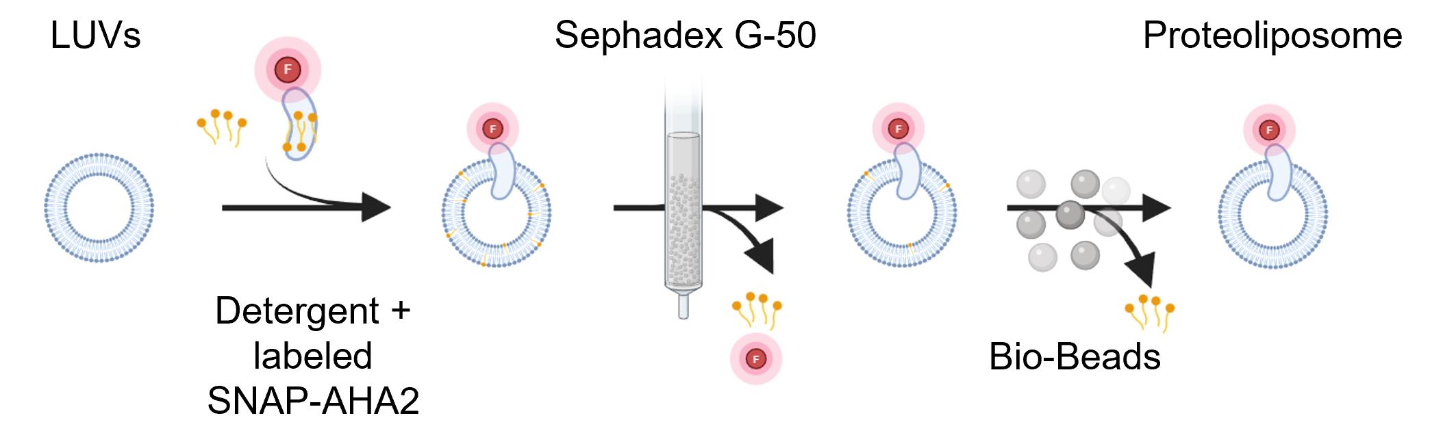
Figure 1. Reconstitution of fluorophore-labeled SNAP-AHA2 into large unilamellar vesicles (LUVs). Preformed LUVs are destabilized by detergent, followed by the addition of detergent-solubilized, Alexa647-labeled SNAP-AHA2. Free Alexa647 dye and most of the detergent are subsequently removed by size-exclusion chromatography using Sephadex G-50. Residual detergent is removed by adsorption onto bio-beads, yielding purified AHA2-containing proteoliposomes.
4. Vortex the sample briefly (3–5 s) to ensure homogeneous mixing and incubate for 5 min at room temperature with gentle end-over-end rotation.
5. Pack a 3 mL disposable syringe containing a drain-disc inlay with Sephadex G-50 gel and centrifuge at 180× g for 5 min at room temperature. Ensure that at least 2 mL of gel remains in the syringe after centrifugation.
6. Load the detergent–proteoliposome mixture (200 μL) onto the column and incubate for 5 min. Centrifuge at 180× g for 8 min at room temperature to remove free Alexa647 dye and most of the detergent (see Figure 1).
7. Collect the proteoliposome fraction by pipetting.
8. To remove residual detergent, add 100 mg of washed bio-beads to the proteoliposome mixture and incubate for 30 min at room temperature with slow end-over-end rotation (see Figure 1).
9. Collect the proteoliposomes by careful pipetting.
10. Protein concentration in proteoliposomes is determined by loading 10 μL of each sample and a protein standard onto the same 10% SDS–PAGE gel, followed by Coomassie blue staining. The stained gels are imaged using a ChemiDoc XRS Imaging System with a white transillumination excitation source and a standard emission filter, using the preconfigured settings for Coomassie-stained gels in Image LabTM. Band intensities are quantified using the gel analysis tool in Image LabTM, and protein concentration in the proteoliposome samples is determined by comparing the band intensities with those of the protein standard run on the same gel [19].
C. Formation of GUVs from the proteoliposomes using PVA-assisted swelling
1. Clean microscope glass slides (thickness: 1.5 mm) with detergent, deionized water, and 70% ethanol, then dry with Kimberly Clark filter wipes.
2. Take the PVA gel (see Recipes) from the 60 °C cabinet and use a 1 mL pipette to distribute 1 mL of the gel evenly, forming a uniform film on cleaned glass slides.
3. Dry PVA film on the heating block at 60 °C for 30 min.
Note: PVA-coated glass slides can be used immediately. Alternatively, they can be stored in a closed container at 4 °C for later use.
4. Cool the glass slide to room temperature for ~5 min and glue an O-ring onto the PVA film side of the glass slide using a vacuum grease (Figure 2A).
5. Use a 2 μL pipette to apply 25–30 drops of 2 μL of proteoliposome solution (5 mM lipids) evenly within the O-ring (Figure 2A, B).
6. Evaporate the solvent under vacuum in a desiccator containing a saturated NaCl solution at 30–100 mbar for 30–60 min in a cold room (4–6 °C) (Figure 2A, B).
Note: To increase the yield of giant vesicles, the sample should be dried thoroughly; however, excessive drying at this step may compromise membrane protein activity. Therefore, drying time, pressure, and temperature should be empirically optimized for each membrane protein.
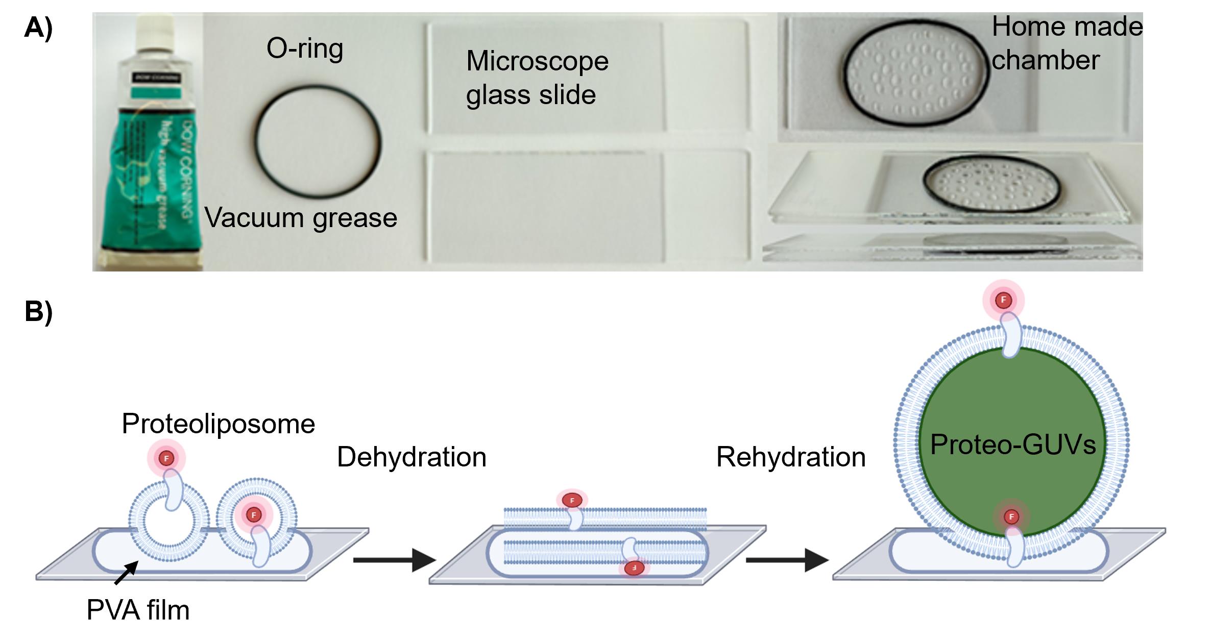
Figure 2. Preparation of proteo-giant unilamellar vesicles (GUVs). (A) Equipment used for proteo-GUV formation in a homemade chamber. (B) Schematic workflow of proteo-GUV formation using the PVA swelling method. Proteoliposomes are applied in 2 μL droplets to PVA-coated glass slides and dehydrated in a NaCl-saturated desiccator at 50–100 mbar for 30–60 min. The dried proteoliposome film is subsequently rehydrated in swelling buffer for 2–4 h, resulting in the formation of proteo-GUVs.
7. Fill the O-ring with 650 μL of swelling buffer and place the second glass slide on top.
8. Seal the chamber with Parafilm to prevent buffer evaporation.
9. Cover the chamber with aluminum foil to protect it from light.
10. Incubate for at least 2–4 h at room temperature.
11. Gently tap each side of the chamber to detach the proteo-GUVs from the gel surface.
12. Transfer the proteo-GUVs to a 2 mL microcentrifuge tube by removing the top glass slide and O-ring. Carefully tilt the chamber and allow the proteo-GUV solution to drip into the tube. Alternatively, use a cut 1 mL pipette tip to gently collect the GUVs into the microcentrifuge tube.
13. Store the proteo-GUVs at room temperature, protected from light, for up to 2–3 days.
Note: Proteo-GUVs and GUVs are extremely fragile. Pipette gently and avoid repeated transfers to minimize rupture. To minimize shear stress, pipette tips can be cut to a wider opening. GUVs are also prone to rupture on bare glass, particularly in the presence of salts in the medium. Coating the coverslip with PLL (poly-L-lysine), PLL-PEG, casein, or supported lipid bilayers (SLBs) can help reduce GUV lysis [20,21].
D. Microscopic observation and analysis of proteo-GUVs
Note: This section describes the observation of the proteo-GUVs under a microscope and the measurement of proton pump activity.
1. Turn on the confocal microscope and computer and open the Leica LAS X software. We used a 63×/1.20 water objective, a pixel size of 2,048 × 2,048 pixels, a 400 Hz unidirectional scanner, and a pinhole of 100 μm (1 AU) with the Leica hybrid photodetector (HyD SMD 2). The λex/λem used for imaging at laser intensity 5% were as follows: Alexa 647 650/655–700 nm and pyranine 488/509–522 nm.
2. Place 20 μL of GUVs (with a cut tip) into 20 μL microscopy buffer (see Recipes) on a coverslip.
Note: Passivate the glass coverslip to prevent GUVs from sticking, spreading, and bursting on the chamber bottom. Cover the glass coverslip with a β-casein or BSA solution, incubate for 5 min, rinse thoroughly with pure water, dry using a stream of air or nitrogen, and finally add the observation buffer.
3. Let the GUVs settle for approximately 5 min before imaging.
Note: Due to the density difference between the sugar solutions inside and outside, vesicles settle at the bottom of the glass slide, which facilitates observation in the brightfield.
4. By moving the microscope stage, find a protein-containing proteo-GUV and take one image.
5. Add 2 mM ATP solution and take images immediately every 30 s from the proteo-GUV.
Note: Add the solution very slowly onto the top of the sample to avoid disturbing the proteo-GUVs. Do not observe the sample for longer than 30 min under the microscope to prevent evaporation during the experiment. Alternatively, a closed chamber can be constructed by gluing a Sticky-Slide 8 Well High (Ibidi, catalog number: 80808) onto the coverslip; however, this setup requires a larger sample volume [8].
Data analysis
A. Data analysis using ImageJ
1. ImageJ was used to analyze the pyranine fluorescence intensity of individual proteo-GUVs before and after ATP addition. Only giant unilamellar vesicles exhibiting an Alexa647 signal from SNAP–AHA were included in the analysis.
2. Open the images in ImageJ by importing the LIF file.
3. Split the channels to separate brightfield, pyranine (pH sensor), and Alexa647 (AHA2) channels, and save each as a TIFF file.
4. Assemble the TIFF images as needed (see Figure 3A).
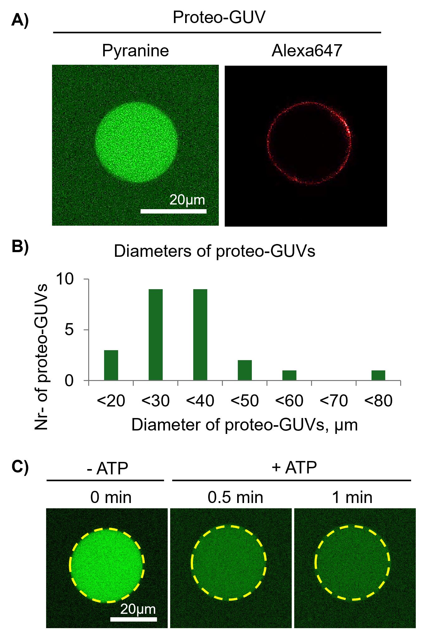
Figure 3. Imaging and analysis of proteo-giant unilamellar vesicles (GUVs) in the absence and presence of ATP. (A) Proteo-GUVs were imaged in the pyranine and Alexa647 channels. (B) Each proteo-GUV diameter was measured using ImageJ and plotted. (C) Pyranine fluorescence intensity was quantified for individual proteo-GUVs by placing a region of interest (ROI; yellow dashed circle) within the GUV lumen and measuring the integrated density per pixel using ImageJ. Proteo-GUVs were analyzed over time before and after ATP addition. Scale bar, 20 μm.
Note: As a control for photobleaching or pyranine dye leakage, fluorescence changes of pyranine can be monitored over time without adding any reagents. In addition, a negative control can be performed by adding ADP instead of ATP, under which conditions the pump cannot hydrolyze ADP and therefore cannot transport protons. Furthermore, proton pump activity can be assessed in the presence of P-type ATPase inhibitors, where activity is expected to be reduced or completely inhibited.
B. Analysis of the pyranine channel
1. Place a region of interest (ROI) within the lumen of the proteo-GUV to measure the pyranine fluorescence intensity by calculating the integrated density per pixel (see Figure 3B).
2. Repeat this procedure for all time points and save the data as an Excel (.xls) file.
3. Plot fluorescence intensity vs. time or normalize the intensity to percent and plot against time.
Validation of protocol
This protocol has been previously used and validated in the following research article:
• Uzun et al. [22]. Functional reconstitution of plant plasma membrane H+-ATPase into giant unilamellar vesicles. Scientific report.
Successful labeling of SNAP–AHA2 and its reconstitution into LUVs were verified by SDS–PAGE, combined with in-gel fluorescence imaging and Coomassie Brilliant Blue staining (Figure 4). After each preparation, at least one proteoliposome sample should be analyzed to confirm labeling efficiency and protein integrity. In this study, AHA2 was used as a model membrane protein. AHA2requires a specific vesicle lipid composition and the presence of K+ ions for activity at pH 7. Therefore, a reconstitution buffer composed of 10 mM MOPS-KOH (pH 7.0) and 50 mM K2SO4 was selected for proteoliposome formation. These buffer conditions were maintained during the subsequent swelling step to generate proteo-GUVs.
Three methods for proteo-GUV generation were evaluated. Electroformation resulted in a low yield of proteo-GUVs, whereas charge-mediated fusion resulted only in the attachment of proteoliposomes to the GUV surface without membrane fusion. In contrast, the swelling method, which is based on gentle partial dehydration followed by rehydration, yielded a higher number of proteo-GUVs and was therefore selected for subsequent experiments. The number of proteo-GUVs obtained was quantified by fluorescence microscopy using fluorophore-labeled SNAP–AHA2.
To ensure reproducibility, each proteo-GUV generation experiment should be performed at least three times using independently prepared proteoliposomes. With respect to functional activity, only a fraction of the generated proteo-GUVs exhibited proton pump activity. Therefore, each proteo-GUV preparation should be functionally validated by ATP addition in at least five individual proteo-GUVs.
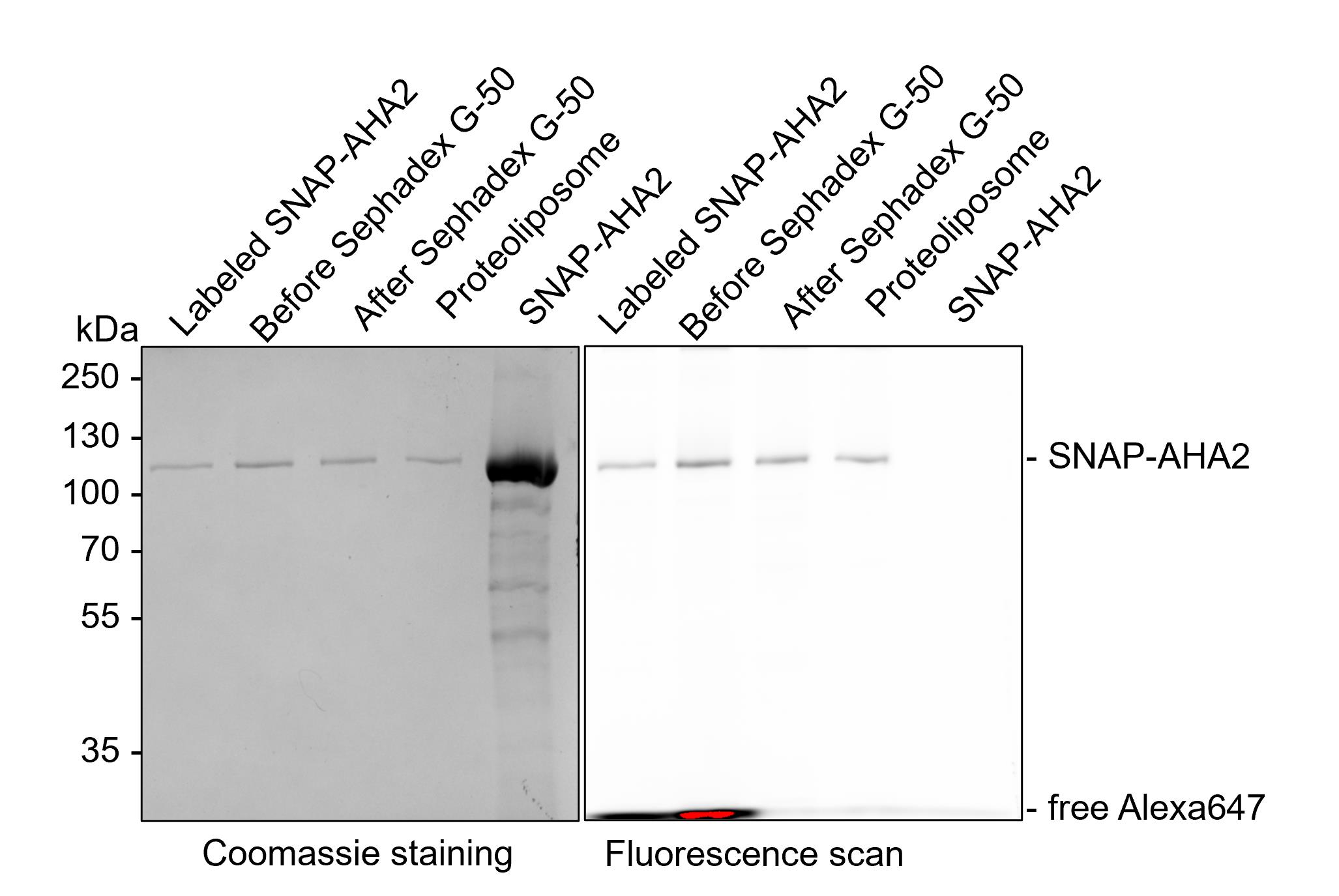
Figure 4. Labeling and reconstitution of AHA2 into large unilamellar vesicles (LUVs). Representative Alexa647 fluorescence images and Coomassie Brilliant Blue–stained SDS–PAGE, acquired using a GelDoc system (Bio-Rad), of samples collected at successive stages of the workflow: before labeling (SNAP–AHA2), after the labeling reaction (Alexa647-labeled SNAP–AHA2), before and after size-exclusion chromatography (before and after Sephadex G-50), and after reconstitution into proteoliposomes (proteoliposomes). Comparison of SNAP–AHA2 and Alexa647-labeled SNAP–AHA2 confirms successful labeling of the proton pump, with bands corresponding to the expected molecular weight of SNAP-AHA2 (119 kDa). The presence of bands of identical molecular weight in all samples demonstrates that the fluorophore remains covalently attached throughout purification and reconstitution and confirms the incorporation of Alexa647-labeled SNAP–AHA2 into proteoliposomes. Free Alexa647 dye was efficiently removed by Sephadex G-50 chromatography.
General notes and troubleshooting
General notes
1. This protocol is optimized for the plant plasma membrane proton pump AHA2. When this protocol is applied to other membrane proteins, the lipid composition of the vesicles and the choice of detergent must be carefully considered. To select an appropriate lipid composition, it is useful to consider the native lipid environment of the protein. In addition, different detergents and detergent-removal strategies may be required. Depending on the properties of the detergent, suitable approaches may include dialysis, gel filtration, or adsorption using polystyrene beads.
2. The buffer composition during reconstitution should also be considered. For AHA2, K2SO4 at pH 7.0 is required, whereas other proteins may require different pH values and/or salt compositions.
3. In this protocol, a lipid-to-protein mass ratio of 67:1 is used. This ratio can be adjusted individually for each protein. To achieve higher activity, the number of incorporated AHA2 per vesicle can be increased. This can be done by increasing the amount of protein added during reconstitution or reducing the LUV concentration, particularly when protein yields are low.
Troubleshooting
Problem 1: No detectable proton transport activity despite successful protein incorporation into LUVs.
Possible cause 1: Loss of protein activity during labeling and/or reconstitution.
Solution: Validate AHA2 activity at multiple stages of the workflow, including the solubilized stage, after labeling, and after reconstitution into proteoliposomes. Protein incorporation efficiency should be assessed by collecting samples after each step and analyzing them by SDS–PAGE. Band intensities can be quantified to identify potential protein loss during the procedure. If activity loss is observed, alternative detergents or detergent-removal methods may be tested; milder detergents like CHAPS and/or approaches such as dialysis can help preserve protein activity. Furthermore, optimization of the lipid composition, protein-to-lipid ratio, detergent-to-lipid ratio, or detergent removal rate may improve protein stability, incorporation efficiency, and functional homogeneity.
Possible cause 2: Inward orientation of the ATP-binding domain of AHA2.
Solution: Verify the orientation of AHA2 within the liposomes using, for example, fluorescence-based methods involving self-labeling enzyme tags combined with membrane-impermeable fluorescent probes, protease protection assays, or antibody accessibility assays performed in the presence and absence of membrane permeabilization [19,23].
Problem 2: Low number of proteo-GUVs.
Possible cause: Insufficient lipid film thickness during swelling.
Solution: Increase the applied volume of proteoliposomes on the PVA-coated glass slide and/or extend the dehydration time to generate a thicker dried lipid film, which can improve GUV formation.
Problem 3: Loss of protein activity after generation of proteo-GUVs.
Possible cause: Excessive dehydration during the swelling procedure.
Solution: Reduce the dehydration time or apply a weaker vacuum during this step to achieve gentler dehydration. Alternatively, add stabilizing agents such as sucrose or trehalose to the proteoliposomes to protect membrane protein activity [24,25].
Problem 4: Proteo-GUVs are leaky to the fluorescent dye (pyranine) or to transported protons.
Possible cause: Insufficient membrane tightness.
Solution: Modify the lipid composition and include sterols to increase membrane tightness and reduce membrane permeability.
Problem 5: Proteo-GUVs move rapidly, making time-lapse imaging difficult.
Possible cause: Lack of surface immobilization.
Solution: Immobilize proteo-GUVs using biotinylated lipids and streptavidin-coated glass slides [8]. Note that immobilization may affect membrane properties and should be carefully evaluated for potential effects on protein activity and membrane integrity.
Acknowledgments
This protocol was adapted from our previous work [22]. Research in the author’s laboratory is funded by grants from the Deutsche Forschungsgemeinschaft (GU 1133/13-1; GU 1133/15-1; INST 213/886-1) and DAAD (57681398). The figures were prepared using Biorender.com.
Authors’ contribution
HDU and TGP designed the study. HDU performed the experiments. HDU and TPG wrote the manuscript and supervised the project. All authors reviewed and approved the final manuscript for submission.
Competing interests
The authors declare no conflicts of interest.
References
- Rigaud, J. L. and Lévy, D. (2003). Reconstitution of Membrane Proteins into Liposomes. Meth Enzymol. 372: 65–86. https://doi.org/10.1016/s0076-6879(03)72004-7
- Sondergaard, T. E., Schulz, A. and Palmgren, M. G. (2004). Energization of Transport Processes in Plants. Roles of the Plasma Membrane H+-ATPase. Plant Physiol. 136(1): 2475–2482. https://doi.org/10.1104/pp.104.048231
- Li, Y., Zeng, H., Xu, F., Yan, F. and Xu, W. (2022). H+-ATPases in Plant Growth and Stress Responses. Annu Rev Plant Biol. 73(1): 495–521. https://doi.org/10.1146/annurev-arplant-102820-114551
- Basu, A., Maity, P., Karmakar, P. and Karmakar, S. (2017). Preparation of Giant Unilamellar Vesicles and Solid Supported Bilayer from Large Unilamellar Vesicles: Model Biological Membranes. J Surf Sci Technol. 32: 85. https://doi.org/10.18311/jsst/2016/7753
- Fenz, S. F. and Sengupta, K. (2012). Giant vesicles as cell models. Integr Biol. 4(9): 982. https://doi.org/10.1039/c2ib00188h
- Walde, P., Cosentino, K., Engel, H. and Stano, P. (2010). Giant Vesicles: Preparations and Applications. ChemBioChem. 11(7): 848–865. https://doi.org/10.1002/cbic.201000010
- Tosaka, T. and Kamiya, K. (2023). Function Investigations and Applications of Membrane Proteins on Artificial Lipid Membranes. Int J Mol Sci. 24(8): 7231. https://doi.org/10.3390/ijms24087231
- Mathiassen, P. and Pomorski, T. (2022). A Fluorescence-based Assay for Measuring Phospholipid Scramblase Activity in Giant Unilamellar Vesicles. Bio Protoc. 12(6): e4366. https://doi.org/10.21769/bioprotoc.4366
- Bhatia, T., Bhatia, T., Cornelius, F., Mouritsen, O. G. and Ipsen, J. H. (2016). Reconstitution of transmembrane protein Na+,K+-ATPase in giant unilamellar vesicles of lipid mixtures involving PSM, DOPC, DPPC and cholesterol at physiological buffer and temperature conditions. Protocol Exchange: e010. https://doi.org/10.1038/protex.2016.010
- Garten, M., Aimon, S., Bassereau, P. and Toombes, G. E. S. (2015). Reconstitution of a Transmembrane Protein, the Voltage-gated Ion Channel, KvAP, into Giant Unilamellar Vesicles for Microscopy and Patch Clamp Studies. J Visualized Exp.: e3791/52281. https://doi.org/10.3791/52281
- Horger, K. S., Liu, H., Rao, D. K., Shukla, S., Sept, D., Ambudkar, S. V. and Mayer, M. (2015). Hydrogel-assisted functional reconstitution of human P-glycoprotein (ABCB1) in giant liposomes. Biochimica et Biophysica Acta (BBA) – Biomembranes. 1848(2): 643–653. https://doi.org/10.1016/j.bbamem.2014.10.023
- Kahya, N., Pécheur, E. I., de Boeij, W. P., Wiersma, D. A. and Hoekstra, D. (2001). Reconstitution of Membrane Proteins into Giant Unilamellar Vesicles via Peptide-Induced Fusion. Biophys J. 81(3): 1464–1474. https://doi.org/10.1016/s0006-3495(01)75801-8
- Bian, T., Autry, J. M., Casemore, D., Li, J., Thomas, D. D., He, G. and Xing, C. (2016). Direct detection of SERCA calcium transport and small-molecule inhibition in giant unilamellar vesicles. Biochem Biophys Res Commun. 481: 206–211. https://doi.org/10.1016/j.bbrc.2016.10.096
- Dezi, M., Di Cicco, A., Bassereau, P. and Lévy, D. (2013). Detergent-mediated incorporation of transmembrane proteins in giant unilamellar vesicles with controlled physiological contents. Proc Natl Acad Sci USA. 110(18): 7276–7281. https://doi.org/10.1073/pnas.1303857110
- Cid, A., Perona, R. and Serrano, R. (1987). Replacement of the promoter of the yeast plasma membrane ATPase gene by a galactose-dependent promoter and its physiological consequences. Curr Genet. 12(2): 105–110. https://doi.org/10.1007/bf00434664
- Lanfermeijer, F. C., Venema, K. and Palmgren, M. G. (1998). Purification of a Histidine-Tagged Plant Plasma Membrane H+-ATPase Expressed in Yeast. Protein Expression Purif. 12(1): 29–37. https://doi.org/10.1006/prep.1997.0788
- Uzun, H., Vázquez-Hernández, M., Bandow, J. and Pomorski, T. (2022). In vitro Assay to Evaluate Cation Transport of Ionophores. Bio Protoc. 12(22): e4552. https://doi.org/10.21769/bioprotoc.4552
- Van Der Linden, I., Herrera, S., Montigny, C., Lenoir, G., Pomorski, T. and Uzun, H. (2025). A Fluorescence-Based Flippase Assay to Monitor Lipid Transport by Drs2-Cdc50. Bio Protoc. 15(1376): e5393. https://doi.org/10.21769/bioprotoc.5393
- Paweletz, L., Veit, S. and Pomorski, T. (2022). A Fluorescence-based Approach Utilizing Self-labeling Enzyme Tags to Determine Protein Orientation in Large Unilamellar Vesicles. Bio Protoc. 12(21): e4542. https://doi.org/10.21769/bioprotoc.4542
- Chandradoss, S. D., Haagsma, A. C., Lee, Y. K., Hwang, J. H., Nam, J. M. and Joo, C. (2014). Surface Passivation for Single-molecule Protein Studies. J Visualized Exp.: e3791/50549. https://doi.org/10.3791/50549
- Lira, R. B., Steinkühler, J., Knorr, R. L., Dimova, R. and Riske, K. A. (2016). Posing for a picture: vesicle immobilization in agarose gel. Sci Rep. 6(1): 25254. https://doi.org/10.1038/srep25254
- Uzun, H. D., Malysenko, E., Justesen, B. H. and Pomorski, T. G. (2025). Functional reconstitution of plant plasma membrane H+-ATPase into giant unilamellar vesicles. Sci Rep. 15(1): 8541. https://doi.org/10.1038/s41598-025-92663-9
- Veit, S., Paweletz, L. C. and Günther Pomorski, T. (2023). Determination of membrane protein orientation upon liposomal reconstitution down to the single vesicle level. Biol Chem. 404(7): 647–661. https://doi.org/10.1515/hsz-2022-0325
- Crowe, L., Reid, D. and Crowe, J. (1996). Is trehalose special for preserving dry biomaterials? Biophys J. 71(4): 2087–2093. https://doi.org/10.1016/s0006-3495(96)79407-9
- Mensink, M. A., Frijlink, H. W., van der Voort Maarschalk, K. and Hinrichs, W. L. (2017). How sugars protect proteins in the solid state and during drying (review): Mechanisms of stabilization in relation to stress conditions. Eur J Pharm Biopharm. 114: 288–295. https://doi.org/10.1016/j.ejpb.2017.01.024
Article Information
Publication history
Received: Jan 16, 2026
Accepted: Mar 18, 2026
Available online: Apr 3, 2026
Published: May 5, 2026
Copyright
© 2026 The Author(s); This is an open access article under the CC BY-NC license (https://creativecommons.org/licenses/by-nc/4.0/).
How to cite
Uzun, H. D. and Pomorski, T. G. (2026). Reconstitution of Active Plant H+-ATPase AHA2 in Giant Unilamellar Vesicles. Bio-protocol 16(9): e5675. DOI: 10.21769/BioProtoc.5675.

 Download Citation in RIS Format
Category
Biochemistry > Protein > Activity
Biochemistry > Lipid > Membrane lipid
Cell Biology > Cell imaging > Confocal microscopy
Do you have any questions about this protocol?
Post your question to gather feedback from the community. We will also invite the authors of this article to respond.
 Post a Question


