


- Protocols

- Articles and Issues
- For Authors
- About
- Become a Reviewer


Quantitative Assessment of Heat Shock-Induced Ferroptosis-Like Cell Death via Electrolyte Leakage in Arabidopsis thaliana Seedlings

Published: Vol 16, Iss 9, May 5, 2026 DOI: 10.21769/BioProtoc.5650 Views: 376
Reviewed by: Noelia ForesiKishwar Jahan ShethiAnonymous reviewer(s)
 PDF
PDF  Ask a question
Original research article
The authors used this protocol in:
Dec 2022
Advertisement



Protocol Collections
Comprehensive collections of detailed, peer-reviewed protocols focusing on specific topics




Related protocols
Abstract
We present a protocol to allow continuous assessment of cell death in Arabidopsis thaliana (L.) seedlings by measuring the release of electrolytes from dying cells upon heat shock. The electrolyte leakage assay is a well-established method to quantify the extent of cell death of plant tissues exposed to pathogen infection, since the activation of the immune response leads to compromised membrane integrity and to the release of ions from the dying cell. This prolonged release of electrolytes is considered a hallmark of regulated cell death in plants. Heat shock in plants induces ferroptosis-like cell death, which can be suppressed either pharmacologically, using inhibitors such as ferrostatin, or genetically through knockout of ferroptosis-related genes. Here, we have adapted the electrolyte leakage assay to quantify cell death in young Arabidopsis seedlings exposed to a heat shock previously shown to induce ferroptosis-like cell death. We also illustrate how this method can be used to assess activation of ferroptosis-like cell death in whole Arabidopsis seedlings using ferrostatin or knockout mutants of potential gene candidates involved in ferroptosis-like cell death.
Key features
• This protocol does not require any technical experience apart from gentle handling of young seedlings and is less labor-intensive than microscopy-based cell death evaluation.
• Builds upon existing methods to quantify the extent of cell death upon immune response in whole seedlings subjected to heat stress.
• Only requires a conductivity meter and allows the assessment of continuous cell death using multiple parallel replicates.
• The protocol demonstrates how heat shock-induced ferroptosis-like cell death can be inhibited pharmacologically or genetically in whole seedlings, supported with quantitative data.
Keywords: Heat shock Ferroptosis-like cell death
Electrolyte leakage
Arabidopsis
Regulated cell death
Ferrostatin
Conductivity meter
Ferroptosis-like cell death
Electrolyte leakage
Arabidopsis
Regulated cell death
Ferrostatin
Conductivity meter
Graphical overview
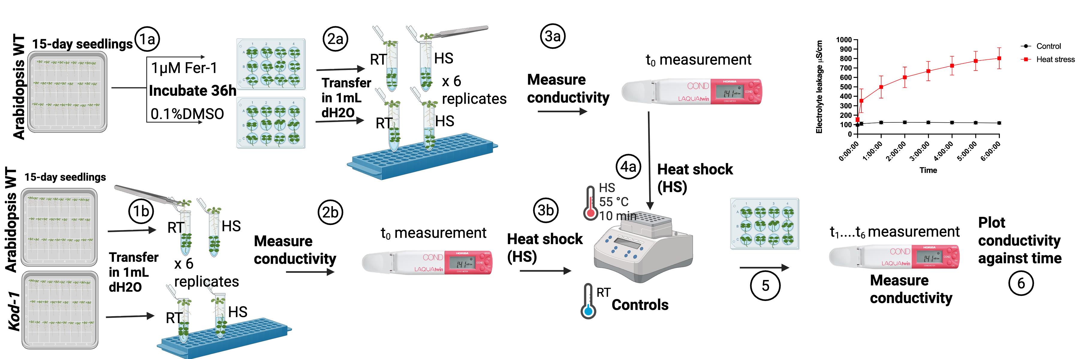
Overview of workflow for quantitative assessment of heat shock-induced cell death by electrolyte leakage assay in Arabidopsis. (1a) Arabidopsis wild-type (WT) seedlings are transferred to a 12-well plate and incubated for 36 h with 1 µM Ferrostatin-1 (Fer-1) or 0.1% DMSO. (2a) Seedlings are transferred to a 2 mL tube containing 1 mL of distilled water. (3a) T0 conductivity measurements are taken from each replicate. (4a) Tubes containing seedlings are incubated at 55 °C for 10 min (HS) or kept at room temperature for 10 min (RT). (1b) Arabidopsis WT and knockout seedlings for the gene of interest (Kiss of death, kod-1 in this case) are transferred to a 2 mL tube containing 1 mL of distilled water. (2b) T0 conductivity measurements are taken from each replicate. (3b) Tubes containing seedlings are incubated at 55 °C for 10 min (HS) or kept at room temperature for 10 min (RT). (5) Conductivity measurements are repeated for 150 min with measurements every 30 min. (6) Data analysis.
Background
Regulated cell death (RCD) is a controlled form of suicide that plant cells activate upon mild homeostatic disturbances, such as pathogen infection and heat stress. Upon biotic or abiotic stress, damaged cells are eliminated to ensure survival of the plant, for instance, to restrict pathogen proliferation from the point of infection [1,2]. RCD also participates during development, contributing to cell differentiation and ensuring the proper development of vegetative and reproductive organs [2,3].
RCD triggered upon pathogen recognition is characterized by compromised membrane integrity, with the concomitant leakage of cytoplasmic electrolytes out of the cell [4]. When dying plants are kept in an aqueous solution, the release of these electrolytes can be used as a proxy for cell death quantification. Current protocols for electrolyte leakage assays use simple and affordable conductivity meters, which allow continuous and repeated measurements of the electrolytes released by dying cells [5–7]. However, to date, these protocols are mainly used for plants exposed to pathogens, and no standardized protocol exists to quantitatively measure cell death induced by heat stress.
Ferroptosis is a form of RCD that is iron and reactive oxygen species (ROS) dependent and results in lipid peroxidation [8]. Ferroptosis-like cell death has recently been described in Arabidopsis upon heat shock and can be suppressed by ferrostatin, which inhibits lipid peroxidation [9]. Ferrostatin is a lipophilic antioxidant that scavenges lipid ROS radicals via its N-cyclohexyl moiety, thereby preventing cell membrane damage and subsequent cell death [10]. Additionally, several genes are upregulated during ferroptosis-like cell death in Arabidopsis, including Kiss of Death (KOD), which encodes a 25 amino acid peptide [9,11]. Ferroptosis-like cell death upon heat shock in Arabidopsis is visualized using the DNA probe Sytox Green [9,12]. In this case, the compromised plasma membrane integrity allows Sytox Green to penetrate the dying cell and to fluoresce upon DNA binding. While this method has been used to demonstrate heat shock–induced ferroptosis-like cell death in Arabidopsis roots, it has limitations: it is labor-intensive, requires a fluorescence or confocal microscope, and does not allow following the progression of cell death over time.
Here, we propose an adapted version of the electrolyte leakage assay to evaluate cell death of entire Arabidopsis seedlings upon heat shock. This new protocol uses a small and affordable conductivity meter and allows the parallel evaluation of several replicates per treatment (with several seedlings per replicate) and for multiple measurements of the same sample over time. We also show how the protocol can be adapted to include inhibitors or mutants to evaluate potential candidate genes involved in heat shock–induced ferroptosis-like cell death, in combination with statistical evaluation.
Materials and reagents
Biological materials
1. Arabidopsis thaliana (L), Colombia wildtype ecotype (Col.0) seeds
2. Kiss of death (kod-1), loss of function mutant seeds (At4g10613: LINE retrotransposon in Col.0; kod-1 T-DNA insertion in the GABI-kat line) [11]
Reagents
1. Murashige & Skoog (MS) basal salts (Sigma-Aldrich, catalog number: M5519)
2. 2-Morpholinoethanesulfonic acid monohydrate (MES) salt (Sigma-Aldrich, CAS number: 145224-94-8)
3. Sucrose (Sigma-Aldrich, CAS number: 57-50-1)
4. Agar (Sigma-Aldrich, CAS number: 9002-18-0)
5. Gellan gum (Sigma-Aldrich, CAS number: 71010-52-1)
6. Ethanol, 96% (VWR, CAS number: 64-17-5)
7. Hydrochloric acid (HCl) (Merck Millipore, CAS number: 7647-01-0)
8. Bleach (Chlorine)
9. Ferrostatin-1 (Fer-1) (Sigma-Aldrich, CAS number: 347174-05-4)
10. Dimethyl Sulfoxide (DMSO) (Thermo-Scientific, CAS number: 67-68-5)
11. Distilled water (dH2O)
Solutions
1. 1/2 MS basal salts medium (see Recipes)
2. Fer-1 solution (20 mM, 1 μM) (see Recipes)
3. Bleach, 50% (see Recipes)
4. Ethanol, 70% (see Recipes)
Recipes
1. 1/2 MS basal salts medium
| Reagent | Final concentration | Quantity or volume |
|---|---|---|
| MS basal salts | 2.2 g/L | 800 mL |
| MES salt | 0.05% | 0.5 g |
| Sucrose | 1% | 10 g |
| Gellan gum* or agar** | 0.8%* or 1%** | 8 g or 10 g |
| dH2O | 1,000 mL |
Gellan gum or agar can be safely used as a gelling agent in all experiments to prepare in-vitro 1/2 MS medium.
Weigh MS basal salts, MES, and sucrose and transfer to a large beaker. Add 800 mL of ultrapure dH2O and stir. Adjust pH to 5.7 with 2 M HCl using a calibrated pH meter. Top up volume to 1,000 mL. Divide the media into two 1,000 mL bottles. Weigh 4 g of gellan gum (or 5 g of agar) and add to each bottle. Autoclave the media at 121 °C for 20 min. Leave the media to cool to 55–60 °C and pour 50 mL aseptically into square plates. Leave the plates to solidify for 20 min in a laminar flow cabinet. Seal and keep plates at 4 °C for up to 1 month.
2. Fer-1 solution (20 mM, 1 μM)
| Reagent | Final concentration | Quantity or volume |
|---|---|---|
| Fer-1 stock | 20 mM in DMSO | 953 μL |
| Fer-1, 1:10 dilution | 2 mM in DMSO | 10 μL |
| DMSO working dilution | 0.1% v/v DMSO | 50 mL |
| Fer-1 working stock | 1 μM in 0.1% DMSO | 15 mL |
Dissolve 5 mg of Fer-1 powder (molecular weight = 262.35 g/mol) in 953 μL of DMSO to make a 20 mM stock solution. Aliquot into 10 μL and freeze at -20 °C. Next, dilute the stock (20 mM) 1:10 by taking 1 μL into 9 μL of DMSO to make 2 mM Fer-1. To prepare 0.1% DMSO, dilute 50 μL of DMSO into 50 mL of ultrapure water. Finally, prepare 1 μM Fer-1 by diluting 7.5 μL of working dilution (2 mM) into 15 mL of 0.1% DMSO.
3. Bleach, 50% v/v
| Reagent | Final concentration | Volume (for 10 mL) |
|---|---|---|
| Bleach, 50% v/v | 50% v/v | 5 mL |
| dH2O | n/a | 5 mL |
| Total | 50% | 10 mL |
4. Ethanol, 70% v/v
| Reagent | Final concentration | Volume (for 100 mL) |
|---|---|---|
| Ethanol, 70% v/v | 70% v/v | 72.9 mL |
| dH2O | n/a | 27.1 mL |
| Total | 70% | 100 mL |
Laboratory supplies
1. 1.5 mL microcentrifuge tubes (Sarstedt, catalog number: 690.001)
2. 2 mL microcentrifuge tubes (Sarstedt, catalog number: 72.695.500)
3. Pipette tips 10 μL (Sarstedt, catalog number: 70.3010)
4. Pipette tips 200 μL (Sarstedt, catalog number: 70.3030.100)
5. Pipette tips 1,000 μL (Sarstedt, catalog number: 70.3050.100)
6. 1,000 mL bottles (VWR, catalog number: 215-1595)
7. 50 mL Falcon tubes (Sarstedt, catalog number: 62.547.254)
8. Square plates 120 × 120 × 17 with vents (Greiner Bio-One, catalog number: 688102)
9. 12-well culture plates (CellStar, catalog number: 665 180)
10. 2,000 mL beaker (VWR, catalog number: 216-1643)
10. Aluminum foil
11. 3M Micropore tape
12. Spoon or spatulas (stainless steel)
13. Tweezers
Equipment
1. Growth chamber (CFL Plant climatics, model: AR-66L2)
2. Sterile hood (Holten LaminAir, model: HB2448)
3. Compact conductivity meter (HORIBA, model: LAQUAtwin-EC-33)
4. Autoclave (TOMY, model: SX-700E)
5. Thermomixer F1.5 (Eppendorf, catalog number: 5384000012)
6. pH meter (Radiometer analytical, model: PHM210)
7. 0.2–2 μL Finnpipette (Thermo Fisher Scientific, catalog number: 4641010N)
8. 2–20 μL Finnpipette (Thermo Fisher Scientific, catalog number: 4641050N)
9. 20–200 μL Finnpipette (Thermo Fisher Scientific, catalog number: 4641080N)
10. 100–1,000 μL Finnpipette (Thermo Fisher Scientific, catalog number: 4641100N)
11. Racks
12. Refrigerator (2–8 °C)
13. Freezer (-20 °C)
14. Cold room (2–8 °C)
15. ELGA water purification system (ELGA LabWater, model: PURELAB Quest)
16. Weighing scale (OHAUS CORP, model: Pioneer PA2102)
Procedure
A. Sterilization and sowing of Arabidopsis seeds
1. Transfer 130–140 Arabidopsis seeds to a 1.5 mL tube.
2. Add 1 mL of 70% ethanol and invert the tube a few times.
3. Allow the seeds to settle down at the bottom of the tube and remove the ethanol.
4. Add 1 mL of 50% bleach.
5. Wait for 5 min inverting the tube every few seconds.
6. Allow the seeds to settle down at the bottom of the tube and remove the bleach.
7. Wash with 1 mL of sterile dH2O.
8. Allow the seeds to settle down at the bottom of the tube and remove the water.
9. Repeat steps A7–8 four more times.
10. Using a 2–20 µL pipette, take one seed at a time and place on the 1/2 MS plate.
11. Repeat step A10 to sow three rows of seeds, with 13–15 seeds per row.
12. Seal plates with micropore tape and cover with aluminum foil.
13. Place plates in a cold room at 4 °C for approximately 48 h.
14. Take plates out of the cold and place them vertically in the growth chamber (16 h light at 21 °C and 8 h dark at 18 °C, 50% RH).
15. Grow seedlings for 15 days.
Notes:
1. One experiment requires 100 Col-0 Arabidopsis seedlings: 50 seedlings for the room temperature control and 50 seedlings for the heat shock treatment. We normally sow 130–140 seeds to account for delayed or no germination of some seeds. We sow 30 seeds per plate.
2. For Kod-1, sow similar amounts of seeds per plate.
3. The experiment with Fer-1 and DMSO (see step B3) requires 200 WT Arabidopsis seedlings.
B. Heat shock of Arabidopsis seedlings
B1. Calibrate the conductivity meter
1. Apply conditioning solution to the sensor and incubate for 10 min.
2. Wash with dH2O and apply calibration buffer 1, 1.41 ms/cm solution.
3. Press CAL.
4. Rinse with dH2O and apply calibration buffer 2, 12.9 ms/cm solution.
5. Press CAL.
6. Finally, rinse with dH2O and start measurements.
B2. Conductivity measurements
Note: This experiment has two treatments (room temperature and heat shock) with ten replicates per treatment, and five seedlings per replicate. In total, each experiment uses 100 seedlings.
1. Switch on the heat block and set it to 55 °C.
2. Transfer 1 mL of dH2O to twenty 2-mL safe lock tubes.
3. Label 10 tubes as RT (room temperature) and 10 tubes as HS (heat shock).
4. Carefully lift the seedlings with a pair of tweezers from under the leaves to avoid damage.
5. Transfer 5 seedlings into each tube as shown in Figure 1C.
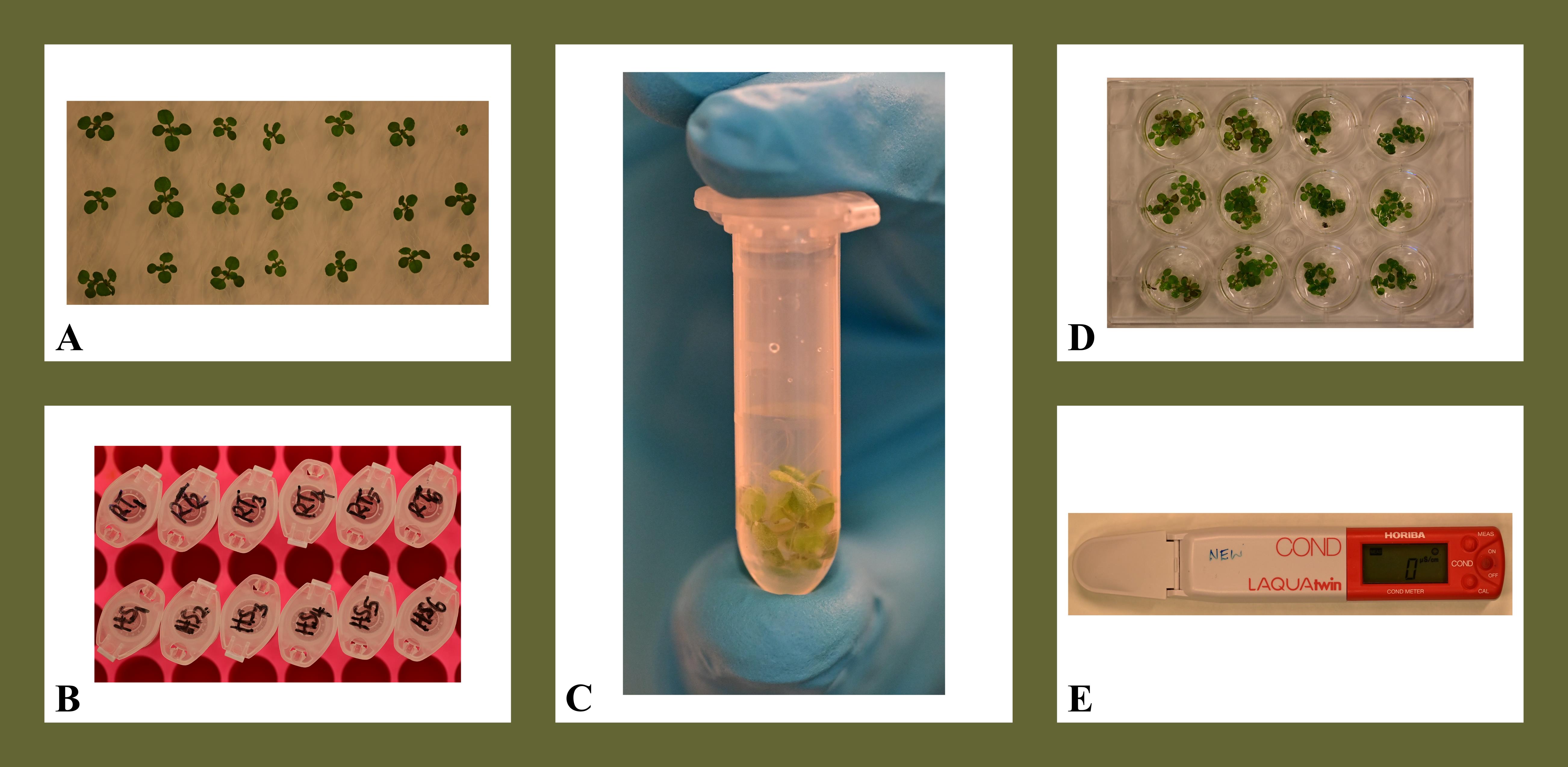
Figure 1. Procedure for quantitative assessment of cell death upon heat shock in Arabidopsis. (A) 15-day-old Arabidopsis seedlings grown in 1/2 MS media. (B) 2 mL tubes, each with 1 mL of Milli Q water to transfer seedlings. (C) 2 mL microcentrifuge tube with 5 seedlings inside. (D) 12-well plate where heat shock and control plants are transferred to. (E) Conductivity meter.
Note: This experiment did not involve handling of toxic chemicals. When working with DMSO and ferrostatin, use gloves and protective wear.
6. Before exposure to heat shock, pipette 120 μL of the water in which the seedlings are submerged onto the sensor of the conductivity meter. This will be the t0 measurement.
7. Pipette back the 120 μL into the respective tube after each measurement.
8. Wash the sensor with dH2O in between each measurement.
9. Repeat the measurements for all 12 tubes.
10. Place the 10 HS tubes containing seedlings on the heat block set at 55 °C for 10 min.
11. Keep the 10 RT tubes at room temperature as controls.
12. After 10 min, remove HS tubes from the heat block.
13. Take measurements immediately after for both HS and RT controls.
14. Transfer the seedlings of each tube into a well of a 12-well plate.
15. Take measurements every 30 min for the next 3 h.
16. Plot a graph of conductivity against time to assess electrolyte leakage over time.
B3. Preincubation with Ferrostatin-1 (Fer-1)
Note: This experiment includes four treatments (DMSO/room temperature, DMSO/heat shock, Fer-1/room temperature, and Fer-1/heat shock) with ten replicates per treatment, and five seedlings per replicate. In total, each experiment uses 200 seedlings.
1. Label four 12-well plates as Fer-1.
2. Label four 12-well plates as DMSO.
3. In two of the 12-well plates labeled Fer-1, pipette 1 mL of 1 μM Fer-1 into 20 wells (keep the other two plates labeled Fer-1 for step B3.15).
4. In two 12-well plates labeled DMSO, pipette 1 mL of 0.1% DMSO into 20 wells (keep the other two plates labeled DMSO for step B3.15).
Caution: DMSO and Fer-1 are toxic chemicals; make sure you wear gloves and a lab coat while preparing stocks and performing this experiment.
5. Carefully lift the seedlings with a pair of tweezers to avoid damage.
6. Transfer 5 seedlings into each well.
7. Submerge the seedlings in Fer-1 or 0.1% DMSO solution and incubate for 36–48 h preceding the heat shock experiment in the growth chamber.
Note: We sometimes observed darkening of the roots during incubation for 48 h, so we decided to adjust the incubation time with Fer-1 for 36 h.
8. Label forty 2-mL microcentrifuge tubes as follows: 10 tubes as DMSO, RT; 10 tubes as DMSO, HS; 10 tubes as Fer-1, RT; and 10 tubes as Fer-1, HS.
9. Pipette 1 mL of dH2O in each of the 40 tubes.
10. Blot dry the seedlings from each well and transfer them into the corresponding tube.
11. Measure initial conductivity (t0) for all tubes.
12. Place the 20 tubes labeled HS (labeled DMSO or Fer-1) in the heat block at 55 °C for 10 min.
13. Keep the tubes labeled RT (labeled DMSO or Fer-1) at room temperature.
14. Repeat conductivity measurements immediately after 10 min for all tubes.
15. Transfer seedlings into respective 12-well plates and incubate for 30 min (plates prepared in steps B3.3 and B3.4).
16. Repeat conductivity measurements every 30 min for a total of 3 h.
17. Plot a graph of conductivity over time to assess electrolyte leakage over time.
B4. Heat shock with Kod-1 mutant seedlings
1. Label 2 mL microcentrifuge tubes as follows: 10 tubes as WT, RT; 10 tubes as WT, HS; 10 tubes as KOD, RT; and 10 tubes as KOD, HS.
2. Add 1 mL of dH2O to each tube.
3. Transfer five seedlings of wild-type Col-0 to the tubes labeled WT.
4. Transfer five seedlings of kod-1 to the tubes labeled KOD.
5. Take conductivity measurements for t0 as explained above (steps B2.6 and B2.7).
6. Transfer HS tubes for wild-type Col.0 and kod-1 to the heat block and heat at 55 °C for 10 min.
7. Leave the controls at room temperature.
8. Take conductivity measurements immediately after 10 min for HS and RT.
9. Transfer seedlings into 12-well plates and incubate for 30 min.
10. Repeat conductivity measurements every 30 min for a total of 3 h.
11. Plot a graph of conductivity over time to assess electrolyte leakage over time.
Data analysis
The data collected corresponds to the exact values displayed by the conductivity meter in mS/cm. The experiment was performed three times, with 6 or 10 replicates each time, for the heat shock and control samples. Statistical analysis to determine significant differences between treatments was performed by one-way ANOVA in GraphPad Prism.
The results showed that after exposure to 55 °C for 10 min, WT Arabidopsis seedlings release electrolytes into the surrounding medium, indicative of cell death activation, as previously reported [9,12] (Figure 2A). The release of electrolytes is partially suppressed when seedlings are preincubated with 1 µM Fer-1 for 36 h prior to the heat shock treatment (Figure 2B). Electrolyte leakage is also reduced in the knockout mutant kod-1, which lacks expression of the gene KOD. KOD codes for a 25 amino acid peptide previously shown to participate in heat shock–induced cell death and to be upregulated upon heat shock (Figure 2C) [9,11].
In conclusion, this updated protocol shows how electrolyte leakage measurements can be used to evaluate the progression of cell death for whole seedlings of Arabidopsis upon heat shock. The protocol can also be used to test pharmacological inhibitors or loss/gain-of-function mutants of potential gene candidates to identify novel signaling components of heat shock–induced cell death.
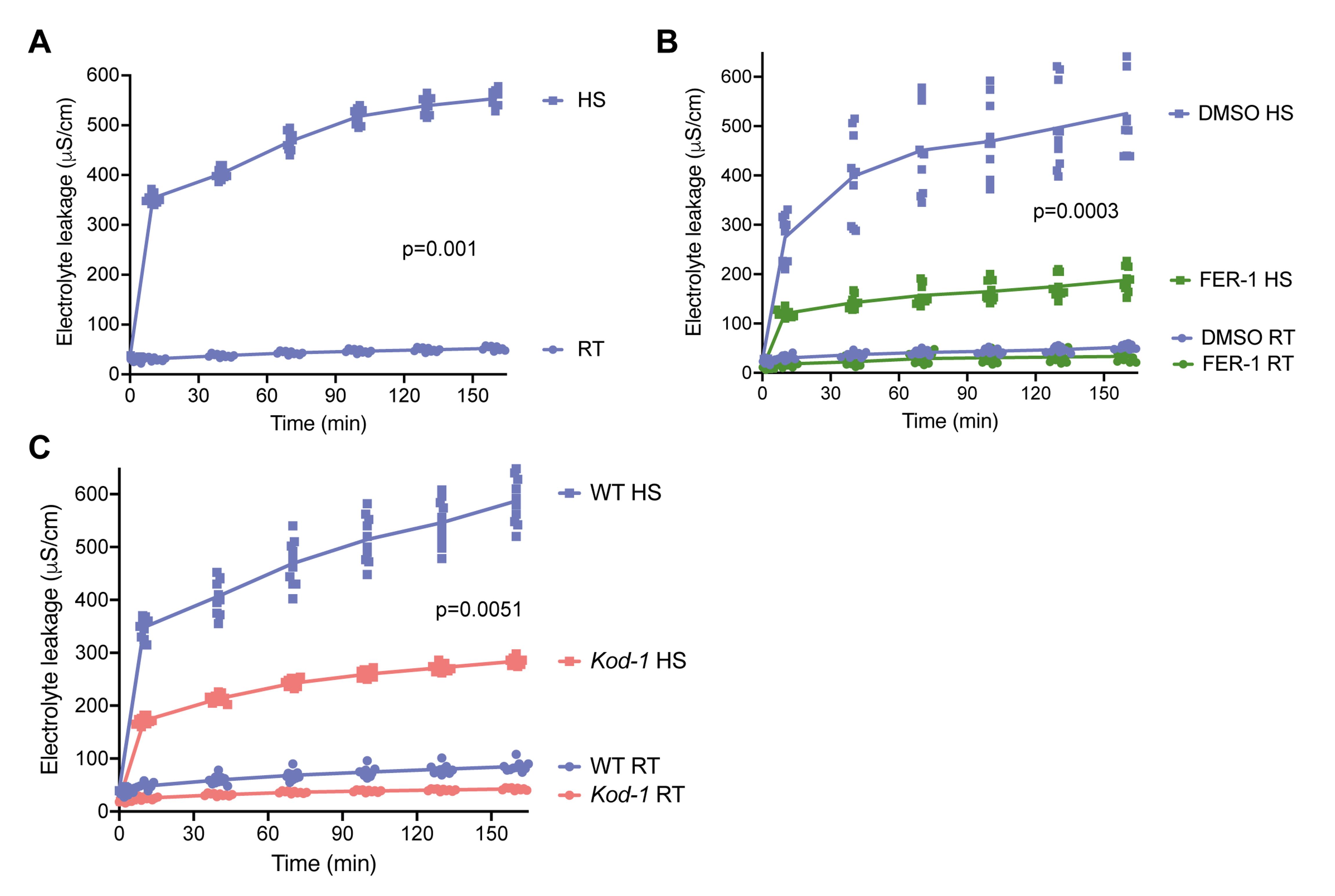
Figure 2. Heat shock induces electrolyte leakage in Arabidopsis seedlings. (A) 15-day-old Arabidopsis seedlings were exposed to 55 °C for 10 min in 1 mL of water [heat shock (HS), squares] or kept at room temperature (RT, circles). Electrolyte leakage was measured before the heat stress, immediately after (10 min), and every 30 min thereafter. (B) Thirty-six hours before the experiment described in A, plants were incubated with 1 μM Ferrostatin1 (Fer-1), and control plants were incubated with 0.1% DMSO. (C) Same experiment as described in A, with wild-type (WT) Arabidopsis and a knock-out mutant of the gene Kiss of Death (kod-1). For all graphs, each dot represents a single replicate with 5 seedlings. All treatments were performed with 10 replicates. Statistical analysis was performed by one-way ANOVA. Significant differences between HS and RT (A), DMSO and Fer-1 (B), or WT and kod-1 upon HS (C) are indicated (p < 0.05).
Validation of protocol
The data analysis section shows an example of data obtained in three different conditions. All experiments were repeated at least three times by different authors with similar results.
The data shown in Figure 2 shows statistically significant values between heat-shock and control Arabidopsis seedlings. In addition, pre-treatment with Fer-1 shows a significant reduction in electrolyte leakage in heat-shock seedlings compared to non-treated seedlings. Lastly, loss of function in the Kod-1 gene significantly reduces electrolyte leakage in seedlings exposed to heat shock.
This protocol or parts of it has been used and validated in the following research article:
• Aguilera et al. [13]. Do photosynthetic cells communicate with each other during cell death? From cyanobacteria to vascular plants. J Exp Bot. 73(22): 7219–7242. https://doi.org/10.1093/jxb/erac363
General notes and troubleshooting
General notes
1. Calibrate the conductivity meter before each experiment. Include a preincubation step with conditioning solution to remove residual salts on the sensor and rinse with ultrapure dH2O. Also, rinse with ultrapure dH2O between measurements.
2. When handling seedlings, carefully lift them using a pair of tweezers and gently transfer them to 12-well plates or 2 mL tubes. Make sure seedlings are submerged at all times, including the roots.
3. Keep the seedlings with gentle agitation between measurements.
4. Remember to return the 120 μL solution to the respective wells to ensure a constant volume is maintained in the entire experiment.
Troubleshooting
Problem 1: Uneven seedling growth.
Possible cause: Too many seeds sown on one plate.
Solution: Space seeds evenly on one plate to allow them space and nutrients to grow evenly.
Problem 2: Conductivity measurements vary too much.
Possible causes: Seedling size variation; seedlings were not handled with care, causing more release of electrolytes due to mechanical stress. The conductivity meter may also harbor residual salts due to insufficient washing.
Solutions: Use seedlings of similar size and handle seedlings with care using a pair of tweezers. Rinse the conductivity meter sufficiently between each measurement. Also, consider increasing the number of replicates to reduce technical variation.
Acknowledgments
Conceptualization, L.G. and D.S.; Investigation, L.G., C.C.S., M.S.C., G.W.; Writing—Original Draft, L.G. and C.C.S.; Writing—Review & Editing, L.G. and D.S.; Funding acquisition, D.S.; Supervision, D.S. We would like to thank Patrick Gallois for providing kod-1 seeds. We would like to thank Maximilian Fuchs for the beautiful Arabidopsis pictures. We would like to thank Liv Ashcroft for lab support. G. Whitmore was a participant of the TRONDBUSS initiative through the STILSA (Students in Life Sciences Abroad) and we gratefully acknowledge the program's support. This work was supported by a Norwegian University of Science and Technology PhD fund to L.G. The Graphical overview was created with Biorender.com. This protocol is adapted and developed from [7,9,11]. This protocol was used in [13].
Competing interests
The authors declare no conflicts of interest.
Ethical considerations
This work did not use human or animal subjects and has no ethical considerations.
References
- Balint-Kurti, P. (2019). The plant hypersensitive response: concepts, control and consequences. Mol Plant Pathol. 20(8): Article 8. https://doi.org/10.1111/mpp.12821
- Huysmans, M., Lema A, S., Coll, N. S. and Nowack, M. K. (2017). Dying two deaths — programmed cell death regulation in development and disease. Curr Opin Plant Biol. 35: 37–44. https://doi.org/10.1016/j.pbi.2016.11.005
- Van Hautegem, T., Waters, A. J., Goodrich, J. and Nowack, M. K. (2015). Only in dying, life: programmed cell death during plant development. Trends Plant Sci. 20(2): Article 2. https://doi.org/10.1016/j.tplants.2014.10.003
- Demidchik, V., Straltsova, D., Medvedev, S. S., Pozhvanov, G. A., Sokolik, A. and Yurin, V. (2014). Stress-induced electrolyte leakage: the role of K+-permeable channels and involvement in programmed cell death and metabolic adjustment. J Exp Bot. 65(5): 1259–1270. https://doi.org/10.1093/jxb/eru004
- Hatsugai, N., Hillmer, R., Yamaoka, S., Hara-Nishimura, I. and Katagiri, F. (2016). The μ Subunit of Arabidopsis Adaptor Protein-2 Is Involved in Effector-Triggered Immunity Mediated by Membrane-Localized Resistance Proteins. Mol Plant-Microbe Interactions®. 29(5): 345–351. https://doi.org/10.1094/MPMI-10-15-0228-R
- Hatsugai, N., Igarashi, D., Mase, K., Lu, Y., Tsuda, Y., Chakravarthy, S., Wei, H., Foley, J. W., Collmer, A., Glazebrook, J., et al. (2017). A plant effector-triggered immunity signaling sector is inhibited by pattern-triggered immunity. EMBO J. 36(18): 2758–2769. https://doi.org/10.15252/embj.201796529
- Hatsugai, N. and Katagiri, F. (2018). Quantification of Plant Cell Death by Electrolyte Leakage Assay. Bio-Protoc. 8(5): e2758. https://doi.org/10.21769/BioProtoc.2758
- Galluzzi, L., Vitale, I., Aaronson, S. A., Abrams, J. M., Adam, D., Agostinis, P., Alnemri, E. S., Altucci, L., Amelio, I., Andrews, D. W., et al. (2018). Molecular mechanisms of cell death: recommendations of the Nomenclature Committee on Cell Death 2018. Cell Death Differ. 25(3): Article 3. https://doi.org/10.1038/s41418-017-0012-4
- Distéfano, A. M., Martin, M. V., Córdoba, J. P., Bellido, A. M., D’Ippólito, S., Colman, S. L., Soto, D., Roldán, J. A., Bartoli, C. G., Zabaleta, E. J., et al. (2017). Heat stress induces ferroptosis-like cell death in plants. J Cell Biol. 216(2): Article 2. https://doi.org/10.1083/jcb.201605110
- Dixon, S. J., Lemberg, K. M., Lamprecht, M. R., Skouta, R., Zaitsev, E. M., Gleason, C. E., Patel, D. N., Bauer, A. J., Cantley, A. M., Yang, W. S., et al. (2012). Ferroptosis: An Iron-Dependent Form of Non-Apoptotic Cell Death. Cell. 149(5): 1060–1072. https://doi.org/10.1016/j.cell.2012.03.042
- Blanvillain, R., Young, B., Cai, Y., Hecht, V., Varoquaux, F., Delorme, V., Lancelin, J.-M., Delseny, M. and Gallois, P. (2011). The Arabidopsis peptide kiss of death is an inducer of programmed cell death. EMBO J. 30(6): 1173–1183. https://doi.org/10.1038/emboj.2011.14
- Distéfano, A. M., Marchetti, F., Zabaleta, E. and Pagnussat, G. C. (2022). Measuring and Perturbing Ferroptosis in Plants. In M. Klemenčič, S. Stael, and P. F. Huesgen (Eds.), Plant Proteases and Plant Cell Death: Methods and Protocols (pp. 185–192). New York, NY: Springer US. https://doi.org/10.1007/978-1-0716-2079-3_15
- Aguilera, A., Distéfano, A., Jauzein, C., Correa-Aragunde, N., Martinez, D., Martin, M. V. and Sueldo, D. J. (2022). Do photosynthetic cells communicate with each other during cell death? From cyanobacteria to vascular plants. J Exp Bot. 73(22): 7219–7242. https://doi.org/10.1093/jxb/erac363
Article Information
Publication history
Received: Dec 9, 2025
Accepted: Feb 26, 2026
Available online: Apr 14, 2026
Published: May 5, 2026
Copyright
© 2026 The Author(s); This is an open access article under the CC BY-NC license (https://creativecommons.org/licenses/by-nc/4.0/).
How to cite
Kago-Gachao, L., Salas, C. C., Campos, M. S., Whitmore, G. and Sueldo, D. (2026). Quantitative Assessment of Heat Shock-Induced Ferroptosis-Like Cell Death via Electrolyte Leakage in Arabidopsis thaliana Seedlings. Bio-protocol 16(9): e5650. DOI: 10.21769/BioProtoc.5650.

 Download Citation in RIS Format
Category
Plant Science > Plant physiology > Ion analysis
Cell Biology > Cell viability > Cell death
Plant Science > Plant physiology > Abiotic stress
Do you have any questions about this protocol?
Post your question to gather feedback from the community. We will also invite the authors of this article to respond.
 Post a Question


