- Protocols

- Articles and Issues
- For Authors
- About
- Become a Reviewer

A Protocol to Induce Brown and Beige Adipocyte Differentiation From Murine and Human Adipose-Derived SVF
(*contributed equally to this work) Published: Vol 15, Iss 23, Dec 5, 2025 DOI: 10.21769/BioProtoc.5522 Views: 1717
Reviewed by: Philipp WörsdörferAnonymous reviewer(s)
Original research article
The authors used this protocol in:

May 2020
Advertisement


Protocol Collections
Comprehensive collections of detailed, peer-reviewed protocols focusing on specific topics


Abstract
Adipose cells vary functionally, with white adipocytes storing energy and brown/beige adipocytes generating heat. Mouse and human subcutaneous white adipose tissue (WAT)-derived stromal vascular fraction (SVF) provides mesenchymal stem cells (MSCs) that can be differentiated into thermogenic adipocytes using pharmacological cocktails. After six days of browning induction, these cells exhibited significant upregulation of thermogenic markers (UCP1, Cidea, Dio2, PRDM16) along with adipogenic genes (PPARγ, aP2), showing enhanced thermogenic potential. This in vitro system offers a practical platform to study adipogenesis and thermogenic regulation.
Key features
• The protocol aims to differentiate murine and human SVF from subcutaneous fat into brown/beige adipocytes for assays.
• This approach employs a straightforward methodology that effectively differentiates SVF cells using optimal chemicals, their concentrations, and durations.
Keywords: Adipocyte differentiationGraphical overview

Background
Obesity, as defined by WHO, is a chronic complex condition characterized by excessive adipose tissue that can adversely affect health [1]. Adipose tissue, often known as "fat," serves as a passive energy reservoir and is involved in various physiological functions, including the regulation of food consumption, energy balance, insulin production, immune response, and the maintenance of body temperature [2,3]. Mammals possess three different types of adipocytes—white, brown, and beige adipocytes—each located in specific anatomical regions and characterized by distinct gene expression profiles [4]. White adipose tissue (WAT) is extensively found in subcutaneous areas and surrounding internal organs. Its cellular structure is characterized by a prominent single lipid droplet, minimal mitochondria, and limited cytoplasm, serving primarily as an energy reservoir. Additionally, WAT functions as an endocrine organ, releasing adipokines like leptin, lipotropin, and TNFα, which play crucial roles in regulating energy metabolism and sustaining physiological balance [5]. Brown adipose tissue (BAT), on the other hand, has many mitochondria and multilocular lipid droplets and is heavily innervated and vascularized. Its major function is thermoregulation, which involves oxidizing fatty acids (FAs) to maintain body temperature [6]. Another form of adipose tissue, known as beige adipose, is found in WAT depots but exhibits brown- like characteristics including multilocular lipid droplets and high mitochondrial density [7,8]. Brown and beige adipocytes are often referred to as “thermogenic adipocytes.” This process is facilitated by uncoupling protein 1 (UCP1) and is referred to as non-shivering thermogenesis [9,10].
Although beige adipocytes exhibit functional similarities to brown adipocytes, these two thermogenic cells differ in their developmental origins. In fact, beige adipocytes originate from the same progenitor cells as white adipocytes [11]. The process by which beige adipocytes are formed is commonly referred to as browning, typically occurring in subcutaneous fat deposits. Collectively, these findings indicate that both extrinsic and intrinsic cellular factors play a role in determining the fate of adipocyte differentiation. In this protocol, we used mouse and human stromal vascular fraction (SVF) isolated from fat depots to identify and evaluate methods for inducing differentiation into brown and beige adipocytes. Slight modifications were introduced in the earlier protocol [12] in our laboratory, where they have been replicated and refined to implement fundamental procedures with reduced toxicity and time interval. It includes the addition of optimized novel chemicals (1 nM T3 and 125 μM Indomethacine), decreased concentration of the earlier used compounds (20 nM insulin, 1 μM dexamethasone, and 0.5 μM IBMX), and shorter time duration for adipocyte differentiation.
Materials and reagents
Biological materials
1. Subcutaneous fat depots (scWAT) of mice
2. Subcutaneous fat depots (scWAT) of human
Reagents
1. Dulbecco’s modified Eagle’s medium (DMEM), high glucose (Corning, catalog number: 10-013-CV), store at 4 °C
2. Fetal bovine serum (FBS) (Thermo Fisher Scientific, catalog number: A5256801), store at -20 °C
3. Penicillin-streptomycin (Pen/Strep) (Thermo Fisher Scientific, catalog number: 15070063), store at -20 °C
4. Phosphate-buffered saline (PBS), 1× (Corning, catalog number: 21-040-CMX12)
5. Collagenase/dispase (Roche, catalog number: 10269638001)
6. TRIzol reagent (Fisher Scientific, catalog number: 15596018)
7. Chloroform (Sigma-Aldrich, catalog number: 472476)
8. Ultrapure DNase/RNase-free distilled water (nuclease-free water) (Fisher Scientific, catalog number: 10-977-023)
9. iScriptTM cDNA Synthesis kit (Bio-Rad, catalog number: 1708890)
10. iQTM SYBR Green Supermix (Bio-Rad, catalog number: 1708880)
11. Insulin (20 nM final concentration) (Sigma-Aldrich, catalog number: I1507), powder storage temperature -20 °C, store at 4 °C after dilution
12. Triiodothyronine (T3) (1 nM final concentration) (Sigma-Aldrich, catalog number: T2877), store at -20 °C
13. Indomethacin (125 μM final concentration) (Sigma-Aldrich, catalog number: I7378), store at -20 °C
14. Dexamethasone (1 μM final concentration) (Sigma-Aldrich, catalog number: D4902), store at -20 °C
15. Rosiglitazone (1 μM final concentration) (Sigma-Aldrich, catalog number: 557366-M), store at -20 °C
16. 3-Isobutyl-1-methylxanthine (IBMX) (0.5 μM final concentration) (Sigma-Aldrich, catalog number: I5879), store at -20 °C
17. Calcium chloride (Sigma-Aldrich, catalog number: C5670)
18. Isopropanol (Sigma-Aldrich, catalog number: 190764)
Solutions
1. Stock solutions: 0.5 mg/mL (87.2 μM) insulin, 1 mM T3, 100 mM Indomethacin, 10 mM Dexamethasone, 0.25 M IBMX, 10 mM Rosiglitazone, and 10 mM CaCl2 (see Recipes)
2. Growth medium (see Recipes)
3. Induction medium (see Recipes)
4. Maintenance medium (see Recipes)
5. Reverse transcription mix (see Recipes)
6. qPCR mix (see Recipes)
7. Washing medium (see Recipes)
Recipes
1. Stock solutions
| Reagent | Final concentration | Quantity or volume |
|---|---|---|
| Insulin | 0.5 mg/mL (87.2 μM) | 1 mg of insulin dissolved in 2 mL of distilled water (pH = 2, add 0.5 μL HCl 16N). Filter the stock solution through a 0.22 μm sterile filter. |
| T3 | 1 mM | 1.3 mg of T3 dissolved in 2 mL of ethanol. Note: T3 requires amber storage due to light sensitivity. |
| Indomethacin | 100 mM | 71.558 mg of Indomethacin in 2 mL of ethanol. To help with solubilization, it may be heated to 75 °C for 1 min using a heating block. |
| Dexamethasone | 10 mM | 7.85 mg of Dexamethasone in 2 mL of ethanol. Note: Dexamethasone requires amber storage due to light sensitivity. |
| IBMX | 0.25 M | 55.56 mg of IBMX in 1 mL of ethanol. To help with solubilization, it may be heated to 75 °C for 1 min using a heating block. |
| Rosiglitazone | 10 mM | 7.15 mg of Rosiglitazone in 2 mL of ethanol. Note: Rosiglitazone requires amber storage due to light sensitivity. |
| CaCl2 | 10 mM | 1.11 mg of CaCl2 (anhydrous) in 1 mL of sterile distilled water. Filter sterilize with 0.22 μm and store at 4 °C. |
Note: Stocks are aliquoted and stored for 2–3 months at -20 °C, except for insulin (working vial), which needs to be stored at 4 °C. Avoid multiple freeze-thaw cycles to maintain the stability of the chemicals.
2. Growth medium
| Reagent | Final concentration | Quantity or volume |
|---|---|---|
| DMEM, high glucose | 90% | 450 mL |
| FBS | 10% | 50 mL |
| Pen/Strep | 1% | 5 mL |
3. Induction medium
| Reagent | Final concentration | Quantity or volume |
|---|---|---|
| Growth medium | 1 mL/well of 12-well plate | |
| Insulin | 20 nM | Diluted from stock in growth medium |
| T3 | 1 nM | Diluted from stock in growth medium |
| Indomethacin | 125 μM | Diluted from stock in growth medium |
| Dexamethasone | 1 μM | Diluted from stock in growth medium |
| IBMX | 0.5 μM | Diluted from stock in growth medium |
| Rosiglitazone | 1 μM | Diluted from stock in growth medium |
| Total | n/a | 1 mL/well of 12-well plate |
4. Maintenance medium
| Reagent | Final concentration | Quantity or volume |
|---|---|---|
| Growth Medium | 1 mL/well of 12-well plate | |
| Insulin | 20 nM | Diluted from stock in growth medium |
| T3 | 1 nM | Diluted from stock in growth medium |
| Total | n/a | 1 mL/well of 12-well plate |
5. Reverse transcription mix (per 1 sample)
| Reagent | Volume |
|---|---|
| Nuclease-free water | Variable |
| iScript reaction mix (5×) | 4 μL |
| iScript RT | 1 μL |
| RNA (1 μg) | Variable |
| Total | 20 μL |
6. qPCR mix (per 1 sample)
| Reagent | Volume |
|---|---|
| iQTM SYBR Green Supermix | 5 μL |
| Forward primer (final concentration 300–500 nM) | 0.5 μL |
| Reverse primer (final concentration 300–500 nM) | 0.5 μL |
| Nuclease-free water | 2 μL |
| cDNA (100–500 ng) | 2 μL |
| Total | 10 μL |
7. Washing medium
| Reagent | Final concentration | Quantity or volume |
|---|---|---|
| PBS | 1× | 9.99 mL |
| Calcium (Ca2+) | 10 μM | 10 μL of 10 mM CaCl2 stock solution |
| Total | 10 mL |
Laboratory supplies
1. Sterile forceps and scissors
2. 70 μm cell strainer (BD Falcon, catalog number: 352350)
3. 12-well cell culture plate (Corning, catalog number: 12-565-321)
4. 0.22 μm sterile filter (NalgeneTM, catalog number: 7262520)
Equipment
1. Heat block (Fisher Scientific, model: 88870001)
2. Tissue culture incubator (37 °C, 5% CO2) (Eppendorf, New Brunswick, model: Galaxy 170R)
3. Water bath (MRC, model: SWBR27-2)
4. Centrifuge (Eppendorf, model: 5430R/refrigerated)
5. Tissue culture hood (Heal force: HF Safe-1200 LC Biosafety Cabinet, Class II, Type II)
6. Inverted microscope (Thermo Fisher Scientific, model: EVOS M3000)
7. Automated cell counter (Thermo Fisher Scientific, model: Countess 3)
8. PCR machine (Thermo Fisher Scientific, model: VeritiTM Thermal Cycler 96 well)
9. NanoDrop Lite Plus (Thermo Fisher Scientific, model: NDLPLUSGL)
10. qPCR machine (Thermo Fisher Scientific, model: QuantStudio 3)
11. Shaking incubator (Helix)
Software and datasets
1. Graphpad Prism 9® software
Procedure
A. Isolation of stromal vascular fractions
1. Wash the dissected scWAT depots in 1× PBS supplemented with 1% Pen/Strep and transfer to another clean Petri dish with forceps. The carryover PBS helps to keep the tissue moist through the mincing process.
2. Mince the tissues with sterile scissors into small pieces until it is smooth. Insufficient mincing will result in larger tissue chunks, extended digestion, and ultimately reduced yield of precursors; prolonged mincing will cause unnecessary mechanical stress and may lower viability and/or adipogenic potential of isolated precursors.
3. Digest 1 g of tissue with 1 mg/mL type IV collagenase at 37 °C for 1 h in a shaking incubator (120 rpm). Stop digestion when the majority of the tissue chunks have disappeared and the solution appears cloudy. Usually, this step takes around 30–40 min.
4. Add 10 mL of washing medium to stop the enzymatic activity in the digestion mixture.
5. Pipette up and down to mix and filter this cell suspension in a 50 mL Falcon tube through a 70 μm cell strainer to eliminate clumps of undigested cells.
6. Centrifuge the filtered suspension at 200× g for 10 min at room temperature.
7. The cells of the SVF pellet down. Carefully remove the supernatant and resuspend the pellet in growth medium.
8. Count the cells using an automated cell counter to ensure accurate and consistent results.
B. Seed cells
1. Day 0: Seed 1 × 10⁵ SVF cells per well in a 12-well plate and culture in 1 mL of growth medium (see Recipes).
2. Change media every 2 days until confluent.
C. Brown/beige differentiation
1. (Day 2) Replace media with 1 mL of induction medium (see Recipes) and culture for 2 days.
2. (Day 4) Refresh with 1 mL of induction medium.
3. (Day 6) After 2 days, replace the media with 1 mL of maintenance medium (see Recipes). Replace maintenance medium after every 2 days.
4. (Day 8) Cells are fully differentiated and ready to use.
D. RNA extraction and cDNA synthesis
1. Discard media from the culture at day 8 of differentiation.
2. Wash cells twice with PBS.
3. Add 500 μL per well of TRIzol and shake the 12-well plates with a plate mixer for 5 min.
4. Collect cell lysate from the wells into 1.5 mL tubes.
5. Add 100 μL of chloroform and shake vigorously.
6. Centrifuge at 12,000× g for 15 min at 4 °C.
7. Transfer the top aqueous layer into new 1.5 mL tubes.
8. Add an equal amount of isopropanol (~250 μL) and shake vigorously.
9. Incubate at 4 °C for 10 min.
10. Centrifuge at 12,000× g for 20 min at 4 °C.
11. Remove supernatant and wash with 75% cold ethanol.
12. Resuspend pellet and centrifuge at 10,000× g for 5 min at 4 °C.
13. Remove supernatant and air-dry the pellet.
14. Add 20 μL of nuclease-free water into the pellet-containing tube.
15. Dry bath at 65 °C for 10 min.
16. Measure total RNA concentration with NanoDrop Lite plus spectrophotometer.
17. Mix RNA (1 μg) with reverse transcription mix (see Recipes) in a 96-well plate and run reverse transcription with the PCR machine (Table 1).
Table 1. Reverse transcription setup
| Step | Temperature | Time | Cycles |
| Incubation | 42 °C | 60 min | 1 |
| Termination | 70 °C | 5 min | 1 |
| Storage | -20 °C | hold |
E. qPCR
1. Dilute the reverse-transcribed cDNA solution (~500 ng), as appropriate for your downstream application, with nuclease-free water.
2. Mix the 2 μL diluted cDNA solution with qPCR mix (see Recipes) in 96-well plates in triplicate (n = 3) for qPCR and run qPCR with the qPCR machine (Table 2).
Table 2. qPCR setup
| Step | Temperature | Time | Cycles |
|---|---|---|---|
| Initial denaturation | 95 °C | 10 min | 1 |
| Denaturation | 95 °C | 15 s | 40 |
| Annealing | 60 °C | 30 s | 40 |
| Extension | 72 °C | 30 s | 40 |
3. Calculate gene expression level according to the ΔΔCt method.
4. Calculate the difference in Ct values (ΔCt) between the gene of interest and the housekeeping gene (36B4).
5. Calculate the difference between the ΔCt of the experimental samples and the ΔCt of the control sample (ethanol).
Data analysis
Here, we describe an in vitro brown/beige differentiation protocol using murine and human SVF. In addition to the cells described here, this protocol has also been applied to the murine preadipocyte cell line C3H10T1/2 [15]. The protocol typically results in 70%–90% of cells exhibiting cytoplasmic lipid droplets, although the proportion of differentiated cells and the size of the lipid droplets largely depend on factors such as the FBS lot and the type, passage, commitment, and confluency of the adipocytes. To illustrate the protocol’s efficiency, we used subcutaneous white adipose tissue (scWAT) from both mice and humans and differentiated the SVF with a brown/beige differentiation cocktail. This cocktail induced the expression of thermogenic genes UCP1, Cidea, Dio2, and PRDM16 in both murine and human SVF (Figure 1). It also upregulated pan-adipocyte markers PPARγ and aP2 (Figure 2). Since classic thermogenic marker UCP1 was induced in brown-like differentiated adipocytes, we further determined their functionality by assessing the mitochondrial genes on day 8. The expression of key brown fat functionality markers such as Elovl3, Mcad, Cpt1α, CytC, and Cox4β was significantly induced, confirming that these cells exhibit a highly functional thermogenic phenotype (Figure 3). The primer sequences used for RT-PCR (5′ to 3′) are summarized in Table S1.

Figure 1. Gene expression analysis of brown/beige selective genes in control and differentiated adipocytes. Relative mRNA levels of UCP1, Cidea, Dio2, and PRDM16 in brown-like differentiated adipocytes of stromal vascular fraction (SVF) derived from subcutaneous white adipose tissue (scWAT) of mouse (a) and human (b) (n = 3). 36B4 mRNA expression was used for normalization. Results are expressed as a mean ± SD; different groups were compared via Student’s t-test or by one-way ANOVA; *p-value < 0.05, **p-value < 0.005, ***p-value < 0.001. Primer sequences are available upon request.
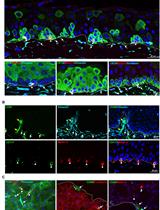
Adipocytes differentiated using this protocol can be applied in multiple experimental settings. In our original research article where this protocol was employed [13–15], we performed gene expression analysis, oxygen consumption measurements, and fluorescence imaging microscopy using the differentiated adipocytes. SVF-derived adipocytes displayed brown/beige-like features, including induction of a thermogenic gene program, increased mitochondrial content, multilocular lipid accumulation (Figure 4), and elevated oxygen consumption rate with high uncoupled respiration. All data, along with information about data processing, statistical analyses, and details of replicates, are provided in the original research article [13].

Figure 2. Expression of adipogenic markers in brown-like differentiated adipocytes. Relative mRNA levels of PPARγ (a, c) and aP2 (b, d) in mouse and human subcutaneous white adipose tissue (scWAT)-derived stromal vascular fraction (SVF) differentiated into brown-like adipocytes (n = 3). 36B4 mRNA expression was used for normalization. Results are expressed as mean ± SD; different groups were compared via Student’s t-test or by one-way ANOVA; *p-value < 0.05, **p-value < 0.005, ***p-value < 0.001. Primer sequences are available upon request.

Figure 3. Expression of the key brown fat functionality markers in control and differentiated adipocytes. Relative mRNA levels of Elovl3, Mcad, Cpt1α, CytC, and Cox4β in brown-like differentiated adipocytes of stromal vascular fraction (SVF) derived from subcutaneous white adipose tissue (scWAT) of mouse (a) and human (b) (n = 3). 36B4 mRNA expression was used for normalization. Results are expressed as mean ± SD; different groups were compared via Student’s t-test or by one-way ANOVA; *p-value < 0.05, **p-value < 0.005, ***p-value < 0.001. Primer sequences are available upon request.

Figure 4. Brightfield microscopy images of brown-like adipocytes differentiated from subcutaneous white adipose tissue (scWAT) -derived stromal vascular fraction (SVF) of mouse and human (day 8). Ethanol was used as control. Magnification, 20×; scale bar represents 100 μm.
Validation of protocol
This protocol or parts of it has been used and validated in the following research article(s):
• Verma et al. [13]. The mRNA levels of heat shock factor 1 are regulated by thermogenic signals via the cAMP-dependent transcription factor ATF3. Journal of Biological Chemistry, 295(18), 5984–5994. DOI: 10.1074/jbc.RA119.012072 (Figure 1c).
• Perie et al. [14]. Transcriptional regulation of ZNF638 in thermogenic cells by the cAMP response element binding protein in male mice. Journal of the Endocrine Society, 3(12), 2326–2340 (Figure 1d).
General notes and troubleshooting
General notes
1. Ensure consistent timing for tissue digestion to avoid over- or under-digestion, which can compromise yield and cell surface marker integrity.
2. Be precise about the ingredients (e.g., buffer or media), quantities, and conditions established for your experiments.
3. Stocks should be aliquoted and stored for 2–3 months at -20 °C, except for insulin (working vial), which needs to be stored at 4 °C. Avoid multiple freeze-thaw cycles to maintain the stability of the chemicals.
4. Prewarm media to 37 °C for 20 min prior to the procedure.
5. SVF isolation and differentiation should be performed in a BSL-2 cabinet, RNA extraction should be performed on ice or in a cold room, and the TRIzol step should be performed in a fume hood.
6. If using this protocol in female mice, optimize digestion and gating parameters accordingly.
Troubleshooting
Problem 1: Low cell yields after digestion.
Possible causes: Incomplete digestion or poor tissue mincing and handling.
Solutions: Ensure uniform mincing of adipose tissue and verify collagenase activity. Extend digestion time slightly if needed.
Problem 2: Low differentiation efficiency (few lipid droplets).
Possible cause: Too low cell confluency at induction.
Solutions: Differentiate at 90% confluency. Use fresh, properly aliquoted induction cocktails (e.g., IBMX, dexamethasone, insulin, rosiglitazone, etc.).
Supplementary information
The following supporting information can be downloaded here:
1. Table S1. Primer sequences
Acknowledgments
Authors’ contribution: Conceptualization, N.V.; Investigation, R.R.Y., N.M., N.V.; Writing—Original Draft, N.V.; Writing—Review & Editing, N.V., R.R.Y., N.M.; Funding acquisition, N.V.; Supervision, N.V.
This work was supported by the Centre of BioMedical Research (CBMR), Lucknow, through an intramural grant (CBMR/IMR/0024/2025) and extramural grant from ICMR (IIRPSG-2025-01-05389) to N.V. This protocol was described and is linked to Verma et al. [13] (DOI: 10.1074/jbc.RA119.012072).
Competing interests
The authors declare no competing interests.
Ethical considerations
All animal work was approved under IACUC protocol ID IA16-01767 by the NYU Grossman School of Medicine, Institutional Animal Care and Use Committee.
References
- World Health Organization. (2025). Obesity and overweight. https://www.who.int/news-room/fact-sheets/detail/obesity-and-overweight
- Sakers, A., De Siqueira, M. K., Seale, P. and Villanueva, C. J. (2022). Adipose-tissue plasticity in health and disease. Cell. 185(3): 419–446. https://doi.org/10.1016/j.cell.2021.12.016
- Scherer, P. E. (2018). The many secret lives of adipocytes: implications for diabetes. Diabetologia. 62(2): 223–232. https://doi.org/10.1007/s00125-018-4777-x
- Mueller, E. (2016). Browning and Graying: Novel Transcriptional Regulators of Brown and Beige Fat Tissues and Aging. Front Endocrinol (Lausanne). 7: e00019. https://doi.org/10.3389/fendo.2016.00019
- Shore, A. M., Karamitri, A., Kemp, P., Speakman, J. R., Graham, N. S. and Lomax, M. A. (2013). Cold-Induced Changes in Gene Expression in Brown Adipose Tissue, White Adipose Tissue and Liver. PLoS One. 8(7): e68933. https://doi.org/10.1371/journal.pone.0068933
- Rosell, M., Kaforou, M., Frontini, A., Okolo, A., Chan, Y. W., Nikolopoulou, E., Millership, S., Fenech, M. E., MacIntyre, D., Turner, J. O., et al. (2014). Brown and white adipose tissues: intrinsic differences in gene expression and response to cold exposure in mice. Am J Physiol Endocrinol Metab. 306(8): E945–E964. https://doi.org/10.1152/ajpendo.00473.2013
- Harms, M. and Seale, P. (2013). Brown and beige fat: development, function and therapeutic potential. Nat Med. 19(10): 1252–1263. https://doi.org/10.1038/nm.3361
- Wu, J., Boström, P., Sparks, L. M., Ye, L., Choi, J. H., Giang, A. H., Khandekar, M., Virtanen, K. A., Nuutila, P., Schaart, G., et al. (2012). Beige Adipocytes Are a Distinct Type of Thermogenic Fat Cell in Mouse and Human. Cell. 150(2): 366–376. https://doi.org/10.1016/j.cell.2012.05.016
- Nedergaard, J., Golozoubova, V., Matthias, A., Asadi, A., Jacobsson, A. and Cannon, B. (2001). UCP1: the only protein able to mediate adaptive non-shivering thermogenesis and metabolic inefficiency. Biochim Biophys Acta-Bioenerg. 1504(1): 82–106. https://doi.org/10.1016/s0005-2728(00)00247-4
- Cannon, B. and Nedergaard, J. (2004). Brown Adipose Tissue: Function and Physiological Significance. Physiol Rev. 84(1): 277–359. https://doi.org/10.1152/physrev.00015.2003
- Ikeda, K., Maretich, P. and Kajimura, S. (2018). The Common and Distinct Features of Brown and Beige Adipocytes. Trends Endocrinol Metab. 29(3): 191–200. https://doi.org/10.1016/j.tem.2018.01.001
- Yu, H., Emont, M., Jun, H., Wu, J. (2018). Isolation and Differentiation of Murine Primary Brown/Beige Preadipocytes. In: Bunnell, B. A., Gimble, J. M. (Eds.). Adipose-Derived Stem Cells. Methods in Molecular Biology, vol 1773. Humana Press, New York, NY. https://doi.org/10.1007/978-1-4939-7799-4_21
- Verma, N., Perie, L. and Mueller, E. (2020). The mRNA levels of heat shock factor 1 are regulated by thermogenic signals via the cAMP-dependent transcription factor ATF3. J Biol Chem. 295(18): 5984–5994. https://doi.org/10.1074/jbc.ra119.012072
- Perie, L., Verma, N., Xu, L., Ma, X. and Mueller, E. (2019). Transcriptional Regulation of ZNF638 in Thermogenic Cells by the cAMP Response Element Binding Protein in Male Mice. J Endocr Soc. 3(12): 2326–2340. https://doi.org/10.1210/js.2019-00238
- Perie, L., Verma, N. and Mueller, E. (2021). The forkhead box transcription factor FoxP4 regulates thermogenic programs in adipocytes. J Lipid Res. 62: 100102. https://doi.org/10.1016/j.jlr.2021.100102
Article Information
Publication history
Received: Aug 20, 2025
Accepted: Oct 19, 2025
Available online: Nov 4, 2025
Published: Dec 5, 2025
Copyright
© 2025 The Author(s); This is an open access article under the CC BY-NC license (https://creativecommons.org/licenses/by-nc/4.0/).
How to cite
Readers should cite both the Bio-protocol article and the original research article where this protocol was used:
- Yadav, R. R., Mishra, N. and Verma, N. (2025). A Protocol to Induce Brown and Beige Adipocyte Differentiation From Murine and Human Adipose-Derived SVF. Bio-protocol 15(23): e5522. DOI: 10.21769/BioProtoc.5522.
- Verma, N., Perie, L. and Mueller, E. (2020). The mRNA levels of heat shock factor 1 are regulated by thermogenic signals via the cAMP-dependent transcription factor ATF3. J Biol Chem. 295(18): 5984–5994. https://doi.org/10.1074/jbc.ra119.012072
Category
Stem Cell > Adult stem cell > Mesenchymal stem cell
Developmental Biology > Cell growth and fate
Do you have any questions about this protocol?
Post your question to gather feedback from the community. We will also invite the authors of this article to respond.