- Protocols

- Articles and Issues
- For Authors
- About
- Become a Reviewer

Phosphoproteomic Analysis and Organotypic Cultures for the Study of Signaling Pathways
(*contributed equally to this work) Published: Vol 14, Iss 4, Feb 20, 2024 DOI: 10.21769/BioProtoc.4941 Views: 2864
Reviewed by: Philipp WörsdörferHsih-Yin TanAnonymous reviewer(s)
Original research article
The authors used this protocol in:

Nov 2022
Advertisement


Protocol Collections
Comprehensive collections of detailed, peer-reviewed protocols focusing on specific topics


Related protocols
Abstract
Signaling pathways are involved in key cellular functions from embryonic development to pathological conditions, with a pivotal role in tissue homeostasis and transformation. Although most signaling pathways have been intensively examined, most studies have been carried out in murine models or simple cell culture. We describe the dissection of the TGF-β signaling pathway in human tissue using CRISPR-Cas9 genetically engineered human keratinocytes (N/TERT-1) in a 3D organotypic skin model combined with quantitative proteomics and phosphoproteomics mass spectrometry. The use of human 3D organotypic cultures and genetic engineering combined with quantitative proteomics and phosphoproteomics is a powerful tool providing insight into signaling pathways in a human setting. The methods are applicable to other gene targets and 3D cell and tissue models.
Key features
• 3D organotypic models with genetically engineered human cells.
• In-depth quantitative proteomics and phosphoproteomics in 2D cell culture.
• Careful handling of cell cultures is critical for the successful formation of theorganotypic cultures.
• For complete details on the use of this protocol, please refer to Ye et al. 2022.
Keywords: Organotypic modelBackground
Signaling pathways are crucial regulators of cell growth and differentiation, playing significant roles in tissue formation and transformation; in many cases, they have been studied in simple 2D cell cultures or in animal studies. While many of the observations from murine models are expected to be applicable to human tissue, there are some inherent difficulties in translating these findings to a human context [1]. Consequently, it is important to use human cell and tissue systems to better understand and translate the functions of different signaling pathways into a human setting. We have previously integrated precise genetic engineering of the human N/TERT-1 and other keratinocyte cell lines to create a genetically traceable starting point to study individual gene products for the formation and transformation of fully differentiated human epidermal tissue [2–4]. The epidermis is a self-renewing, stratified tissue composed of proliferating basal cells and non-proliferating suprabasal cells [5]. Their properties are influenced by the underlying extracellular matrix (ECM) and dermal fibroblasts via intense bidirectional crosstalk mediated by secreted factors [6,7]. This creates the possibility to study both endogenous changes in cells and crosstalk between cell types in a controlled manner [2–4,8]. Therefore, in vitro human skin development is a relevant model to interrogate the various functions of signaling pathways, particularly when combined with proteomics and phosphoproteomics.
Quantitative liquid chromatography–mass spectrometry (LC–MS)-based proteomics and phosphoproteomics have emerged as the preferred methods to study cell signaling [9]. Isobaric labeling methods, like tandem mass tags (TMT), provide relative quantitation of identified proteins and phosphopeptides in multiple samples [10]. Combining the use of genetic engineering, proteomics, and phosphoproteomics with 3D organotypic human tissue models is an important tool in examining signaling pathways, as showed by our group [11]. In that study, we showed the potential different roles between Smad4-dependent and Smad4-independent TGF-β pathways and their role in diverse cellular behaviors such as human tissue homeostasis.
Taken together, these methods can be used as tools for characterizing signaling in human tissue formation and homeostasis. The described protocol details the creation of 3D organotypic cultures (protocol: organotypic tissue culture) that imitate in vivo tissue and its use to examine signaling pathways. Post incubation, the cultures are processed for histology or other analyses. In combination, parallel proteomics and phosphoproteomics analyses (protocol: Proteomics) of 2D cellular cultures, including cell lysis, protein digestion, phosphopeptide enrichment, and LC–MS analysis, allow for a detailed molecular investigation of the affected signaling pathways. The method provides novel insight into cell signaling, relevant for drug development and the understanding of disease pathology. The method can furthermore be used for drug testing and toxicology studies and minimizes the use of animal models.
Materials and reagents
Biological materials
For organotypic tissue culture
MRC-5 (ATCC, catalog number: CCL-171)
N/TERT-1 (James G. Rheinwald, Harvard Institute of Medicine)
For proteomics
Amine (NH2) functional microparticles, MagReSyn® Amine [ReSyn Biosciences (Pty) Ltd., South Africa]
Hydroxyl terminated beads, MagReSyn® Hydroxyl [ReSyn Biosciences (Pty) Ltd., South Africa]
Magnetic microparticles with chelated Ti4 + metal ions for highly specific phosphopeptide enrichment, MagReSyn® Ti-IMAC HP [ReSyn Biosciences (Pty) Ltd., South Africa]
Reagents
For organotypic tissue culture
Penicillin/streptomycin (Thermo Fisher, Gibco, catalog number: 151401)
DMEM (Thermo Fisher, Gibco, catalog number: 41966029)
Keratinocyte-SFM (Thermo Fisher, Gibco, catalog number: 17005034)
TrypLE (Thermo Fisher, Gibco, catalog number: 12605028)
Bovine pituitary extract (BPE) (Thermo Fisher, Gibco, catalog number: 13028014)
Fetal calf serum (FCS) (HyClone, catalog number: SH30071.03HI)
EGF (Thermo Fisher, Gibco, catalog number: PHG0314)
Insulin (Sigma-Aldrich, catalog number: 11070-73-8)
Hydrocortisone (Sigma-Aldrich, catalog number: H0888)
Triiodothyronine (T3) (Sigma-Aldrich, catalog number: T2752)
L-Glutamine (Thermo Fisher, Gibco, catalog number: A2916801)
Hams F12 (Thermo Fisher, Gibco, catalog number: 11765-054)
Rat tail collagen I (made in own lab [12])
Adenine (Sigma-Aldrich, catalog number: A8626)
Hydrochloric acid (HCl) (Sigma-Aldrich, catalog number: H1758)
Dulbecco’s phosphate buffered saline (PBS) (Sigma-Aldrich, catalog number: D5652)
Bovine serum albumin (BSA) (Sigma-Aldrich, catalog number: A7979)
Sodium hydroxide (NaOH) (Sigma-Aldrich, catalog number: 655104)
Calcium chloride (CaCl2·2H2O) (Sigma-Aldrich, catalog number: C7902)
Gentamycin sulphate (Sigma-Aldrich, catalog number: G1264)
Cholera enterotoxin (Sigma-Aldrich, catalog number: C8180)
10× minimum essential media (10× MEM) (Thermo Fisher, Gibco, catalog number: 11430-030)
Sodium bicarbonate (Thermo Fisher, Gibco, catalog number: 15750037)
Tissue-Tek® (SAKURA, catalog number: 4583)
Formalin solution neutral buffered, 10% (Sigma-Aldrich, catalog number: HT501128)
Sucrose (Sigma-Aldrich, catalog number: S0389)
Isopentane (Sigma-Aldrich, catalog number: PHR1661)
For proteomics
Lysis buffer, composed of 2% sodium dodecyl sulfate (SDS), 5 mM tris(2-carboxyethyl)phosphine (TCEP), 5.5 mM chloroacetamide (CAA), and 100 mM Tris pH 8.5; used for cell lysis
Acetonitrile (ACN), LC grade (SupelcoTM Analytical, catalog number: 75-05-8), used in various concentrations throughout the protocol, namely 70% for dilution of lysates, 95% for washing proteins, and 40% and 60% for elution on SepPak. ACN is also used for TMT-labeling
Ethanol (anhydrous) (Sigma-Aldrich, catalog number: 459836), used in 70% concentration for washing proteins after aggregation
50 mM ammonium bicarbonate (ABC) (Sigma-Aldrich, catalog number: 09830), used for protein digestion and for resuspending TMT-labeled peptides
50% trifluoroacetic acid (TFA), HPLC (SupelcoTM Analytical, catalog number: TX1276), used for acidification of samples post digestion
1 M TEAB, pH 8 (Thermo ScientificTM, catalog number: 90114), used to achieve a final concentration of 100 mM in TMT-labeling
50% hydroxylamine (Thermo ScientificTM, catalog number: 90115), used to quench the TMT-labeling reaction
0.1% formic acid (v/v) in water (LC–MS grade) (Thermo ScientificTM, catalog number: 85170), used for resuspension of each TMT-labeled sample before pooling
Loading buffer, composed of 80% ACN, 5% TFA, and 1 M glycolic acid (Sigma-Aldrich, catalog number: 8.04104); used for resuspending peptides for phosphopeptide enrichment
1% ammonia (ammonia solution 25%) (Supelco, catalog number: 105428), used for elution of phosphopeptides
5 mM ABC (buffer A) and 100% ACN (buffer B), used in high pH reversed-phase peptide fractionation
PierceTM BCA Protein Assay kit (Thermo ScientificTM, catalog number: 23225)
TMTproTM 16plex label reagent set (Thermo ScientificTM, catalog number: A44520)
LysC, Lysyl endopeptidase (Fujifilm Wako, catalog number: 129-02541)
Trypsin, Sequencing grade modified trypsin (Promega, catalog number: V511X)
Sep-Pak C18 1 cc Vac Cartridge (Waters, catalog number: WAT054955)
Acquity CSH C18 1.7 μm, 1 mm × 150 mm column (Waters, catalog number: 186006935)
Solutions
Adenine (see Recipes)
Cholera enterotoxin (CT) (see Recipes)
Hydrocortisone (HC) (see Recipes)
Epidermal growth factor (EGF) (see Recipes)
Triiodothyronine (T3) (see Recipes)
Calcium chloride (CaCl2) (see Recipes)
Insulin (1,000× for 5 µg/mL) (see Recipes)
Sodium bicarbonate (71.2 mg/mL milli q water) (see Recipes)
Keratinocyte serum free medium (K-SFM) complete (see Recipes)
Organotypic culture medium (see Recipes)
DMEM 10% FBS (DMEM-10) (see Recipes)
Recipes
For organotypic tissue culture
Adenine
Prepare 2.4 mg/mL of adenine in HCl (this is a 100× stock):
Dissolve 243 mg of adenine in 100 mL of 0.05 M HCl.
Stir for approximately 1 h at room temperature (RT).
Filter sterilize with 0.2 μm filters.
Divide into 5 mL aliquots.
Store at -20 °C.
Cholera enterotoxin (CT)
Prepare 10 µM solution of cholera enterotoxin (this is 1,000× stock solution for 100 pM):
Add 1.18 mL of Milli-Q water to a 1 mg vial of cholera enterotoxin to prepare a 10-5 M solution.
Dilute this in 110 mL of PBS.
Filter sterilize with 0.2 µm filters.
Aliquot 2 mL per cryovial.
Store at -20 °C.
Note: MWCT = 84.000 Da
Hydrocortisone (HC)
Prepare 0.2 mg/mL hydrocortisone (this is a 500× stock for 0.4 µg/mL):
Dissolve a 25 mg vial in 5 mL of 100% ethanol. This solution is 5 mg/mL, which is 12.5× for 0.4 µg/mL. This can be stored at -20 °C indefinitely.
Add 2 mL of this concentrated stock to 48 mL of sterile PBS + 0.1% BSA.
Filter-sterilize with 0.2-µm filters.
Aliquot 2 mL into cryovials.
Store at -20 °C.
Epidermal growth factor (EGF)
Prepare 10 µg/mL of EGF (this is a 50,000× stock for 0.2 ng/mL):
Dissolve 100 µg of EGF in 10 mL of PBS + 0.1% BSA.
Filter sterilize with 0.2 µm filters.
Aliquot 1 mL into cryovials.
Store at -20 °C.
Triiodothyronine (T3)
Prepare 20 nM (2 × 10⁻⁸ M) of T3 [this is a 1,000× stock for 20 pM (2 × 10-11 M)]:
Measure 6.8 mg of T3 and dissolve in 7.5 mL of 0.02N NaOH.
Add 42.5 mL PBS (this is 100,000× for 20 pM T3).
Store at -20 °C in 10 mL aliquots.
To make the 1000× stock, dilute 0.4 mL of the 100,000× 1:100 into 39.6 mL of PBS.
Filter sterilize with 0.2 µm filters.
Divide into 2 mL aliquots.
Store at -20 °C.
Calcium chloride (CaCl2)
Prepare using calcium dihydrate CaCl2·2H2O (this is a 1,000× stock for 0.3 mM):
Dissolve 4.4 g of CaCl2·2H2O in 100 mL of Milli Q water.
Stir with magnetic bar to dissolve completely.
Filter sterilize with 0.2 µm filters.
Aliquot 2 mL per cryovial.
Store at -20 °C.
Insulin (1,000× for 5 µg/mL)
Use insulin from bovine pancreas.
Dissolve 100 mg of insulin in 20 mL of 0.005 M HCl in a 50 mL blue-capped tube.
Filter sterilize through a celluloseacetate, lowprotein binding 0.2 µM filter.
Aliquot 500 µL per tube.
Store at -20 °C.
Sodium bicarbonate (71.2 mg/mL milliQ water)
For organotypic cultures
Keratinocyte serum free medium (K-SFM) complete
Reagent Final concentration Quantity Keratinocyte-SFM n/a 500 mL CaCl2 (1,000×) 0.3 mM 500 µL BPE (16.8 mg/mL) 25 µg/mL 750 µL EGF (10 µg/mL) 0.2 ng/mL 10 µL Total n/a 501.26 mL Optional: Penicillin/Streptomycin can be added to K-SFM as well (100 U/mL).
Organotypic culture medium
Reagent Final concentration Quantity DMEM n/a 375 mL F12 n/a 125 mL Pen/Strep 100 U/mL 5.14 mL Adenine (2.4 mg/mL stock) 24 µg/mL 5.14 mL HC (0.2 mg/mL stock) 0.4 µg/mL 1 mL CT (10 µM stock) 100 pM 0.514 mL T3 (1,000×) 20 pM 0.514 mL Insulin (1,000×) 5 µg/mL 500 µL FCS 0.3% 1.54 mL Total 514.3 mL DMEM 10% FBS (DMEM-10)
Reagent Final concentration Quantity DMEM n/a 445 mL FBS 10% 50 mL Pen/Strep 100 U/mL 5 mL Total 500 mL
Laboratory supplies
For organotypic tissue culture
Deep-well plates (Corning, catalog number: 355467)
Cell culture insert, PET membrane, 3.0 µm pore size (Corning, catalog number: 353092)
Airlift pads (cut in circles and autoclaved) (Whatman, GE Healthcare, catalog number: 10382461)
Cell culture Petri dishes or flasks from any brand (we use NUNC, Falcon, and Corning)
Equipment
For organotypic tissue culture
CO2 incubator at 37 °C (any brand can be used)
Centrifuge (any brand, for 15 mL tubes and a minimum of 100 g)
Freezers (-20 °C and -80 °C)
Equipment and software for proteomics
KingFisherTM Flex purification system (Thermo Scientific, catalog number: 5400610)
SpeedVac (Eppendor, model: VacufugeTM Concentrator)
Sonicator (Fisherbrand, model: 120 Sonic Dismembrator)
Spectrophotometer (Thermo Scientific, model: NanoDropTM 2000 C)
HPLC system [Thermo Scientific, model: UltiMateTM 3000 Standard (SD)]
Evosep ONE (Evosep)
Mass spectrometer (Thermo Scientific, model: Orbitrap ExplorisTM 480)
Proteome Discoverer software v2.4 (Thermo Scientific)
Chromeleon software v7.2 (Thermo Fisher Scientific)
Procedure
Part I: Organotypic tissue culture
Preparing cells for organotypic cultures (day 1–5)
Fibroblast cultures
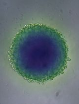
Culture fibroblasts (we use MRC-5 cells) until you have an 80%–90% confluent T75 flask. You will need 1.8 million fibroblasts for one tray of organotypic cultures. For most fibroblasts, you will need to seed 0.8 million fibroblasts three days in advance (see Figure 1).

Figure 1. Fibroblasts suitable for organotypic collagen gelsTrypsinize cells for 5 min and neutralize with an equal volume of DMEM with 10% FCS. Count the cells (only viable cells) and spin down.
Resuspend fibroblasts at 8×10⁵ per milliliter of DMEM-10.
Leave cells on ice while mixing the collagen gel.
Casting organotypic cultures
Note: Make sure you have enough fibroblasts ready at this point. You will need 1.8 million cells.
Acellular collagen gels
Note: All solutions must be ice cold or placed on ice.
For additional guidance watch Video 1.
 Video 1. How to make an organotypic culture
Prepare a deep-well plate (6-well size) by adding one cell culture insert into each well. Use sterile forceps.
In a 50 mL tube on ice, mix the following components without making air bubbles:
650 µL of 10× MEM
70 µL of L-glutamine (100× stock of 200 mM)
10 µL of gentamicin sulphate (40 mg/mL)
725 µL of fetal calf serum (FCS)
725 µL of sodium bicarbonate (71.2 mg/mL)
5.5 mL of rat tail collagen (4 mg/mL in 0.05% acetic acid) (homemade, see [12])
850 µL of DMEM with 10% FCS
Add a few drops of 1 M NaOH to neutralize.
Carefully pipette 1 mL of the acellular gel mixture from the previous step into the culture insert and make sure it covers the whole bottom of the insert. Leave to polymerize in the incubator while preparing the fibroblasts and the cellular gel. The gel will be polymerized after 10–15 min.
Making of cellular gel
In a 50 mL tube on ice, mix the following components (avoid making air bubbles):
1.7 mL of 10× MEM
170 µL of L-glutamine (100× stock of 200 mM)
25 µL of gentamicin sulphate (40 mg/mL)
1.9 mL of FCS
525 µL of sodium bicarbonate (71.2 mg/mL)
14.4 mL of rat tail collagen (1 mg/ml in 0.05% acetic acid) (homemade)
2.1 mL of human fibroblasts (1.8 million cells) suspended in 10% calf serum DMEM
Mix gently and add a few drops of 1 M NaOH to neutralize the solution (the color should change to orange/light pink).
Quickly add 3 mL to the culture inserts on top of the acellular gel. Avoid air bubbles.
Place the tray in the incubator for approximately 30 min; the gels will be fully polymerized.
Fill DMEM 10% FCS in the well (18 mL) and 2 mL on top of the gel. Do not spray. See Figure 2 and Video 2.

Figure 2. Casted organotypic collagen gels, ready for media to be added Video 2. How to fill media on organotypic cultures
Maturing collagen gels
Four to six days after the gels are contracted and appear macroscopically as a slightly shrunken, white disk, they are ready for keratinocytes.
Note: In our hands, this results in good organotypic cultures, possibly due to active deposition and reorganization of the ECM by fibroblasts.
Seeding keratinocytes on the casted collagen gels with fibroblasts (timing 2 h)
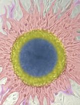
Trypsinize N/TERT-1 cells to be seeded (WT and validated knock-outs) and adjust concentration to 1 × 10⁷ cells/mL (Figure 3).
Note: 3 × 10⁵ cells need to be plated for each gel.
Aspirate medium from the bottom of each well in the deep-well plate.
Remove medium from the culture insert with a p1000 pipette.
Critical: Avoid keeping the pipette too close to the gel inside the insert, as you may aspirate the gel.
Add 30 µL of cell suspension (from step E1) in the center of the gel (Video 3).
Video 3. Adding keratinocytes to organotypic culturesAdd organotypic culture medium (see Recipes) at the bottom of the well in the deep-well plate until it reaches the bottom of the culture insert (∼9 mL).
Incubate at 37 °C for 20 min to let the cells adhere.
Add 2 mL of organotypic culture medium on top of the adhered cells and ∼16 mL of organotypic culture medium outside the insert and carefully place in the incubator. Do not spray as the keratinocytes will then detach.
Incubate for four days before lifting the gels.

Figure 3. Keratinocyte colonies. A. Colonies well-suited for organotypic collagen gels. B. Colonies not suited for organotypic cultures.
Air-liquid interface lifting of the keratinocyte. Timing: 30 min (Video 4)
Video 4. Placing airlift padsOn day 4 after seeding the keratinocytes, carefully aspirate medium from the culture inserts and from the plate wells.
Place the insert into a sterile 100 mm dish using sterile forceps.
Add two sterile airlift pads to the base of each well in the deep-well plate and place back the insert into the well over the pads.
Add approximately 9 mL of organotypic culture medium in the deep wells until the pads are completely wet.
Critical: The level of the medium should cover the pads and not be higher.
Adding inhibitors to organotypic cultures (Day 4–14)
For experiments using different inhibitors, culture medium is supplemented with inhibitors in the desired concentration when the inserts are raised to the air-liquid interface. Concentration of the inhibitor is replenished with subsequent medium changes. Note that for some inhibitors, concentration needs to be raised compared to the use on 2D cultured cells. Add the desired inhibitor directly to the media in the well. Replenish with every media change.
Maturing of organotypic cultures
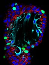
Incubate the plate at 37 °C in a humidified incubator and change media in the deep wells every other day. Gels are ready to harvest in 10 days (Figure 4).

Figure 4. Organotypic culture ready to be fixed. Arrow points to the concavity in the contracted collagen gel where keratinocytes are growing.Note: Organotypic cultures can now be fixed for histology or processed for western blot, RNAseq, or proteomics.
Formalin-fixed paraffin-embedded (FFPE) section processing
Cut the membranes from the insert and place the 3D cultures in a histology cassette in 10% formalin for >24 h followed by paraffin embedding.
Frozen sections
Note: The best morphology is obtained when the 3D cultures are incubated in 2 M sucrose (in dH2O) for >48 h before embedding in Tissue-Tek and freezing in isopentane on dry ice.
Cut the membranes from the insert and place the 3D cultures in a 2 M sucrose solution for >48 h.
Freeze in isopentane on dry ice and keep at -80 °C until sectioning.
Note: Sections cut at 4–6 μm give better staining results.
Part II: Proteomic analyses in 2D cell culture
Note: Steps K–P, Sample preparation for proteomics and phosphoproteomics and mass spectrometry analysis (2–3 days)
Cell lysis
Cultivate N/TERT-1 cells in 2D until they reach 80% confluence. Approximately 1 million cells are needed.
Lyse the cells by adding lysis buffer (500 μL) and boil the lysates for 10 min at 95 °C.
Further lyse by micro-tip probe sonication for 2 min, pulsing 1 s on and 1 second off at 60% amplitude.
Measure the protein concentrations using a BCA protein assay kit.
Protein digestion
Note: Digest protein extracts overnight using the KingFisher Flex robot following the protein aggregation capture (PAC) method [13], adapted for a 96-well plate format (Figure 5).
Dilute the lysates to a final concentration of 70% ACN.
Add magnetic hydroxyl terminated beads for protein aggregation using a 1:2 protein/bead ratio. The amine beads can be replaced by hydroxyl-terminated beads.
Wash proteins three times in 95% ACN and twice in 70% ethanol.
Digest in 300 μL of 50 mM ABC with LysC and trypsin at enzyme/protein ratios of 1:500 and 1:250, respectively, for 12 h at 37 °C.
Acidify samples with 50% TFA to a final concentration of 1% TFA.
Clean the peptide mixtures on SepPak C18 1 cc Vac cartridge and elute with 300 μL of 40% ACN followed by 300 μL of 60% ACN.
Evaporate the ACN in a SpeedVac.
Estimate the final peptide concentration by measuring absorbance at 280 nm on a NanoDrop 2000 C spectrophotometer.

Figure 5. Layout of the KingFisher plates for protein digestion
TMT labeling
Note: Label peptides from each sample with TMTpro 16-plex Label Reagent. After labeling, 10 µg of pooled peptides is reserved for proteome samples and the rest is used for phosphopeptide enrichment.
Add 1 M TEAB to each sample to reach a final concentration of 100 mM, followed by ACN to 50% final concentration.
Incubate the labeled peptides with TMT reagents for 1 h at RT.
Quench the reaction by adding 1% hydroxylamine (1:1 hydroxylamine/TMT) for 15 min at RT.
Pool the samples and evaporate the ACN by SpeedVac.
Phosphopeptide enrichment
Resuspend TMT-labeled peptides in 200 μL of loading buffer (Figure 6).

Figure 6. Layout of the KingFisher plates for phosphopeptide enrichmentUse Ti-IMAC beads in a 1:2 peptide/beads ratio and wash them in 500 μL of loading buffer for 5 min.
Incubate the beads with the peptides for 20 min.
Wash the beads successively with loading buffer, 80% ACN, 1% TFA, and finally 10% ACN and 0.2% TFA for 2 min each.
Elute the phosphopeptides in 200 μL of 1% ammonia for 10 min.
Repeat the enrichment process on the unbound remnant from the initial enrichment step with new elution buffers and beads.
Micro-flow high-pH reversed-phase peptide fractionation
Resuspend TMT-labeled peptides in 50 mM ABC.
Fractionate the peptides using a reversed-phase Acquity CSH C18 column on an UltiMate 3000 HPLC system with the Chromeleon software.
Follow the provided multi-step gradient.
Collect and concatenate to 12 fractions for phosphoproteome and 46 fractions for proteome, acidify with 10% formic acid, and dry with SpeedVac.
LC–MS/MS
Analyze all samples by LC–MS/MS using the Evosep One system with pre-programmed gradients.
MS settings:
Spray voltage to 1.8 kV
Heated capillary temperature at 275 °C
Funnel RF level at 40
Acquire data in profile mode with the full MS mass range set at 350–1,400 with an AGC target at 300%
Set full MS resolution at 60,000 at m/z 200 with a maximum injection time of 25 ms
Set the HCD fragment spectra resolution at 45,000 with an injection time of 86 ms and a Top10 method
Set AGC target value at 200% and the intensity threshold at 2E5
Isolation window set as at 0.8 m/z
Normalized collision energy at 32%
Data analysis
Data analysis (proteomics)
All raw data files are analyzed using the Proteome Discoverer 2.4 software (developed by Thermo Fisher Scientific). The human SwissProt FASTA database, with 20,355 entries as of March 2019, was used as the reference protein database.
Trypsin was selected as the digestion enzyme. Up to two missed cleavages were permitted. TMTpro is specified as a fixed modification on lysine and peptide N terminus, and methionine oxidation is specified as a variable modification. In addition, phosphorylation is set as a variable modification on serine, threonine, and tyrosine residues in phosphoproteome samples.
Validation of protocol
Organotypic tissue culture
The human skin organotypic model with N/TERT-1 cells (WT cells and genetic engineered cells), or any other keratinocyte cell type or cancer cell type we have used, is very robust. In our laboratory, we have made more than 400 organotypic cultures with WT cells and more than 4,000 organotypic cultures during the last decades. The outcome varies with +/- 1 cell layer counted on >100 organotypic wildtype cell cultures. Furthermore, the phenotype of the 3D organotypic models is highly similar to the normal human skin (see Ye et al. [11], figure 7 below).

Figure 7. Comparison of normal human skin with 3D organotypic skin cultures. Top row: staining of human skin with differentiation markers [involucrin (INV), filaggrin (Flg), loricrin (Lor), keratin 10 (K10), and keratin 1 (K1)] and the proliferation marker Ki67. Lower row: the same staining in N/TERT-1 WT organotypic cultures. Statistical analysis, columns to the right, using one-way ANOVA followed by Dunnett’s multiple comparison test. The organotypic cultures show the same distribution of these markers as the normal skin, but the basal lamina will be straight compared to the wavy nature of the normal human skin.
General notes and troubleshooting
General notes
The 3D organotypic culture with keratinocytes and fibroblasts is very robust and reproducible.
The media is cheaper than premade media bought from different companies, although it is a little time-consuming to make all the stock solutions the first time.
The fact that we use 6-well plates makes it easy to have enough material for many different assays and also have enough good tissue for immunohistochemistry.
Troubleshooting
| Problem | Potential solution 1 | Potential solution 2 |
Collagen gels do not contract after point D | Good-quality collagen and fresh growing fibroblasts are pivotal for establishing good-quality organotypic cultures. | Collagen gels may be left for up to seven days in the incubator before seeding keratinocytes, giving the fibroblasts more time to reorganize the ECM. |
| Fibroblasts should not have been cultured for more than two weeks and should be actively dividing. The passage number varies between fibroblast lines. | ||
Keratinocytes do not form stratified layers | Keratinocytes should not have been cultured for more than three weeks and should be actively dividing. Do not shake the culture dishes during incubations, especially within the first 4 h after seeding the cells. Any passage number can be used of the N/TERT-1 cells as they are immortalized. | Do not dry out the gel while lifting to the air-liquid interphase. Do not add too much media at this step as well (9 mL is usually enough; the media must not overflow into the inserts). |
| Avoid opening the incubator too often while growing the organotypic cultures. In our experience, organotypic cultures tend to fail more often in a very busy incubator (opened many times during the day). | The organotypic cultures are stable for up to three weeks after lifting. |
Acknowledgments
The work was supported by the European Commission (GlycoSkin H2020-ERC), European Commission (Imgene H2O20), Lundbeck Foundation, The Danish Research Councils (Sapere Aude Research Leader grant to H.H.W.), Danish National Research Foundation (DNRF107), the Friis Foundation (H.H.W AND S.D), Danish Strategic Research Council, and the program of excellence from the University of Copenhagen (CDO2016). We thank Karin Uch Hansen, Birgit Poulsen, Karen Biré, and Louise Rosgaard Duus for the excellent technical assistance. We acknowledge the Core Facility for Integrated Microscopy, Faculty of Health and Medical Sciences, University of Copenhagen. We would like to thank James G. Rheinwald, Harvard Institute of Medicine, for providing the N/TERT-1 cells.
This work was described in Ye et al. [11] and Dabelsteen et al. [2].
Competing interests
The authors have no financial or non-financial competing interests concerning this manuscript.
Ethical considerations
The two human cell lines used in this study were either bought at ATCC or provided by James G. Rheinwald, Harvard Institute of Medicine where the use for academic purposes is approved.
References
- Nestle, F. O., Di Meglio, P., Qin, J. Z. and Nickoloff, B. J. (2009). Skin immune sentinels in health and disease. Nat. Rev. Immunol. 9(10): 679-691. https://doi.org/10.1038/nri2622.
- Dabelsteen, S., Pallesen, E. M. H., Marinova, I. N., Nielsen, M. I., Adamopoulou, M., Romer, T. B., Levann, A., Andersen, M. M., Ye, Z., Thein, D., et al. (2020). Essential Functions of Glycans in Human Epithelia Dissected by a CRISPR-Cas9-Engineered Human Organotypic Skin Model. Dev. Cell. 54(5): 669-684 e667. https://doi.org/10.1016/j.devcel.2020.06.039.
- Bagdonaite, I., Pallesen, E. M. H., Ye, Z. L., Vakhrushev, S. Y., Marinova, I. N., Nielsen, M. I., Kramer, S. H., Pedersen, S. F., Joshi, H. J., Bennett, E. P., et al. (2020). O-glycan initiation directs distinct biological pathways and controls epithelial differentiation. Embo. Reports 21(6). https://doi.org/10.15252/embr.201948885.
- Nielsen, M. I., de Haan, N., Kightlinger, W., Ye, Z., Dabelsteen, S., Li, M., Jewett, M. C., Bagdonaite, I., Vakhrushev, S. Y. and Wandall, H. H. (2022). Global mapping of GalNAc-T isoform-specificities and O-glycosylation site-occupancy in a tissue-forming human cell line. Nat. Commun. 13(1): 6257. https://doi.org/10.1038/s41467-022-33806-8.
- Blanpain, C. and Fuchs, E. (2009). Epidermal homeostasis: a balancing act of stem cells in the skin. Nat Rev. Mol. Cell. Biol. 10(3): 207-217. https://doi.org/10.1038/nrm2636.
- Briggaman, R. A. (1982). Epidermal-dermal interactions in adult skin. J. Invest. Dermatol. 79 Suppl 1: 21s-24s. https://doi.org/10.1111/1523-1747.ep12544628.
- Smola, H., Stark, H. J., Thiekotter, G., Mirancea, N., Krieg, T. and Fusenig, N. E. (1998). Dynamics of basement membrane formation by keratinocyte-fibroblast interactions in organotypic skin culture. Exp. Cell. Res. 239(2): 399-410. https://doi.org/10.1006/excr.1997.3910.
- Aasted, M. K. M., Groen, A. C., Keane, J. T., Dabelsteen, S., Tan, E., Schnabel, J., Liu, F., Lewis, H. S., Theodoropulos, C., Posey, A. D., et al. (2023). Targeting Solid Cancers with a Cancer-Specific Monoclonal Antibody to Surface Expressed Aberrantly O-glycosylated Proteins. Mol. Cancer. Ther. 22(10): 1204-1214. https://doi.org/10.1158/1535-7163.MCT-23-0221.
- Olsen, J. V., Blagoev, B., Gnad, F., Macek, B., Kumar, C., Mortensen, P. and Mann, M. (2006). Global, in vivo, and site-specific phosphorylation dynamics in signaling networks. Cell 127(3): 635-648. https://doi.org/10.1016/j.cell.2006.09.026.
- Li, J., Van Vranken, J. G., Vaites, L. P., Schweppe, D. K., Huttlin, E. L., Etienne, C., Nandhikonda, P., Viner, R., Robitaille, A. M. and Thompson, A. H. (2020). TMTpro reagents: a set of isobaric labeling mass tags enables simultaneous proteome-wide measurements across 16 samples. Nat. Methods. 17(4): 399-404. https://www.nature.com/articles/s41592-020-0781-4.
- Ye, Z., Kilic, G., Dabelsteen, S., Marinova, I. N., Thøfner, J. F., Song, M., Rudjord-Levann, A. M., Bagdonaite, I., Vakhrushev, S. Y. and Brakebusch, C. H. (2022). Characterization of TGF-β signaling in a human organotypic skin model reveals that loss of TGF-βRII induces invasive tissue growth. Sci. Signal. 15(761): eabo2206. https://www.science.org/doi/10.1126/scisignal.abo2206
- Rajan, N., Habermehl, J., Coté, M. F., Doillon, C. J. and Mantovani, D. (2006). Preparation of ready-to-use, storable and reconstituted type I collagen from rat tail tendon for tissue engineering applications. Nat Protoc 1(6): 2753-2758. https://doi.org/10.1038/nprot.2006.430.
- Batth, T. S., Tollenaere, M. A. X., Ruther, P., Gonzalez-Franquesa, A., Prabhakar, B. S., Bekker-Jensen, S., Deshmukh, A. S. and Olsen, J. V. (2019). Protein Aggregation Capture on Microparticles Enables Multipurpose Proteomics Sample Preparation. Mol Cell Proteomics 18(5): 1027-1035. https://doi.org/10.1074/mcp.TIR118.001270.
Article Information
Copyright
© 2024 The Author(s); This is an open access article under the CC BY-NC license (https://creativecommons.org/licenses/by-nc/4.0/).
How to cite
Readers should cite both the Bio-protocol article and the original research article where this protocol was used:
- Ye, Z., Wandall, H. H. and Dabelsteen, S. (2024). Phosphoproteomic Analysis and Organotypic Cultures for the Study of Signaling Pathways. Bio-protocol 14(4): e4941. DOI: 10.21769/BioProtoc.4941.
- Ye, Z., Kilic, G., Dabelsteen, S., Marinova, I. N., Thøfner, J. F., Song, M., Rudjord-Levann, A. M., Bagdonaite, I., Vakhrushev, S. Y. and Brakebusch, C. H. (2022). Characterization of TGF-β signaling in a human organotypic skin model reveals that loss of TGF-βRII induces invasive tissue growth. Sci. Signal. 15(761): eabo2206. https://www.science.org/doi/10.1126/scisignal.abo2206
Category
Cell Biology > Cell isolation and culture > 3D cell culture
Cell Biology > Cell signaling
Molecular Biology > Protein
Do you have any questions about this protocol?
Post your question to gather feedback from the community. We will also invite the authors of this article to respond.