- Protocols

- Articles and Issues
- For Authors
- About
- Become a Reviewer

Analysis of Heterocyst and Akinete Specific Glycolipids in Cyanobacteria Using Thin-layer Chromatography
Published: Vol 12, Iss 6, Mar 20, 2022 DOI: 10.21769/BioProtoc.4355 Views: 2497
Reviewed by: Dennis J NürnbergAmberley D. StephensKumiko Okazaki
Original research article
The authors used this protocol in:

Apr 2021
Advertisement


Protocol Collections
Comprehensive collections of detailed, peer-reviewed protocols focusing on specific topics


Abstract
Several filamentous cyanobacteria like Nostoc differentiate specialized cells in response to changes in environmental factors, such as low light or nutrient starvation. These specialized cells are termed heterocysts and akinetes. Under conditions of nitrogen limitation, nitrogen-fixing heterocysts form in a semi-regular pattern and provide the filament with organic nitrogen compounds. Akinetes are spore-like dormant cells, which allow survival during adverse unfavorable conditions. Both cell types possess multilayered thick envelopes mainly composed of an outermost polysaccharide layer and inner layers of glycolipids, that are important for stress adaptation. To study these envelope glycolipids, a method for the isolation, separation and analysis of lipids from heterocysts and akinetes is essential. The present protocol describes a method involving the extraction of lipids from cyanobacteria using solvents and their separation and visualization on silica plates, to render analysis simple and easy. This protocol is relevant for studying mutants that are defective in glycolipid layer formation and for the comparison of glycolipid composition of heterocysts and akinetes under different environmental stresses.
Keywords: CyanobacteriaBackground
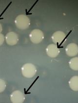
Filamentous cyanobacteria, such as Anabaena variabilis ATCC 29413 and Nostoc punctiforme ATCC 29133, are widely used as model organisms for the study of multicellularity and their capability to differentiate heterocysts and akinetes. Under favorable conditions, these photosynthetic Gram-negative bacteria grow in the form of filaments, which are composed of hundreds of vegetative cells. In response to insufficient supply of organically combined nitrogen, about 10% of semi-randomly spaced cells can differentiate into nitrogen-fixing heterocysts, which provide the filaments with nitrogen (Figure 1A) (Fay, 1992; Muro-Pastor and Maldener, 2019). Akinetes are spore-like resting cells that differentiate from the vegetative cells in response to diverse environmental conditions, including changes in light intensity and quality, temperature, and nutrient deficiency (Figure 1B).

Figure 1. Light micrographs showing cell differentiation in A. variabilis. (A) Vegetative filament containing heterocyst, indicated by black arrowhead. (B) Akinetes induced under low light condition. Scale bars, 10 µm.
Both cell types—heterocysts and akinetes—are characterized by the presence of a thick multilayered envelope, mainly composed of exopolysaccharides and glycolipids (HGLs) (Maldener et al., 2014; Perez et al., 2016; Garg and Maldener, 2021b). The glycolipids, which form laminated layers in the heterocyst and akinete envelopes, are of great interest due to their different functions in these two cell types. In heterocysts, they act as an oxygen diffusion barrier to protect the oxygen labile enzyme nitrogenase from oxygen (Fay, 1992; Wolk et al., 1994), while in akinetes, glycolipids protect the cells from various stress factors, such as freezing, desiccation, and lysozyme attack (Garg and Maldener, 2021a).
Mutants with aberrantly formed heterocyst envelopes are unable to grow without alternative sources of combined nitrogen. Many of those lose the ability to synthesize HGLs or to transport them beyond the cell wall to the heterocyst envelope (Ernst et al., 1992; Fiedler et al., 1998; Fan et al., 2005). The akinete envelope shares some structural similarities with the heterocyst envelope, suggesting that akinetes are the evolutionary ancestors of heterocysts (Wolk et al., 1994; Perez et al., 2018). To better understand this evolutionary relationship, a prerequisite is the elucidation of the composition of these special cells’ envelopes. Additionally, mutants showing phenotypes related to survival under environmental stress or starvation could be checked for the correct laminated layer composition using the protocol presented here.
Here, we use the thin-layer chromatography (TLC) protocol that was previously established by Nichols and Wood in 1968; with this method, they identified glycolipids, which are specific for the heterocyst forming cyanobacteria (Nichols and Wood, 1968). Later, the Wolk group showed that these glycolipids constitute the laminated layer in the envelope of heterocysts (Winkenbach et al., 1972). The two major heterocyst specific glycolipids in Anabaena species are characterized as 1-α-glucosyl-3,25-hexacosanediol (HG26-diol) and its 3-ketotautomer (HG26-keto-ol). Their separation via TLC is dependent on their polarity. We utilized this method with a few modifications for the analysis of the akinete specific glycolipids (Perez et al., 2018; Garg and Maldener, 2021a). We present a reproducible and reliable TLC protocol to analyze the glycolipids present in the heterocyst and akinete envelopes. This protocol includes the easy extraction of lipids using regular solvents, such as methanol and chloroform, and their clear separation and visualization on the TLC plate, which can be scanned directly. Furthermore, this method allows the extraction of specific glycolipids from the TLC plate in a manner that subsequent analysis of their chemical composition by HPLC-MS analysis is possible, as previously described (Perez et al., 2018; Shvarev et al., 2018).
Materials and Reagents
50 mL of cyanobacterial liquid cultures containing heterocysts or akinetes
Pipette tips (10 µL, 200 µL, 1,000 µL) (LTS, RAININ, Mettler Toledo. Gießen)
1.5 mL microcentrifuge tubes
50 mL conical tubes
Soft graphite pencil
30 cm ruler
Paper tape (masking tape)
Methanol ≥99.9% (HPLC grade) (Sigma, Merck, Darmstadt)
Chloroform (HPLC grade) (Roth, Karlsruhe)
Aluminium plate coated with silica gel (Macherey-Nagel, catalog number: 818033)
Acetic acid (Sigma, Merck, Darmstadt)
MilliQ water
25% Sulfuric acid (H2SO4) (Carl Roth, catalog number: 0967.1)
Equipment
Pipettes (P1000, P200, P20 and P10) (RAININ, Mettler Toledo)
100 mL Erlenmeyer flasks
50 mL glass beaker
Centrifuges:
Centrifuge (Eppendorf, 5417C) for microcentrifuge tubes
Centrifuge (Eppendorf, Centrifuge 5804 R) for conical tubes
Vortex Genie® 2 (Scientific Industries, Inc., catalog number: 6235684)
Fume hood
Hair dryer
Incubator or oven for TLC plate development (This can be any incubator that reaches the desired temperature of 180°C)
Flat-bed Scanner (e.g., EPSON Perfection V550 Photo)
Developing Chamber for TLC (Macherey-Nagel GmbH & Co. KG, catalog number: 9003500)
TLC sprayer with Erlenmeyer flask (e.g., DWK Life Sciences KimbleTM KontesTM Reagent Sprayers Head Only)
Software
Scanner software (e.g., EPSON Scan)
Procedure
Cell culture
The protocol requires cyanobacterial cultures grown in the presence of NO3- (filaments with vegetative cells only) and without NO3- (filaments containing heterocysts), or the cultures that were induced to form akinetes (Perez et al., 2016).
Thin-layer chromatography (TLC)
Day 1. Sample preparation
Take 3 × 1 mL of each culture and estimate the chlorophyll a concentration as described (Zavřel et al., 2015).
Note: To obtain a good visible lipid pattern in the TLC plates, the sample should contain 20–50 µg Chl a.
Centrifuge the estimated volume of culture at 3,000 × g and room temperature (RT) for 10 min (the cells were pelleted at equal Chl a amount in each sample).
Add 30 mL of methanol:chloroform (1:1) to the pellet and mix well with a vortex mixer. This step will result in the extraction of lipids.
Note: Always work under the fume hood when using organic solvents.
Centrifuge at 3,000 × g for 10 min and transfer the supernatant to a 50 mL glass flask.
Let it evaporate overnight under a fume hood. You may accelerate evaporation by applying a stream of nitrogen vapor. This would be necessary if detailed analysis is intended, such as precise identification and quantification of different glycolipid species by high performance liquid chromatography coupled to mass spectrometry (HPLC-MS).
Day 2Sample collection
Add 200 µL of chloroform to the dried extract in the glass flask, dissolve the pellet by pipetting, collect the lipids and transfer to 1.5-mL microcentrifuge tubes. Repeat this step one more time to collect all the extract from the flask (This step should be done quickly because the chloroform evaporates very fast).
Let the sample dry in the fume hood until a volume of 20–40 µL remains in the tube.
Preparation of developing chamber
Always prewash the chamber with methanol before doing an experiment.
Mix the solvents (chloroform:methanol:acetic acid:water) in the chamber, at a ratio of 23:4:2.7:1 (170 mL of chloroform, 30 mL of methanol, 20 mL of acetic acid, and 7.4 mL of MilliQ water).
Close the lid and seal the tank with paper tape. Do not move the chamber.
Before using the solvent system (solvent mixture), equilibrate the chamber with it for 1 h.
Spotting of samples and development of the TLC
Note: This can be done while the chamber is being equilibrated.
Using a soft pencil, mark the origin line (the line at which you will spot the samples) at 2 cm and the development line at 15 cm (predetermined solvent front height) from the bottom of the TLC plate.
Notes:
Always handle the TLC plate with gloves on. Never touch the surface of the plate even with gloves.
Mark only the outer edges of the TLC plate, because the pencil may interfere with the samples and solvent.
Use a micropipette (P20) to spot 5–10 µL of the samples on the origin line, at a distance of at least 1 cm from each other.
After spotting, use a hair drier in low heat mode to dry the spots.
Repeat steps b. and c. until the entire sample is used up and dried.
Quickly place the TLC plate in the chamber to prevent solvent vapor elusion from the chamber.
Let the samples run until they reach the development line (~1.5 h).
Take the plate out from the chamber, and let it dry under the fume hood for 10–20 min (do not use a hair drier).
Note: Always remove the remainder of the solvent from the chamber and transfer it to the organic waste.
When the plate is completely dry, spray the whole plate with H2SO4 (25%) with a TLC sprayer, from a distance of 5–10 cm under the fume hood (spraying close to the plate can obfuscate the result).
Note: Use lab apron, gloves, and surgical mask.
Allow the plate to dry under the fume hood for a short time (2–5 min).
Place the plate into an oven at 180°C for 60–120 s (until you see the bands).
Use a flat-bed scanner to record the result (see Figure 2).
Data analysis

Figure 2. Thin-layer chromatogram of lipid extracts from different cell extracts of A. variabilis (1–3) and N. punctiforme (4–6). (1, 4) Vegetative filaments grown with nitrate. (2, 5) Heterocysts containing filaments grown without nitrate. (3) Akinetes induced by low light, or (6) phosphate starvation for 2 months. The blue arrow indicates one heterocyst-specific glycolipid (HG26-diol). The red arrow indicates an unknown lipid in the akinete extract of A. variabilis. Figure adapted from Perez et al. (2018) .Acknowledgments
This protocol is an adapted version of the method described by Nichols and Wood (1968) and Winkenbach et al. (1972).
R.G. and R.P. were supported by the grant GRK1708 from the Deutsche Forschungsgemeinschaft (DFG, German Research Foundation).
Competing interests
We declare no conflict of interests.
References
- Ernst, A., Black, T., Cai, Y., Panoff, J. M., Tiwari, D. N. and Wolk, C. P. (1992). Synthesis of nitrogenase in mutants of the cyanobacterium Anabaena sp. strain PCC 7120 affected in heterocyst development or metabolism. J Bacteriol 174(19): 6025-6032.
- Fan, Q., Huang, G., Lechno-Yossef, S., Wolk, C. P., Kaneko, T. and Tabata, S. (2005). Clustered genes required for synthesis and deposition of envelope glycolipids in Anabaena sp. strain PCC 7120. Mol Microbiol 58(1): 227-243.
- Fay, P. (1992). Oxygen relations of nitrogen fixation in cyanobacteria. Microbiol Rev 56(2): 340-373.
- Fiedler, G., Arnold, M., Hannus, S. and Maldener, I. (1998). The DevBCA exporter is essential for envelope formation in heterocysts of the cyanobacterium Anabaena sp. strain PCC 7120. Mol Microbiol 27(6): 1193-1202.
- Garg, R. and Maldener, I. (2021a). The dual role of the glycolipid envelope in different cell types of the multicellular cyanobacterium Anabaena variabilis ATCC 29413. Front Microbiol 12: 645028.
- Garg, R. and Maldener, I. (2021). The Formation of Spore-Like Akinetes: A Survival Strategy of Filamentous Cyanobacteria. Microb Physiol 31(3): 296-305.
- Maldener, I., Summers, M. L., and Sukenik., A. (2014). Cellular differentiation in filamentous cyanobacteria. In: The Cell Biology of Cyanobacteria. Flores, E. and Herrero, A. (Eds.). Caister Academic Press. 263-291.
- Muro-Pastor, A. M. and Maldener, I. (2019). Cyanobacterial heterocysts. eLS (Wiley). 1-10.
- Nichols, B. W. and Wood, B. J. B. (1968). New glycolipid specific to nitrogen-fixing blue-green algae. Nature 217(5130): 767-768.
- Perez, R., Forchhammer, K., Salerno, G. and Maldener, I. (2016). Clear differences in metabolic and morphological adaptations of akinetes of two Nostocales living in different habitats. Microbiology (Reading) 162(2): 214-223.
- Perez, R., Wormer, L., Sass, P. and Maldener, I. (2018). A highly asynchronous developmental program triggered during germination of dormant akinetes of filamentous diazotrophic cyanobacteria. FEMS Microbiol Ecol 94(1). doi: 10.1093/femsec/fix131.
- Shvarev, D., Nishi, C. N., Wörmer, L. and Maldener, I. (2018). The ABC transporter components HgdB and HgdC are important for glycolipid layer composition and function of heterocysts in Anabaena sp. PCC 7120. Life (Basel) 8(3): 26.
- Winkenbach, F., Wolk, C. P. and Jost, M. (1972). Lipids of membranes and of the cell envelope in heterocysts of a blue-green alga. Planta 107(1): 69-80.
- Wolk, C. P., Ernst, A., and Elhai, J. (1994). Heterocyst metabolism and development. The Molecular Biology of Cyanobacteria (Springer Netherlands), 769-823.
- Zavřel, T., Sinetova, M. A. and Červený, J. (2015). Measurement of chlorophyll a and carotenoids concentration in cyanobacteria. Bio-protocol 5(9): e1467.
Article Information
Copyright
© 2022 The Authors; exclusive licensee Bio-protocol LLC.
How to cite
Garg, R., Perez, R. and Maldener, I. (2022). Analysis of Heterocyst and Akinete Specific Glycolipids in Cyanobacteria Using Thin-layer Chromatography. Bio-protocol 12(6): e4355. DOI: 10.21769/BioProtoc.4355.
Category
Biochemistry > Lipid > Lipid isolation
Microbiology > Microbial physiology > Adaptation
Cell Biology > Cell structure > Cell surface
Do you have any questions about this protocol?
Post your question to gather feedback from the community. We will also invite the authors of this article to respond.