- Protocols

- Articles and Issues
- For Authors
- About
- Become a Reviewer

Isolation of Human Blood Progenitor and Stem Cells from Peripheral Blood by Magnetic Bead


Published: Vol 2, Iss 21, Nov 5, 2012 DOI: 10.21769/BioProtoc.281 Views: 16757
Original research article
The authors used this protocol in:
Mar 2012
Advertisement


Protocol Collections
Comprehensive collections of detailed, peer-reviewed protocols focusing on specific topics


Related protocols
Abstract
The antigen CD34 is a well-known marker present on human progenitor and stem cells. This protocol explains the isolation of CD34+ cells from peripheral blood using magnetic bead separation technique. The approximate abundance of CD34+ cells in blood is 0.1% of mononuclear cells.
Keywords: HematopoiesisMaterials and Reagents
- CD34+ cells
- Peripheral blood sample (at least 50 ml)
- Dextran solution (Sigma-Aldrich, catalog number: D1037-500G )
- PBS (Life Technologies, Invitrogen™, catalog number: 14190-094 )
- Ficoll human (PAA Laboratories GmbH, catalog number: P04-60500 )
- Fetal calf serum (FCS) (Hyclone, catalog number: SV30160.03 )
- CD34 MicroBeads and FcR blocking reagent (Miltenyi Biotec, catalog number: 130-046-702 )
- APC mouse anti-human CD34 antibody (BD Biosciences, catalog number: 555824 )
- EDTA
- 2% dextran solution
- 1 L 2% dextran solution (see Recipes)
Equipment
- Centrifuges
- Auto MACS Pro separator (Miltenyi Biotec)
- 30 μm nylon mesh (Miltenyi Biotec, catalog number: 130-041-407 )
- Tissue culture hood
- Flow cytometer
- Vacuum filter unit (22 μm, GP Millipore Express PLUS membrane)
Procedure
- Peripheral blood mononuclear cells (PBMCs) separation
- Under tissue culture hood, add equal volume of 2% dextran solution and blood sample and incubate it at room temperature for 45 min (elimination of majority of erythrocytes).
- Collect the supernatant and centrifuge at 1,600 rpm (ROTANA 460RF, rotor 5624 Hettich) for 10 min.
- Discard the supernatant and re-suspend the pellet, containing lymphocytes and granulocytes, in 10 ml PBS with 0.1% EDTA.
- Add 7.5 ml ficoll solution to a suitable centrifuge tube and carefully top it with 10 ml cell suspension from step A3.
- Centrifuge at 2,200 rpm for 20 min.
- After centrifugation there are three phases: Upper one containing plasma and platelets. The whitish layer containing the mononuclear cells and the lower phase and pellet containing the granulocytes.
- Discard upper aqueous phase containing plasma and collect the whitish layer containing PBMCs (around 2 ml) and re-suspend it in 50 ml of PBS with 0.1% EDTA.
- Count the PBMCs.
- Centrifuge the PBMCs at 1,200 rpm for 10 min and re-suspend 1 x 10⁸ cells in 300 μl of PBS with 0.1% EDTA. If you have 5 x 10⁷ cells, you need to resuspend in 150 μl of PBS with 0.1% EDTA.
- Under tissue culture hood, add equal volume of 2% dextran solution and blood sample and incubate it at room temperature for 45 min (elimination of majority of erythrocytes).
- CD34 magnetic labeling
- Add CD34 MicroBeads and FcR blocking reagent (100 μl each for 1 x 10⁸ cells) to 300 μl of PBMCs suspension. The FcR blocking reagent is used to avoid non specific labeling.
- Mix well and incubate at 4 °C for 30 min (or overnight with 30% FCS).
- Add CD34 MicroBeads and FcR blocking reagent (100 μl each for 1 x 10⁸ cells) to 300 μl of PBMCs suspension. The FcR blocking reagent is used to avoid non specific labeling.
- Magnetic separation with auto MACS separator
- Add 10 ml PBS 0.1% EDTA to magnetic beads labeled cells and centrifuge them at 1,200 rpm for 10 min.
- Discard supernatant and re-suspend the cells in PBS with 0.1% EDTA (500 μl for 1x 10⁸ cells).
- Filter the cells using 30 μm nylon mesh to exclude the cell clumps and rinse the tube and filter with additional 500 μl PBS with 0.1% EDTA to collect maximum cells.
- Pass the cells through auto MACS separator as explained in user manual (CD34 MicroBead kit human).
- After separation, collect CD34+ cells and determine cell number.
- Wash the cells with 5-10 ml PBS 0.1% EDTA at 1,200 rpm for 10 min at room temperature.
- Once washed re-suspend cells in appropriate cell culture medium or freeze them for later use.
- Add 10 ml PBS 0.1% EDTA to magnetic beads labeled cells and centrifuge them at 1,200 rpm for 10 min.
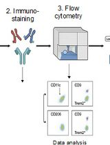
- CD34+ cells purity assessment
- Take 10 x 103 cells from step C 4 and re-suspend them in 100 μl of PBS.
- Add 1 μl of APC-conjugated mouse anti-human CD34 antibody.
- Incubate for 15-20 min at 4 °C in dark.
- Wash the cells with 500 μl PBS at 1,200 rpm for 10 min.
- Discard the supernatant and re-suspend the cells in 300 μl PBS.
- Pass them through a flow cytometer and analyze the cells for APC labeling excluding dead cells and debris using scatter signals.
- Take 10 x 103 cells from step C 4 and re-suspend them in 100 μl of PBS.
Recipes
- 1 L 2% dextran solution
Add 9 g NaCl and 20 g dextran to beaker containing 800 ml deionized H2O
Dissolve NaCl and dextran using magnetic stirrer and magnetic stirrer bar (approx. 2 h)
Add deionized H2O to make up the volume to 1 L
Under tissue culture hood, filter the 2% dextran solution using 22 μm pore vacuum filter
Conserve the solution at 4 °C
Acknowledgments
The protocol was previously published in Nakatake et al. (2012). This work was supported by grants from ” Association pour la Recherche sur le Cancer (projet libre 2012), Agence Nationale de la Recherche, programme Jeunes Chercheuses et Jeunes Chercheurs, Laboratory of Excellence Globule Rouge-Excellence is funded by the program “Investissements d’avenir.” HS was supported by fellowships from la Ligue Nationale Contre le Cancer.
References
- Nakatake, M., Monte-Mor, B., Debili, N., Casadevall, N., Ribrag, V., Solary, E., Vainchenker, W. and Plo, I. (2012). JAK2(V617F) negatively regulates p53 stabilization by enhancing MDM2 via La expression in myeloproliferative neoplasms. Oncogene 31(10): 1323-1333.
Article Information
Copyright
© 2012 The Authors; exclusive licensee Bio-protocol LLC.
How to cite
Hasan, S. and Plo-Azevedo, I. (2012). Isolation of Human Blood Progenitor and Stem Cells from Peripheral Blood by Magnetic Bead . Bio-protocol 2(21): e281. DOI: 10.21769/BioProtoc.281.
Category
Stem Cell > Adult stem cell > Hematopoietic stem cell
Cell Biology > Cell isolation and culture > Cell isolation
Do you have any questions about this protocol?
Post your question to gather feedback from the community. We will also invite the authors of this article to respond.