- Protocols

- Articles and Issues
- For Authors
- About
- Become a Reviewer

Rat Aortic Ring Model to Assay Angiogenesis ex vivo


Published: Vol 5, Iss 20, Oct 20, 2015 DOI: 10.21769/BioProtoc.1622 Views: 11735
Reviewed by: Ivan Zanoni
Original research article
The authors used this protocol in:
Jan 2015
Advertisement


Protocol Collections
Comprehensive collections of detailed, peer-reviewed protocols focusing on specific topics


Related protocols
Abstract
Angiogenesis is a multifactorial event which requires the migration, proliferation, differentiation and structure rearrangement of endothelial cells. This angiogenic process has been commonly studied using in vitro assays such as Boyden chamber assay, wound healing assay and tube formation assay. These assays mainly use monolayers of endothelial cells which are modified by repeated passages and are fully proliferative, a situation far away from physiology. In addition, not only endothelial cells are involved in this process but surrounding cells (such as pericytes, smooth muscle cells, fibroblasts) and the supporting matrix are also major players.
The three-dimensional ex vivo aortic ring model recapitulates the complexities of angiogenesis and combines the advantages of in vitro and in vivo models. The aortic ring is cultivated in a chemically defined culture environment. Microvessels which grow in this system are lumenized vessels with surrounding supporting cells and are essentially indistinguishable from microvessels formed during angiogenesis in vivo. The efficacy of pro-or anti-angiogenic factors can be determined in the absence of serum molecules which may otherwise interfere with the substances being tested (Nicosia and Ottinetti, 1990). However, this system requires access to fresh rat tissue but several samples can be prepared from one aorta.
Materials and Reagents
- Aluminium foils
- Cell culture plates 48 well (VWR International, catalog number: 734-2157 )
- Petri dishes (VWR International, catalog number: 734-2374 )
- LEW/Ss NHsd males rats 8-10 weeks old (Harlan Laboratories)
- Endothelial Cell Growth Medium (Lonza, catalog number: CC-3162 ) + 1% penicillin/streptomycin (P/S) (Thermo Fisher Scientific, GibcoTM, catalog number: 15140-122 )
- Collagen R (2 mg/ml) (SERVA Electrophoresis GmbH, catalog number: 47254 )
- Sodium bicarbonate (NaHCO3) (Sigma-Aldrich, catalog number: S-5761 )
- 10x MEM (Thermo Fisher Scientific, GibcoTM, catalog number: 21430-020 )
- NaOH (Merck Millipore Corporation, catalog number: 1.06498 )
Equipment
- Dissecting microscope (Motic Stereomicroscope K-400L)
- Laminar flow hood
- Inverted light microscopes
- 50 ml Beaker
- 25 mm magnetic bar
- Magnetic stirrer
- Bleue extra gillette razor blades
- Scissors (Fine Science Tools, catalog number: 14090-11 )
- Vannas-Tübingen spring scissors (Fine Science Tools, catalog number: 15003-08 )
- 2 Microdissection forceps (VWR International, catalog number: 734-0448 )
- Dissecting boards and pins
Procedure
The day before
Wrapped dissecting board in 2 aluminium foils
Sterilize: Beaker, magnetic bar, blades and dissecting materials
Put in a fridge: 200 µl tips box, Cell culture plates 48 well
On the day
- Aortic ring preparation
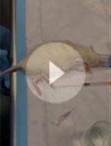
- Sacrifice rat by CO2 inhalation and decapitation.
- Fix the rat on his back on a dissecting board.
- Disinfect skin with 80% ethanol.
- Make a Y-shape incision.
- Open the abdominal cavity.
- Cut the sternal plate and the diaphragm with scissors to expose the thoracic cavity.
- Displace the intestines, liver, stomach and spleen to the right side.
- Remove the lungs.
- The aorta is visible along the vertebral column.
- Excise the aorta starting at the point where abdominal aorta divides into two arteries up to the aortic arch using scissors placed parallel to the vertebral column and tweezers (do not stretch and mechanically damage the aorta).
- Transfer the aorta to a Petri dish filled with serum-free EBM-2 medium supplemented with 1% P/S on ice. All of the following steps will be performed under sterile conditions.
- Prior to pinning the aorta to the dissecting board remove the first sheet of aluminium foil and make sure that the aorta doesn’t dry by regularly adding serum-free EBM-2 medium + P/S.
- Under a dissecting microscope, remove the periaortic fibroadipose tissue, stumps of collateral vessels and blood clots with Vannas-Tübingen spring scissors and microdissection forceps.
- When the aorta is clean, transfer it to a clean Petri dish containing cold serum-free EBM-2 medium and keep it on ice.
- Prepare the collagen mix before cross-sectioning the thoracic aorta.
- Whilst in the Petri dish, Cross-section the thoracic aorta into rings that are 1 to 2 mm in length (20 to 25 rings/aorta) using a razor blade.
- Discard the aortic arch and the abdominal part of the aorta.
- Replace the medium 5 times with cold serum-free EBM-2 medium in the Petri-dish to rinse the rings.
- Sacrifice rat by CO2 inhalation and decapitation.
- Embedding of aortic rings in collagen gels
- Homogenously spread 200 µl of collagen solution onto the bottom of each well in a pre-chilled 48-well plate using pre-chilled tips.
- Put the plate into a CO2 incubator (37 °C) for 30 min to allow polymerization.
- Take a ring with a forceps.
- Dry the ring by carefully dabbing it on the side of an empty Petri dish.
- Place the ring in the middle of the well at the top of the polymerized collagen.
- Repeat this process for all the rings, but be as quick as possible to make sure the rings do not dry out.
- Add a second layer of 200 µl collagen mix at the top of the aorta ring using pre-chilled tips.
- Place the plate back in the incubator.
- After 30 min, add 500 µl of serum-free EBM-2 medium supplemented or not with the drug of interest.
- Homogenously spread 200 µl of collagen solution onto the bottom of each well in a pre-chilled 48-well plate using pre-chilled tips.
- Quantification
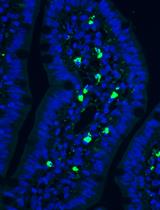
Quantification of microvessel outgrowth is done manually from pictures obtained after 7-8 days of culture with a 2.5x magnification on an inverted microscope. The quantification involves counting the number of vessels and branch points (Nicosia and Ottinetti, 1990). A visualization of the counting process is presented in Aplin et al. (2008).
Representative data

Figure 1. Representative pictures of one aortic ring at different times in culture (top left to bottom left) and pictures of aortic rings treated with a growing concentration of an angiogenic agent (from left to right) at 8 days post-isolation
Notes
- Some isolated cells and vessels start to be present around 3-4 days. A well-developed network is visible around 7 to 9 days before regression.
- In this study, commercial collagen matrice has been used but it can be replaced by fibrin or Matrigel. One should note, however, that non-endothelial cells including fibroblasts have been shown to organize into networks in Matrigel (Donovan et al., 2001).
Recipes
- Collagen solution
The collagen solution is prepared in a 50 ml beaker with pre-chilled reagents under slow agitation to avoid air bubbles.- Mix 1 volume 10x MEM with 1.5 volumes 186 mM NaHCO3 until the color of the solution changes to orange.
- Add 7.5 volumes 2 mg/ml collagen R.
- Add few drops of 1 M NaOH to reach pH 7.4 (checked by the color indicator in the solution).
- Mix 1 volume 10x MEM with 1.5 volumes 186 mM NaHCO3 until the color of the solution changes to orange.
Acknowledgments
This protocol was adapted from Nicosia and Ottinetti (1990) and Blacher et al. (2001). This study was supported by the Society for Research on Cardiovascular Diseases and The Ministry of Culture, Higher Education and Research of Luxembourg.
References
- Aplin, A. C., Fogel, E., Zorzi, P. and Nicosia, R. F. (2008). The aortic ring model of angiogenesis. Methods Enzymol 443: 119-136.
- Blacher, S., Devy, L., Burbridge, M. F., Roland, G., Tucker, G., Noel, A. and Foidart, J. M. (2001). Improved quantification of angiogenesis in the rat aortic ring assay. Angiogenesis 4(2): 133-142.
- Donovan, D., Brown, N. J., Bishop, E. T. and Lewis, C. E. (2001). Comparison of three in vitro human 'angiogenesis' assays with capillaries formed in vivo. Angiogenesis 4(2): 113-121.
- Nicosia, R. F. and Ottinetti, A. (1990). Growth of microvessels in serum-free matrix culture of rat aorta. A quantitative assay of angiogenesis in vitro. Lab Invest 63(1): 115-122.
Article Information
Copyright
© 2015 The Authors; exclusive licensee Bio-protocol LLC.
How to cite
Ernens, I., Lenoir, B., Devaux, Y. and Wagner, D. R. (2015). Rat Aortic Ring Model to Assay Angiogenesis ex vivo. Bio-protocol 5(20): e1622. DOI: 10.21769/BioProtoc.1622.
Category
Cancer Biology > Angiogenesis > Animal models > Cell migration
Immunology > Animal model > Rat
Do you have any questions about this protocol?
Post your question to gather feedback from the community. We will also invite the authors of this article to respond.