- Protocols

- Articles and Issues
- For Authors
- About
- Become a Reviewer

Ex vivo Human Natural Killer (NK) Cell Stimulation and Intracellular IFNγ and CD107a Cytokine Staining

Published: Vol 5, Iss 12, Jun 20, 2015 DOI: 10.21769/BioProtoc.1501 Views: 19278
Reviewed by: Thomas J. BartoshAnonymous reviewer(s)
Original research article
The authors used this protocol in:

Dec 2013
Advertisement


Protocol Collections
Comprehensive collections of detailed, peer-reviewed protocols focusing on specific topics


Related protocols
Abstract
Natural killer (NK) cells comprise 5–20% of peripheral blood mononuclear cells (PBMC) in humans. In addition to their fundamental roles in the defense against viral infections and tumor surveillance, NK cells help shape adaptive immune responses through their production of cytokines. NK cells are traditionally identified as CD3neg, CD14neg, CD19neg lymphocytes expressing CD56. Using a combination of markers that includes CD56 and CD7 greatly increases the ability to define the phenotype and function of NK cell subsets. Two key markers of NK cell function are the production of IFNγ and the release of cytotoxic granules measured by the expression of CD107a. Here we describe a method to assess IFNγ and CD107a expression in NK cells following stimulation with target cells or cytokines. This method can be used to assess the general functional capacity of NK cells in peripheral blood mononuclear cells from a wide range of study participants.
Keywords: NK cellMaterials and Reagents
- Alexa700-conjugated mouse anti-human CD7 clone 124-1D1 (eBioscience, catalog number: 56-0079-42 )
- K562 cell line (Kindly provided by Dr. Lewis L. Lanier, University of California, San Francisco, USA)
Note: These can also be purchased from ATCC, catalog number CCL-243 . - Phycoerythrin (PE)-Texas Red (ECD)-conjugated mouse anti-human CD3 clone UCHT1 (Beckman Coulter, catalog number: IM2705U )
- ECD-conjugated mouse anti-human CD14 clone RMO52 (Beckman Coulter, catalog number: IM2707U )
- PE-Cy7-conjugated mouse anti-human CD56 clone NCAM16.2 (BD Biosciences, catalog number: 335791 )
- Pacific Blue-conjugated mouse anti-human CD16 clone 3G8 (BD Biosciences, catalog number: 558122 )
- APC-Cy7-conjugated mouse anti-human CD19 clone SJ25C1 (BD Biosciences, catalog number: 557791 )
- Fluorescein isothiocyanate (FITC)-conjugated mouse anti-human CD107a clone H4A3 (BD Biosciences, catalog number: 555800 )
- APC-conjugated mouse anti-human IFNγ clone B27 (BD Biosciences, catalog number: 554702 )
- Human IgG (Sigma-Aldrich, catalog number: I4506 )
- Anti–mouse immunoglobulin G–coated compensation beads (BD Biosciences, catalog number: 552843 )
- Amine Aqua Reactive Dye (AARD) (Life Technologies, catalog number: L34957 )
- 96 well U bottom plate (Corning, catalog number: 353077 )
- 96 well V bottom plate (Corning, catalog number: 3894 )
- RPMI (Life technologies, catalog number: 11875)
- L-Glutamine 200 mM (100x) (Life technologies, catalog number: 25030 )
- Penicillin (10,000 Units/ml)-Streptomycin (10,000 μg/ml) (Life technologies, catalog number: 15140 )
- Fetal bovine serum (Hyclone, catalog number: SH30071 )
- Buffy coats from Stanford Blood Center used to obtain Peripheral Blood Mononuclear Cells (PBMC)
- Ficoll-Paque Premium (GE Healthcare, catalog number: 17-5442-03 )
- Cryopreserved PBMC samples from San Francisco based HIV-1 infected cohorts SCOPE and OPTIONS
- Recombinant IL-12 (Peprotech, catalog number: 200-12 )
- Recombinant IL-18 (MBL & Biological Laboratories, catalog number: B001-5 )
- Brefeldin A from Penicilium brefeldianum (Sigma-Aldrich, catalog number: B7651 )
- BD golgi stop protein transport inhibitor containing monensin (BD Biosciences, catalog number: 554724 )
- Phosphate buffered saline (PBS) (Corning, catalog number: 21-040-CV )
- Ethylenediaminetetraacetic Acid (EDTA) (Teknova, catalog number: E0306 )
- Bovine Serum Albumin (BSA) (Gemini BioProducts, catalog number: 700-100P )
- 16% Paraformaldehyde (PFA) (Electron Microscopy Science, catalog number: 15710 )
- BD FACS Permeabilizing solution 2 (BD Biosciences, catalog number: 340973 )
- Cell Culture Grade Water (HyClone, catalog number: SH30529.02 )
- Deoxyribonuclease (DNase) I (Sigma-Aldrich, catalog number: DN25 )
- 15 ml conicals (Thermo Fisher Scientific, catalog number: 05-539-5 )
- Trypan blue in PBS (0.4% w/v) (Corning, catalog number: 25-900-CI )
- Complete media (see Recipes)
- FACS buffer (see Recipes)
- Paraformaldehyde recipe (see Recipes)
Equipment
- Biosafety cabinet (Nuaire, model: 407FM600 )
- 37 °C water bath (Cole Parmer)
- Calibrated single-channel and multi-channel pipettes
- Pipet-aid
- Tips (10 μl, 20 μl, 200 μl, 1,000 μl)
- Centrifuge (Beckman Coulter, Allegra 6R , rotor GH-3.8)
- 37° Celsius Incubator (Thermo Forma, model: 3110 )
- Nikon Optiphot microscope for counting cells
- Hemacytometer for counting cells (Hausser Scientific, catalog number: 1490 )
- 4-laser (405 nm, 488 nm, 532 nm and 633 nm) BD LSR-II
Software
- FlowJo Single Cell Analysis software
Procedure
- Recovery of Peripheral Blood Mononuclear Cells (PBMCs) from cryostorage.
- Warm complete media in a 37 °C water bath prior to thawing.
- Transfer frozen vials of cryopreserved PBMC from a cryofreezer into a 37 °C water bath and gently move the tube back and forth in the water, allowing the contents of the vial to thaw until there is only a small amount left frozen. Do not leave the cryovial unattended during the thawing process. Thawing will take approximately 1-2 min.
- In a biosafety cabinet, remove the liquid from the cryovial and place into a 15 ml conical tube using a 2 ml serological pipette and pipettor.
- Slowly add 10 ml of pre-warmed (37 °C) media dropwise into the 15 ml conical tube. Gently flick the tube as you are adding media to ensure that the liquid is well mixed.
- Cap the tube and centrifuge at 330 x g for 5 min in a pre-cooled 4 °C centrifuge.
- Decant supernatant, break up the pellet by flicking the tube, and resuspend in 10 ml of complete media containing DNase (0.01 mg/ml) to count cells.
- Mix 10 μl of cells with 10 μl of Trypan blue. Take 10 μl of that mixture and place onto a hemacytometer for counting.
- Centrifuge cells at 330 x g for 5 min at 4 °C, decant supernatant, break up the cell pellet by flicking the tube, and resuspend in complete media containing DNase at a concentration of 5 million cells per ml.
- Warm complete media in a 37 °C water bath prior to thawing.
- Plate 500,000 PBMC per well in 100 μl by single channel pipette in a 96 well U bottom plate.
Note: Alternative protocol for stimulation: Thawed PBMC can be incubated overnight (16-20 h) at 37 °C and 5% CO2 in complete media supplemented with 200 IU/ml human recombinant IL-2 (NCI BRB Preclinical Repository). This significantly increases the responsiveness of the NK cells to stimulation with target cells; however, culturing overnight in IL-2 can also alter the phenotype of the NK cells. One needs to consider what the desired readout for the experiment is in choosing between the main protocol and this alternative approach. - Add FITC-conjugated anti-CD107a antibody using a single channel pipette at a dilution of 1:50 (i.e. 4 μl in 200 μl culture conditions) to all wells of a 96 well plate containing cells.
Notes:- The dilution of this antibody should be titrated for use with a particular cytometer.
- The final volume in each well should be 200 μl. There will be 100 μl of PBMC and either 100 μl of target cells (K562) or 100 μl of cytokine mixture.
- The dilution of this antibody should be titrated for use with a particular cytometer.
- For target cell stimulation (i.e. K562), count and suspend 500,000 K562 cells in 1 ml of complete media containing DNase and add 50,000 target cells (100 μl) using a single channel pipette to appropriate wells.
- For cytokine stimulation, add IL-12 and IL-18 at a final concentration of 10 ng per cytokine per well. Add 1 μl of each cytokine to 998 μl complete media containing DNase. Add 100 μl per well using a single channel pipette to appropriate wells for stimulation.
- Mix all wells with a multichannel pipette by carefully pipetting up and down 3 times being careful not to tocause bubbles.
- Place plate in incubator for 1 h at 37 °C and 5% CO2.
- After 1 h, centrifuge plate at 330 x g in a 25 °C centrifuge.
- Remove 50 μl supernatant from each well being careful not to disturb cell pellet. To not disturb the pellet, tilt the plate at a 45-60° angle, and using a multichannel pipette, place tip to the side of the well and remove liquid slowly. The supernatant can be discarded.
- Prepare a master mix containing Brefeldin A (BFA) and GolgiStop (monensin) mixture.
- Dilute GolgiStop 7.8 μl into 42.2 μl complete media containing 0.01mg/ml DNase.
- Make a master mix of: 20 μl 10 mg/ml Brefeldin A (BFA)
20 μl of diluted GolgiStop mixture from step 10a.
4,960 μl complete media containing DNase - Plate 50 μl master mix into each well using a single channel pipette. Mix well using a multichannel pipette.
- Dilute GolgiStop 7.8 μl into 42.2 μl complete media containing 0.01mg/ml DNase.
- Incubate another 5 h at 37 °C, 5% CO2.
- Transfer cells to a 96 well V bottom plate. Centrifuge plate at 330 x g for 5 min at 4 °C and remove supernatant. Flick supernatant into a container to be discarded. To flick, turn the plate over and give one firm shake.
- Resuspend cells in 200 μl FACS buffer using a multichannel pipette and centrifuge plate at 330 x g for 5 min at 4 °C.
- Wash plate again adding 200 μl FACS buffer per well using a multichannel pipette. Centrifuge plate at 330 x g for 5 min at 4 °C. Flick supernatant into a container to be discarded. To stop the protocol overnight at this point, see step To continue, skip to step 16.
- For time considerations, the protocol can now be stopped here overnight. Resuspend cells in 200 μl FACS buffer using a multichannel pipette, place in the refrigerator overnight, and stain the next day. Prior to staining on Day 2, centrifuge plate at 330 x g for 5 min at 4 °C. Flick out supernatant. Blot the plate on paper towels. Then continue with step 16.
- Surface stain cells.
- Make up a master mixture of surface antibodies diluted in FACS buffer that have previously been titrated for your flow cytometer. Staining is performed in 50 μl volume for 500,000 cells.
- We have found the following combination works well for our BD LSR-II.
Antigen Fluorophore Clone Dilutions CD3 ECD UCHT1 1:50 CD14 ECD RMO52 1:400 CD19 APCCy7 SJ25C1 1:100 CD7 Alexa700 124-1D1 1:50 CD16 Pacific Blue 3G8 1:100 CD56 PECy7 NCAM16.2 1:100 IgG (100 μg/mL)1:10 Live/Dead Marker Amine Aqua Reactive Dye (AARD) 1:200 FACS buffer - The use of fluorescence minus one (FMOs) is recommended. These can be made using the exact same mixture of antibodies but omitting one antibody. We focus on FMOs for CD107a and IFNγ, although FMOs can be used for all fluorophores initially to set appropriate gating of cytometry data.
- Make sure to have centrifuged the plate at 330 x g for 5 min at 4 °C and flicked out the supernatant prior to adding the surface stain. Aliquot 50 μl of antibody cocktail into each well using a single channel pipette. Mix well by pipetting up and down using a multichannel pipette. Allow plate to incubate on ice for 30 min to stain.
- We have found the following combination works well for our BD LSR-II.
- Make up a master mixture of surface antibodies diluted in FACS buffer that have previously been titrated for your flow cytometer. Staining is performed in 50 μl volume for 500,000 cells.
- After 30 min, add 150 μl FACS buffer to all wells using a multichannel pipette, centrifuge plate at 515 x g for 5 min at 4 °C. To improve recovery of cells from this step forward, a faster centrifuge speed is used.
- Flick supernatant into a container to be discarded and blot the plate on paper towels.
- Fix cells by adding 100 μl per well of 2% paraformaldehyde in PBS on ice for 20 min using a multichannel pipette.
- Spin at 915 x g in a 4 °C centrifuge for 5 min.
- Flick supernatant into a container to be discarded and blot on paper towels.
- Permeabilize cells using BD FACS Permeabilizing solution 2.
- Perm2 is supplied as a 10x solution. Dilute to 1x using cell culture grade water.
- Add 75 μl of the 1x Perm2 solution per well using a multichannel pipette and pipet up and down to resuspend cell pellet.
- Incubate plate for 10 min only at room temperature.
- Shorter incubations will reduce intracellular staining while incubating longer than 10 min will significantly affect cellular integrity and result in loss of sample.
- Perm2 is supplied as a 10x solution. Dilute to 1x using cell culture grade water.
- Wash once by adding 125 μl FACS buffer to each well using a multichannel pipette.
- Centrifuge plate at 915 x g in a 4 °C centrifuge for 5 min.
- Flick supernatant into a container to be discarded and blot the plate on paper towels.
- Intracellularly stain cells.
- Make up an antibody master mix with APC-conjugated anti-IFNγ antibody diluted into FACS buffer (do not dilute into Perm2 buffer).
- Aliquot 50 μl of antibody cocktail into each well by single channel pipette. Mix well by pipetting up and down using a multichannel pipette. Allow plate to incubate on ice for 30 min to stain.
- Make up an antibody master mix with APC-conjugated anti-IFNγ antibody diluted into FACS buffer (do not dilute into Perm2 buffer).
- Wash once by adding 150 μl FACS buffer to each well using a multichannel pipette. Centrifuge plate at 915 x g in a 4 °C centrifuge for 5 min.
- Flick supernatant into a container to be discarded and blot plate on paper towels.
- To fix, add 50 μl per well of 2% paraformaldehyde diluted with PBS using a multichannel pipette. Cells should be fixed a minimum of 10 min on ice, but samples can be stored at 4 °C for up to 2 days without significant changes in fluorescent staining. The fixative does not need to be washed out prior to analysis.
- Run cells on BD LSR-II.
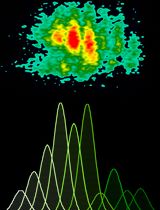
- Data are analyzed using FlowJo Single Cell Analysis software. Gates for IFNγ and CD107a expression are set using FMOs on a control sample and applied to all other samples collected. An example of the gating strategy and results of IFNγ and CD107a expression following media only (i.e. no stimulation), K562 target cell stimulation and IL-12 + IL-18 stimulation are shown (Figure 1).

Figure 1. Representative gating strategy to identify NK cells and responses to stimulation. NK cells are identified as live lymphocytes that are CD3, CD14 and CD19 negative, but express CD7, CD56 and CD16. K562 target cell stimulation induces expression of both CD107a and IFNγ. IL-12 + IL-18 cytokine stimulation induces a robust IFNγ response from NK cells, however these cytokines do not result in degranulation as measured by CD107a expression.
Recipes
- Complete media (make fresh each time)
RPMI
10% Fetal Bovine Serum
2 mM L-glutamine
Penicillin (100 Units/ml)/Streptomycin (100 μg/ml)
DNase 0.01 mg/ml - FACS buffer (shelf life 1 month)
Phosphate buffer saline
0.5% Bovine serum albumin
2 mM Ethylenediaminetetraacetic Acid - Paraformaldehyde (PFA) recipe
Dilution of PFA made fresh each time
Dilute 16% stock PFA in PBS to a final concentration of 2%
Acknowledgments
This protocol has been adapted from the publications by Milush et al. (2009 and 2013). This research was supported, in part, by the Department of Health and Human Services funding under NIH Grant number 5T32HL007185 to JMM.
References
- Milush, J. M., Lopez-Verges, S., York, V. A., Deeks, S. G., Martin, J. N., Hecht, F. M., Lanier, L. L. and Nixon, D. F. (2013). CD56negCD16(+) NK cells are activated mature NK cells with impaired effector function during HIV-1 infection. Retrovirology 10: 158.
- Milush, J. M., Long, B. R., Snyder-Cappione, J. E., Cappione, A. J., 3rd, York, V. A., Ndhlovu, L. C., Lanier, L. L., Michaelsson, J. and Nixon, D. F. (2009). Functionally distinct subsets of human NK cells and monocyte/DC-like cells identified by coexpression of CD56, CD7, and CD4. Blood 114(23): 4823-4831.
Article Information
Copyright
© 2015 The Authors; exclusive licensee Bio-protocol LLC.
How to cite
York, V. A. and Milush, J. M. (2015). Ex vivo Human Natural Killer (NK) Cell Stimulation and Intracellular IFNγ and CD107a Cytokine Staining. Bio-protocol 5(12): e1501. DOI: 10.21769/BioProtoc.1501.
Category
Immunology > Immune cell isolation > Lymphocyte
Immunology > Immune cell function > Cytotoxicity
Immunology > Immune cell staining > Flow cytometry
Do you have any questions about this protocol?
Post your question to gather feedback from the community. We will also invite the authors of this article to respond.