


- Protocols

- Articles and Issues
- For Authors
- About
- Become a Reviewer


Immunostaining Protocol: P-Smad2 (Xenograft and Mice)

Published: Vol 4, Iss 9, May 5, 2014 DOI: 10.21769/BioProtoc.1119 Views: 9249
Reviewed by: Lin Fang
 PDF
PDF  Ask a question
Original research article
The authors used this protocol in:
Nov 2012
Advertisement




Protocol Collections
Comprehensive collections of detailed, peer-reviewed protocols focusing on specific topics




Related protocols
Abstract
Metastasis depends on a gene program expressed by the tumor microenvironment upon TGF-beta stimulation. CRC (Colorectal cancer) cell lines did not induce robust stromal TGF-beta responses when injected into nude mice as shown by lack of p-SMAD2 accumulation in tumor-associated stromal cells. To enforce high TGF-beta signaling in xenografts, we engineered CRC cell lines to secrete active TGF-beta. Subcutaneous tumors obtained from HT29-M6TGF-β, KM12L4aTGF-β cells and SW48TGF-β cells contained abundant p-SMAD2+ stromal cells.
Materials and Reagents
- Paraffin sections (subcutaneous tumors samples or liver metastasis from nude mice respectively injected subcutaneously or intrasplenic with CRC cells)
- XILOL
Note: Xylol also referred to as xylene or dimethylbenzene is a solvent used in histology as a clearing agent to remove paraffin from dried microscope slides prior to staining.
- MilliQ H2O
- Wash buffer (Dako, catalog number: K800721 )
- Rabbit anti-P-Smad2 (Cell Signaling Technology, catalog number: 3108 )
- BrightVision poly-HRP anti- Rabbit (Immunologic, catalog number: DPVR-110HRP )
- Envision FLEX antibody diluent (Dako, catalog number: K8006 )
- Peroxidase Blocking Solution (Dako, catalog number: S202386 )
- ImmPACT DAB (Vector Laboratories, catalog number: SK-4105 )
- DPX mounting media (Sigma-Aldrich, catalog number: 06522 )
- Hematoxylin
- Citrate buffer (pH 6) (see Recipes)
Equipment
- Oven
- Immunostaining apparatus
- Autoclave
Procedure
- Stove samples at 65 °C just before starting the immunostaining technique. Remove the samples from the oven when the wax present in sections is completely undone.
- De-waxing and rehydration: Place slides in a rack to perform following washes (bath).
- XILOL: 10 min
- XILOL: 10 min
- XILOL: 5 min
- 100% EtOH: 10 min
- 100% EtOH: 5 min
- 96% EtOH: 5 min
- 90% EtOH: 10-15 times
- 80% EtOH: 10-15 times
- 70% EtOH: 10-15 times
- 50% EtOH: 10-15 times
- 25% EtOH: 10-15 times
- H2O MilliQ: 10-15 times
- XILOL: 10 min
- Antigen retrieval.
- Citrate Buffer (pH 6)
- Time: 20 min autoclave (121 °C)
- Citrate Buffer (pH 6)
- 3 washes 5 min with 1 ml 1x wash buffer.
- Blocking endogenous peroxidase.
- 200 ml Peroxidase Blocking Solution
- Time: 10 min
- 200 ml Peroxidase Blocking Solution
- 3 washes 5 min with 1ml 1x wash buffer.
- Incubation with primary antibody.
- Antibody: Rabbit anti-P-Smad2
- Dilution 1/200 in Envision FLEX antibody diluent
- 200 µl/sample
- O/N 4 °C
- Antibody: Rabbit anti-P-Smad2
- 3 washes 5 min with 1 ml 1x wash buffer.
- Incubation with antibody BrightVision.
- Antiboby: BrightVision anti-Rabbit
- 150 µl/sample
- Time: 45 min at room temperature
- Antiboby: BrightVision anti-Rabbit
- 3 washes 5 min with 1 ml 1x wash buffer.
- Revealed with ImmPACT DAB.
- 200 µl/sample
- Time: 10 min
- 200 µl/sample
- 3 washes 5 min with distilled water.
- Hematoxilin (1 ml) counterstaining, time: 2 min.
- Rinse in distilled water bath.
- Rinse in TAP water bath.
- Dehydration and mounting with DPX.
Representative data
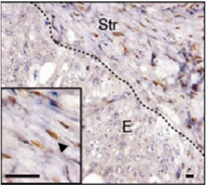
Figure 1. p-SMAD2 staining (arrowhead) in liver metastasis generated after intrasplenic injection of CRC cells. E: epithelial cells, Str: stromal cells. Scale bars = 10 μm
Recipes
- Citrate buffer (pH 6)
Citrate 5.8 g sodium citrate
Set pH 6 with citric acid
2 L MilliQ
Acknowledgments
A.C. and D.V.F.T hold a Juan de la Cierva postdoctoral fellowship, E.E. an FPI PhD fellowship (both from Spanish Ministry of Science and Innovation). This work has been supported by grants from Instituto de Salud Carlos III FEDER (RD09/0076/00036) and the “Xarxa de Bancs de tumors sponsored by Pla Director d’Oncologia de Catalunya (XBTC), to E.B. from the European Research Council (Starting grant - 208488) and Consolider programmes (MICINN), to E.S. and A.C. by the Spanish Ministry of Science and Innovation, to JM by NIH grant CA34610 and to RM by grants PS09/00965 (MICINN) and NanoCoMets (CIBERBBN).
References
- Calon, A., Espinet, E., Palomo-Ponce, S., Tauriello, D. V., Iglesias, M., Cespedes, M. V., Sevillano, M., Nadal, C., Jung, P., Zhang, X. H., Byrom, D., Riera, A., Rossell, D., Mangues, R., Massague, J., Sancho, E. and Batlle, E. (2012). Dependency of colorectal cancer on a TGF-beta-driven program in stromal cells for metastasis initiation. Cancer Cell 22(5): 571-584.
Article Information
Copyright
© 2014 The Authors; exclusive licensee Bio-protocol LLC.
How to cite
Calon, A., Espinet, E., Palomo-Ponce, S., Tauriello, D. V. F., Iglesias, M., Céspedes, M. V., Sevillano, M., Nadal, C., Jung, P., Zhang, X. H., Byrom, D., Riera, A., Rossell, D., Mangues, R., Massague, J., Sancho, E. and Batlle, E. (2014). Immunostaining Protocol: P-Smad2 (Xenograft and Mice). Bio-protocol 4(9): e1119. DOI: 10.21769/BioProtoc.1119.

 Download Citation in RIS Format
Category
Cancer Biology > Invasion & metastasis > Animal models > Cell isolation and culture
Cell Biology > Cell imaging > Fluorescence
Biochemistry > Protein > Immunodetection > Immunostaining
Do you have any questions about this protocol?
Post your question to gather feedback from the community. We will also invite the authors of this article to respond.
 Post a Question


