- Protocols

- Articles and Issues
- For Authors
- About
- Become a Reviewer

Cyclic Nucleotide (cAMP and cGMP) Assays and Capture ELISA for Quantitative Analysis of Plasmodium falciparum Blood-stage Egress

Published: Vol 4, Iss 5, Mar 5, 2014 DOI: 10.21769/BioProtoc.1055 Views: 11897
Reviewed by: Anonymous reviewer(s)
Original research article
The authors used this protocol in:
May 2013
Advertisement


Protocol Collections
Comprehensive collections of detailed, peer-reviewed protocols focusing on specific topics


Abstract
Upon rupture of Plasmodium falciparum (P. falciparum) schizonts in vitro (an event known as egress), merozoites are released into the culture medium. The merozoites invade fresh red blood cells, a process that involves shedding of a microneme protein called apical membrane antigen-1 (AMA1) from the merozoite surface. This shedding, which takes place even in the absence of invasion, is therefore a surrogate marker for the degree of egress taking place in a culture, and can be measured using a specific capture ELISA to quantify AMA1 levels in culture supernatants (Collins et al., 2013). The assay uses a monoclonal antibody specific for AMA1 (called 4G2dc1) (Kocken et al., 1998; Collins et al., 2009) to capture and immobilize the protein from culture supernatants, then uses a specific rabbit polyclonal antiserum to detect the immobilized antigen. A phosphatase-conjugated goat anti-rabbit antibody is finally used to quantify the binding of the second antibody. Egress is absolutely dependent upon the activity of a parasite cGMP-dependent protein kinase, PKG, and so is influenced by levels of intracellular cGMP (Collins et al., 2013). This is regulated by the interplay between guanylate cyclases and phosphodiesterases. The latter enzymes may also degrade cAMP, so it may also be informative to measure intracellular cAMP levels.
Materials and Reagents
- Plasmodium falciparum schizonts
- DetectX Direct cGMP or cAMP immunoassay kit (Arbor Assays, catalog number: K020-H1 or K019-H1 )
- Protein-free RPMI 1640 (Life Technologies, InvitrogenTM)
- Albumax
- Zaprinast (Sigma-Aldrich, catalog number: Z0878 )
Note: Make up as a 50 mM stock solution in DMSO and stored at -20 °C.
- PKG inhibitor compound 1 {4-[2-(4-fluorophenyl)-5-(1-methylpiperidine-4-yl)-1H-pyrrol-3-yl] pyridine} (Merck KGaA)
Note: Make up as a 10 mM stock solution in DMSO and stored at -20 °C.
- Anti-AMA1 monoclonal antibody (clone 4G2dc1) (Kocken et al., 1998)
- Complete RPMI 1640 culture medium (Blackman, 1994)
- Tween 20
- Rabbit polyclonal anti-AMA1 serum (Collins et al., 2009)
- Phosphatase-conjugated goat anti-rabbit IgG (whole molecule) (Sigma-Aldrich, catalog number: A3687 )
- Phosphatase substrate (Sigma-Aldrich, catalog number: S0942 )
- Phosphate-buffered saline (PBS)
- Sodium azide [10% (w/v) stock solution in water]
- Dry ice-ethanol mix for freezing
- Gelatine-tween stock solution (blocking buffer) (see Recipes)
- 0.1 M Sodium carbonate/bicarbonate (pH 9.6) (see Recipes)
- Diethanolamine buffer (pH 9.8) (see Recipes)
Equipment
- Centrifuge
- 96-well Immulon plates (Nunc®)
- ELISA reader
- Incubator
Procedure
- Assay of cGMP levels in the malaria parasite
- For measurement of intracellular cyclic nucleotide levels, Percoll-enriched schizonts were suspended at a 5% haematocrit in gassed, protein-free RPMI 1640 at 37 °C. Standard procedures were used to culture the parasites and enrich schizonts (Blackman, 1994; Yeoh et al., 2007). The suspension was divided into aliquots each containing 2 x 10⁸ schizonts (2 x 10⁸ schizonts corresponds to ~20 μl packed cells).
- Duplicate zero time samples (each containing 2 x 10⁸ schizonts) were pelleted by centrifugation (1 min, 13,000 x g) and snap-frozen in a dry ice-ethanol mix.
- Further duplicate samples were then taken at various time intervals following addition of 75 µM zaprinast, zaprinast plus the 2.5 µM PKG inhibitor compound 1, or vehicle only (DMSO), and similarly processed. Zaprinast is an inhibitor of parasite phosphodiesterase (which degrades cGMP and cAMP), and so results in an increase in cGMP levels, inducing egress.
- All samples were stored at -70 °C until required for assay.
- cGMP and cAMP levels in schizont extracts were measured using a DetectX Direct cGMP or cAMP immunoassay kit , precisely as directed by the manufacturer.
- For measurement of intracellular cyclic nucleotide levels, Percoll-enriched schizonts were suspended at a 5% haematocrit in gassed, protein-free RPMI 1640 at 37 °C. Standard procedures were used to culture the parasites and enrich schizonts (Blackman, 1994; Yeoh et al., 2007). The suspension was divided into aliquots each containing 2 x 10⁸ schizonts (2 x 10⁸ schizonts corresponds to ~20 μl packed cells).
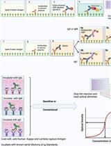
- Capture ELISA for quantitative analysis of P. falciparum blood-stage egress
- Coat 96-well Immulon plates with 100 μl/well of a stock solution of purified monoclonal antibody 4G2dc1 made up at 2.5 μg/ml in carbonate/bicarbonate buffer (pH 9.6).
- Seal the plates and incubate overnight in a humidified atmosphere at 4 °C. This and all subsequent incubations are done whilst stationary.
- Wash the plates once with PBS 0.5% (v/v) Tween 20 (PBS/T), then block by adding 250 μl/well of a 1:4 dilution of gelatine-Tween stock solution, and incubating for 2 h at room temperature.
- Prepare serial 2-fold dilutions of test culture supernatant samples in a separate 96-well plate. Dilute the samples into RPMI 1640 culture medium containing 0.5% Albumax. For negative control wells, use RPMI Albumax medium only. Add samples to the washed ELISA plates, then seal and incubate the plates for 1 h at room temperature.
- Wash plates 3x in PBS/T.
- Make up a solution of rabbit polyclonal anti-AMA1 serum diluted 1:1,000 in PBS/T. Add 100 μl/well, then seal and incubate the plates a further 1 h at room temperature.
- Wash plates 3x with PBS.
Note: No Tween at this stage, as it interferes with the alkaline phosphatase activity in the next step of the assay.
- Add 100 μl/well phosphatase-conjugated goat anti-rabbit IgG (whole molecule), diluted 1:30,000 in PBS. Seal the plates, then incubate 1 h at room temperature.
- Wash plates 5x with PBS (no tween) then add 50 μl/well substrate (1 tablet of phosphatase substrate, in 5 ml diethanolamine buffer). Incubate in the dark at 37 °C for at least 30 min. Determine OD405 using an ELISA reader.
- Use the data to produce titration curves of antigen levels in the various culture supernatants.
- Coat 96-well Immulon plates with 100 μl/well of a stock solution of purified monoclonal antibody 4G2dc1 made up at 2.5 μg/ml in carbonate/bicarbonate buffer (pH 9.6).
Recipes
- Gelatine-tween stock solution (blocking buffer)
5 g Gelatine
2.5 ml Tween-20
150 μl 10% (w/v) sodium azide
500 ml PBS
Warm at 37 °C to dissolve the gelatine
Use at 1: 4 dilution in PBS
Stored stock at 4 °C
May require warming before coaxing out of the bottle
- 0.1 M Sodium carbonate/bicarbonate (pH 9.6)
Make up the two component buffers first
These are
7.155 g Na2CO3.10H2O made up to 250 ml water
2.1 g NaHCO3 made up to 250 ml water
Mix the above buffers to obtain a buffer with a pH of 9.6
As an approximate measure, this requires ~37 ml 0.1 M Na2CO3.10H2O plus 73 ml 0.1 M NaHCO3
- Diethanolamine buffer (pH 9.8)
48.5 ml Diethanolamine
982 μl1 M MgCl2
2.5 ml10% (w/v) sodium azide
Add 400 ml water then adjust pH to 9.8 with 1 M HCl before making up to a total of 500 ml with water
Stored in the dark at 4 °C
Acknowledgments
This method is adapted from an original method described in Collins et al. (2013). The authors are grateful to Alan Thomas (Biomedical Primate Research Centre, Rijswijk, The Netherlands) for the gift of monoclonal antibody 4G2dc1. This work was supported by the UK Medical Research Council (U117532063), and received funding from the European Community's Seventh Framework Programme (FP7/2007-2013) under grant agreement N° 242095 (EviMalAR).
References
- Blackman, M. J. (1994). Purification of Plasmodium falciparum merozoites for analysis of the processing of merozoite surface protein-1. Methods Cell Biol 45: 213-220.
- Collins, C. R., Withers-Martinez, C., Hackett, F. and Blackman, M. J. (2009). An inhibitory antibody blocks interactions between components of the malarial invasion machinery. PLoS Pathog 5(1): e1000273.
- Collins, C. R., Hackett, F., Strath, M., Penzo, M., Withers-Martinez, C., Baker, D. A. and Blackman, M. J. (2013). Malaria parasite cGMP-dependent protein kinase regulates blood stage merozoite secretory organelle discharge and egress. PLoS Pathog 9(5): e1003344.
- Kocken, C. H., van der Wel, A. M., Dubbeld, M. A., Narum, D. L., van de Rijke, F. M., van Gemert, G. J., van der Linde, X., Bannister, L. H., Janse, C., Waters, A. P. and Thomas, A. W. (1998). Precise timing of expression of a Plasmodium falciparum-derived transgene in Plasmodium berghei is a critical determinant of subsequent subcellular localization. J Biol Chem 273(24): 15119-15124.
- Yeoh, S., O'Donnell, R. A., Koussis, K., Dluzewski, A. R., Ansell, K. H., Osborne, S. A., Hackett, F., Withers-Martinez, C., Mitchell, G. H., Bannister, L. H., Bryans, J. S., Kettleborough, C. A. and Blackman, M. J. (2007). Subcellular discharge of a serine protease mediates release of invasive malaria parasites from host erythrocytes. Cell 131(6): 1072-1083.
Article Information
Copyright
© 2014 The Authors; exclusive licensee Bio-protocol LLC.
How to cite
Readers should cite both the Bio-protocol article and the original research article where this protocol was used:
- Hackett, F., Collins, C. R., Strath, M. and Blackman, M. J. (2014). Cyclic Nucleotide (cAMP and cGMP) Assays and Capture ELISA for Quantitative Analysis of Plasmodium falciparum Blood-stage Egress. Bio-protocol 4(5): e1055. DOI: 10.21769/BioProtoc.1055.
- Collins, C. R., Hackett, F., Strath, M., Penzo, M., Withers-Martinez, C., Baker, D. A. and Blackman, M. J. (2013). Malaria parasite cGMP-dependent protein kinase regulates blood stage merozoite secretory organelle discharge and egress. PLoS Pathog 9(5): e1003344.
Category
Microbiology > Microbial biochemistry > Protein > Immunodetection
Biochemistry > Protein > Immunodetection > ELISA
Biochemistry > Other compound > cAMP
Do you have any questions about this protocol?
Post your question to gather feedback from the community. We will also invite the authors of this article to respond.