Improve Research Reproducibility A Bio-protocol resource
- Protocols

- Articles and Issues
- For Authors
- About
- Become a Reviewer

Past Issue in 2012
Volume: 2, Issue: 16
Biochemistry
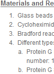
Co-immunoprecipitation in Yeast

This protocol describes investigation of protein-protein interactions in baker yeast by co-immunoprecipitation (CoIP). CoIP is a technique to identify physiologically relevant protein-protein interactions in the cell. The interesting protein can be isolated out of solution using antibody that specifically binds to that particular protein (antigene protein). The partner proteins that are bound to a specific target protein can be co-immunoprecipitated together with an antigen. These protein complexes can then be analyzed to identify new binding partners, binding affinities, the kinetics of binding and the function of the target protein. Here I describe the protocols that allow to immunoprecipitate different protein complexes, for example NAC complex (Panasenko et al., 2009), Ccr4-Not complex (Panasenko and Collort, 2011), ribosomes (Panasenko and Collort, 2012) and investigate their partners. For each CoIP I used the different lysis buffer, as indicated below in recipes.
![Radiolabeling of Chlorophyll by [14C]Glutamic Acid in vivo and Relative Quantification of Labeled Chlorophyll by Using Thin Layer Chromatography (TLC)](https://en-cdn.bio-protocol.org/imageup/arcimg/249.jpg?t=1776202521)
Radiolabeling of Chlorophyll by [14C]Glutamic Acid in vivo and Relative Quantification of Labeled Chlorophyll by Using Thin Layer Chromatography (TLC)
This is an accurate method to assess the rate of chlorophyll biosynthesis in vivo in cyanobacteria. Given that labeled glutamate is used as the very early precursor of chlorophyll together with a short pulse of labeling (30 min), this method provides information about the metabolic flow through the whole chlorophyll biosynthetic pathway on a short timescale.
Cancer Biology
Senescence Associated β-galactosidase Staining

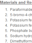
Detection of senescent cells using a cytochemical assay was first described in 1995 (Dimri et al., 1995). The identification of senescent cells is based on an increased level of lysosomal β-galactosidase activity (Kurz et al., 2000). Cells under normal growth condition produce acid lysosomal β- galactosidase, which is localized in the lysosome. The enzymatic activity can be detected at the optimal pH 4.0, using the chromogenic substrate 5-bromo-4-chloro-3-indolyl β D-galactopyranoside (X-gal) (Miller, 1972). In comparison, upon senescence, the lysosomal mass is increased, leading to production of a higher level of β-galactosidase, termed senescence-associated β-galactosidase (SA-β-gal) (Kurz et al., 2000). The abundant senescence-associated enzyme is detectable over background despite the less favorable pH conditions (pH 6.0) (Dimri et al., 1995). The SA-β gal positive cells stain blue-green, which can be scored under bright-field microscopy. In this assay it is best to avoid over-confluency of the cells, or cells that have undergone too many passages, as these conditions can cause false positive results.
Microbiology
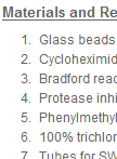
Ribosome Fractionation in Yeast
This protocol describes yeast ribosome fractionation in the gradient of sucrose. During the cyclic process of translation, a small (40S) and large (60S) ribosomal subunit associate with mRNA to form an 80S complex (monosome). This ribosome moves along the mRNA during translational elongation, and then dissociates into the 40S and 60S subunits on termination. During elongation by one ribosome, further ribosomes can initiate translation on the same mRNA to form polysomes. The mass of each polysomal complex is determined primarily by the number of ribosomes it contains. Hence, the population of polysomes within the cell can be size-fractionated by sucrose density gradient centrifugation on the basis of the loading of ribosomes on the mRNA. Several compounds help to maintain or to disrupt the polysomes (Figure 1).
Plant Science
RNA Extraction from RNase-Rich Senescing Leaf Samples

Isolation of intact, full-length high quality RNAs is essential for RNA sequencing, reverse transcription PCR analysis of gene expression as well as RNA gel blot analysis. This simple yet easy protocol is developed to meet this need; in addition to regular samples, this protocol is especially good for isolating RNAs from RNase-rich samples such as senescing leaves and ripening fruits (from which RNAs isolated using standard method are generally degraded to certain degree). The total RNA yield varies from 900 μg total RNA/g non-senescing leaves to 200 μg total RNA/g senescent leaves.