- Protocols

- Articles and Issues
- For Authors
- About
- Become a Reviewer

Past Issue in 2025
Volume: 15, Issue: 20
Cancer Biology
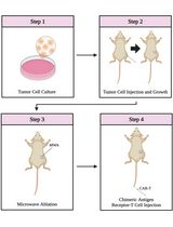
Combining Microwave Ablation With CAR-T-Cell Therapy in Tumor-Bearing Mouse Models

Cell Biology
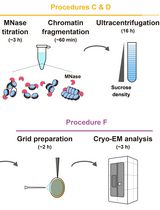
Preparation of Chromatin Fragments From Human Cells for Cryo-EM Analysis

Studying Cargo Transport Using RudLOV
Generation of Insulin-Producing Alpha TC1-6 Cells Using EpiCRISPR System for Targeted DNA Methylation
Immunology
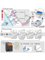
High-Dimensional Phospho-CyTOF Characterization of T-Cell Activation Responses in Whole Blood
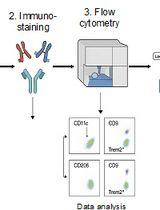
Identification and Sorting of Adipose Inflammatory and Metabolically Activated Macrophages in Diet-Induced Obesity
Molecular Biology
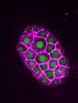
SunTag-Based Single-Molecule Translation Imaging in Caenorhabditis elegans
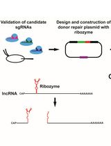
Ribozyme-Mediated Knockdown of lncRNA Gene Expression in Drosophila
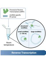
Efficient circRNA Detection Using the Processive Reverse Transcriptase uMRT

Plant Science
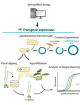
Effective Gene Silencing in Plants by Synthetic Trans-Acting siRNAs Derived From Minimal Precursors


Direct Plant Regeneration From Immature Male Inflorescence of Banana (Musa spp.)
Stem Cell
Rapid and Simplified Induction of Spinal Motor Neurons From Human Induced Pluripotent Stem Cells