Past Issue in 2024

Volume: 14, Issue: 4

left

Dec

20

Dec

5

Nov

20

Nov

5

Oct

20

Oct

5

Sep

20

Sep

5

Aug

20

Aug

5

Jul

20

Jul

5

Jun

20

Jun

5

May

20

May

5

Apr

20

Apr

5

Mar

20

Mar

5

Feb

20

Feb

5

Jan

20

Jan

5

right

Bioinformatics and Computational Biology

CoCoNat: A Deep Learning–Based Tool for the Prediction of Coiled-coil Domains in Protein Sequences

CoCoNat: A Deep Learning–Based Tool for the Prediction of Coiled-coil Domains in Protein Sequences

MM Matteo Manfredi
CS Castrense Savojardo
PM Pier Luigi Martelli
RC Rita Casadio
1644 Views
Feb 20, 2024
Coiled-coil domains (CCDs) are structural motifs observed in proteins in all organisms that perform several crucial functions. The computational identification of CCD segments over a protein sequence is of great importance for its functional characterization. This task can essentially be divided into three separate steps: the detection of segment boundaries, the annotation of the heptad repeat pattern along the segment, and the classification of its oligomerization state. Several methods have been proposed over the years addressing one or more of these predictive steps. In this protocol, we illustrate how to make use of CoCoNat, a novel approach based on protein language models, to characterize CCDs. CoCoNat is, at its release (August 2023), the state of the art for CCD detection. The web server allows users to submit input protein sequences and visualize the predicted domains after a few minutes. Optionally, precomputed segments can be provided to the model, which will predict the oligomerization state for each of them. CoCoNat can be easily integrated into biological pipelines by downloading the standalone version, which provides a single executable script to produce the output.Key features• Web server for the prediction of coiled-coil segments from a protein sequence.• Three different predictions from a single tool (segment position, heptad repeat annotation, oligomerization state).• Possibility to visualize the results online or to download the predictions in different formats for further processing.• Easy integration in automated pipelines with the local version of the tool.Graphical overview

Biological Engineering

Unlocking Bio-Instructive Polymers: A Novel Multi-Well Screening Platform Based on Secretome Sampling

Unlocking Bio-Instructive Polymers: A Novel Multi-Well Screening Platform Based on Secretome Sampling

SF Shirin Fateh
RA Reem A. Alromaihi
AG Amir M. Ghaemmaghami
MA Morgan R. Alexander
1957 Views
Feb 20, 2024
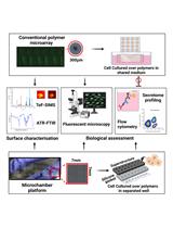
Biomaterials are designed to interact with biological systems to replace, support, enhance, or monitor their function. However, there are challenges associated with traditional biomaterials’ development due to the lack of underlying theory governing cell response to materials’ chemistry. This leads to the time-consuming process of testing different materials plus the adverse reactions in the body such as cytotoxicity and foreign body response. High-throughput screening (HTS) offers a solution to these challenges by enabling rapid and simultaneous testing of a large number of materials to determine their bio-interactions and biocompatibility. Secreted proteins regulate many physiological functions and determine the success of implanted biomaterials through directing cell behaviour. However, the majority of biomaterials’ HTS platforms are suitable for microscopic analyses of cell behaviour and not for investigating non-adherent cells or measuring cell secretions. Here, we describe a multi-well platform adaptable to robotic printing of polymers and suitable for secretome profiling of both adherent and non-adherent cells. We detail the platform's development steps, encompassing the preparation of individual cell culture chambers, polymer printing, and the culture environment, as well as examples to demonstrate surface chemical characterisation and biological assessments of secreted mediators. Such platforms will no doubt facilitate the discovery of novel biomaterials and broaden their scope by adapting wider arrays of cell types and incorporating assessments of both secretome and cell-bound interactions.Key features• Detailed protocols for preparation of substrate for contact printing of acrylate-based polymers including O2 plasma etching, functionalisation process, and Poly(2-hydroxyethyl methacrylate) (pHEMA) dip coating.• Preparations of 7 mm × 7 mm polymers employing pin printing system.• Provision of confined area for each polymer using ProPlate® multi-well chambers.• Compatibility of this platform was validated using adherent cells [primary human monocyte–derived macrophages (MDMs)) and non-adherent cells (primary human monocyte–derived dendritic cells (moDCs)].• Examples of the adaptability of the platform for secretome analysis including five different cytokines using enzyme-linked immunosorbent assay (ELISA, DuoSet®).Graphical overview

Biophysics

Resolving the In Situ Three-Dimensional Structure of Fly Mechanosensory Organelles Using Serial Section Electron Tomography

Resolving the In Situ Three-Dimensional Structure of Fly Mechanosensory Organelles Using Serial Section Electron Tomography

LS Landi Sun
JM Jana Meissner
JH Jianfeng He
LC Lihong Cui
TF Tobias Fürstenhaupt
XL Xin Liang
2370 Views
Feb 20, 2024
Mechanosensory organelles (MOs) are specialized subcellular entities where force-sensitive channels and supporting structures (e.g., microtubule cytoskeleton) are organized in an orderly manner. The delicate structure of MOs needs to be resolved to understand the mechanisms by which they detect forces and how they are formed. Here, we describe a protocol that allows obtaining detailed information about the nanoscopic ultrastructure of fly MOs by using serial section electron tomography (SS-ET). To preserve fine structural details, the tissues are cryo-immobilized using a high-pressure freezer followed by freeze-substitution at low temperature and embedding in resin at room temperature. Then, sample sections are prepared and used to acquire the dual-axis tilt series images, which are further processed for tomographic reconstruction. Finally, tomograms of consecutive sections are combined into a single larger volume using microtubules as fiducial markers. Using this protocol, we managed to reconstruct the sensory organelles, which provide novel molecular insights as to how fly mechanosensory organelles work and are formed. Based on our experience, we think that, with minimal modifications, this protocol can be adapted to a wide range of applications using different cell and tissue samples.Key features• Resolving the high-resolution 3D ultrastructure of subcellular organelles using serial section electron tomography (SS-ET).• Compared with single-axis tilt series, dual-axis tilt series provides a much wider coverage of Fourier space, improving resolution and features in the reconstructed tomograms.• The use of high-pressure freezing and freeze-substitution maximally preserves the fine structural details.Graphical overview

Cell Biology

Phosphoproteomic Analysis and Organotypic Cultures for the Study of Signaling Pathways

Phosphoproteomic Analysis and Organotypic Cultures for the Study of Signaling Pathways

ZY Zilu Ye
HW Hans H. Wandall
SD Sally Dabelsteen
1949 Views
Feb 20, 2024
Signaling pathways are involved in key cellular functions from embryonic development to pathological conditions, with a pivotal role in tissue homeostasis and transformation. Although most signaling pathways have been intensively examined, most studies have been carried out in murine models or simple cell culture. We describe the dissection of the TGF-β signaling pathway in human tissue using CRISPR-Cas9 genetically engineered human keratinocytes (N/TERT-1) in a 3D organotypic skin model combined with quantitative proteomics and phosphoproteomics mass spectrometry. The use of human 3D organotypic cultures and genetic engineering combined with quantitative proteomics and phosphoproteomics is a powerful tool providing insight into signaling pathways in a human setting. The methods are applicable to other gene targets and 3D cell and tissue models.Key features• 3D organotypic models with genetically engineered human cells.• In-depth quantitative proteomics and phosphoproteomics in 2D cell culture.• Careful handling of cell cultures is critical for the successful formation of theorganotypic cultures.• For complete details on the use of this protocol, please refer to Ye et al. 2022.

Immunology

Addressing the Role of Conventional CD8αβ+ T Cells and CD4+ T Cells in Intestinal Immunopathology Using a Bone Marrow–Engrafted Model

Addressing the Role of Conventional CD8αβ+ T Cells and CD4+ T Cells in Intestinal Immunopathology Using a Bone Marrow–Engrafted Model

AA Amneh Aoudi
OL Ossama Labiad
RI Ramdane Igalouzene
OM Ousséma Mejri
MS Maxime Sanchez
SS Saïdi Soudja
2054 Views
Feb 20, 2024
Inflammatory bowel disease (IBD) is characterized by an aberrant immune response against microbiota. It is well established that T cells play a critical role in mediating the pathology. Assessing the contribution of each subset of T cells in mediating the pathology is crucial in order to design better therapeutic strategies. This protocol presents a method to identify the specific effector T-cell population responsible for intestinal immunopathologies in bone marrow–engrafted mouse models. Here, we used anti-CD4 and anti-CD8β depleting antibodies in bone marrow–engrafted mouse models to identify the effector T-cell population responsible for intestinal damage in a genetic mouse model of chronic intestinal inflammation.Key features• This protocol allows addressing the role of CD4+ or CD8αβ+ in an engrafted model of inflammatory bowel disease (IBD).• This protocol can easily be adapted to address the role of other immune cells or molecules that may play a role in IBD.

Neuroscience

Generation of Human Induced Pluripotent Stem Cell (hiPSC)-Derived Astrocytes for Amyotrophic Lateral Sclerosis and Other Neurodegenerative Disease Studies

Generation of Human Induced Pluripotent Stem Cell (hiPSC)-Derived Astrocytes for Amyotrophic Lateral Sclerosis and Other Neurodegenerative Disease Studies

KS Katarina Stoklund Dittlau
AC Abinaya Chandrasekaran
KF Kristine Freude
LV Ludo Van Den Bosch
5111 Views
Feb 20, 2024
Astrocytes are increasingly recognized for their important role in neurodegenerative diseases like amyotrophic lateral sclerosis (ALS). In ALS, astrocytes shift from their primary function of providing neuronal homeostatic support towards a reactive and toxic role, which overall contributes to neuronal toxicity and cell death. Currently, our knowledge on these processes is incomplete, and time-efficient and reproducible model systems in a human context are therefore required to understand and therapeutically modulate the toxic astrocytic response for future treatment options. Here, we present an efficient and straightforward protocol to generate human induced pluripotent stem cell (hiPSC)-derived astrocytes implementing a differentiation scheme based on small molecules. Through an initial 25 days, hiPSCs are differentiated into astrocytes, which are matured for 4+ weeks. The hiPSC-derived astrocytes can be cryopreserved at every passage during differentiation and maturation. This provides convenient pauses in the protocol as well as cell banking opportunities, thereby limiting the need to continuously start from hiPSCs. The protocol has already proven valuable in ALS research but can be adapted to any desired research field where astrocytes are of interest.Key features• This protocol requires preexisting experience in hiPSC culturing for a successful outcome.• The protocol relies on a small molecule differentiation scheme and an easy-to-follow methodology, which can be paused at several time points.• The protocol generates >50 × 106 astrocytes per differentiation, which can be cryopreserved at every passage, ensuring a large-scale experimental output.Graphical overview
A Versatile Pipeline for High-fidelity Imaging and Analysis of Vascular Networks Across the Body

A Versatile Pipeline for High-fidelity Imaging and Analysis of Vascular Networks Across the Body

SV Stephen Vidman
ED Elliot Dion
AT Andrea Tedeschi
2028 Views
Feb 20, 2024
Structural and functional changes in vascular networks play a vital role during development, causing or contributing to the pathophysiology of injury and disease. Current methods to trace and image the vasculature in laboratory settings have proven inconsistent, inaccurate, and labor intensive, lacking the inherent three-dimensional structure of vasculature. Here, we provide a robust and highly reproducible method to image and quantify changes in vascular networks down to the capillary level. The method combines vasculature tracing, tissue clearing, and three-dimensional imaging techniques with vessel segmentation using AI-based convolutional reconstruction to rapidly process large, unsectioned tissue specimens throughout the body with high fidelity. The practicality and scalability of our protocol offer application across various fields of biomedical sciences. Obviating the need for sectioning of samples, this method will expedite qualitative and quantitative analyses of vascular networks. Preparation of the fluorescent gel perfusate takes < 30 min per study. Transcardiac perfusion and vasculature tracing takes approximately 20 min, while dissection of tissue samples ranges from 5 to 15 min depending on the tissue of interest. The tissue clearing protocol takes approximately 24–48 h per whole-tissue sample. Lastly, three-dimensional imaging and analysis can be completed in one day. The entire procedure can be carried out by a competent graduate student or experienced technician.Key features• This robust and highly reproducible method allows users to image and quantify changes in vascular networks down to the capillary level.• Three-dimensional imaging techniques with vessel segmentation enable rapid processing of large, unsectioned tissue specimens throughout the body.• It takes approximately 2–3 days for sample preparation, three-dimensional imaging, and analysis.• The user-friendly pipeline can be completed by experienced and non-experienced users.Graphical overview
A Simple Immunofluorescence Method to Characterize Neurodegeneration and Tyrosine Hydroxylase Reduction in Whole Brain of a Drosophila Model of Parkinson’s Disease

A Simple Immunofluorescence Method to Characterize Neurodegeneration and Tyrosine Hydroxylase Reduction in Whole Brain of a Drosophila Model of Parkinson’s Disease

RC Rahul Chaurasia
MA Mohamad Ayajuddin
GR Girish S. Ratnaparkhi
SL Shashidhara S. Lingadahalli
SY Sarat C. Yenisetti
1253 Views
Feb 20, 2024
Dopaminergic (DAergic) neurodegeneration in the substantia nigra pars compacta of the human brain is the pathological feature associated with Parkinson’s disease (PD). Drosophila also exhibits mobility defects and diminished levels of brain dopamine on exposure to neurotoxicants mimicking PD. Our laboratory demonstrated in a Drosophila model of sporadic PD that there is no decrease in DAergic neuronal number; instead, there is a significant reduction in tyrosine hydroxylase (TH) fluorescence intensity (FI). Here, we present a sensitive assay based on the quantification of FI of the secondary antibody (ab). As the FI is directly proportional to the amount of TH synthesis, its reduction under PD conditions denotes the decrease in the TH synthesis, suggesting DAergic neuronal dysfunction. Therefore, FI quantification is a refined and sensitive method to understand the early stages of DAergic neurodegeneration. FI quantification is performed using the ZEN 2012 SP2 single-user software; a license must be acquired to utilize the imaging system to interactively control image acquisition, image processing, and analysis. This method will be of good use to biologists, as it can also be used with little modification to characterize the extent of degeneration and changes in the level of degeneration in response to drugs in different cell types. Unlike the expensive and cumbersome confocal microscopy, the present method will be an affordable option for fund-constrained neurobiology laboratories.Key features• Allows characterizing the incipient DAergic and other catecholaminergic neurodegeneration, even in the absence of loss of neuronal cell body.• Great alternative for the fund-constrained neurobiology laboratories in developing countries to utilize this method in different cell types and their response to drugs/nutraceuticals.Graphical overview