Past Issue in 2024

Volume: 14, Issue: 3

left

Dec

20

Dec

5

Nov

20

Nov

5

Oct

20

Oct

5

Sep

20

Sep

5

Aug

20

Aug

5

Jul

20

Jul

5

Jun

20

Jun

5

May

20

May

5

Apr

20

Apr

5

Mar

20

Mar

5

Feb

20

Feb

5

Jan

20

Jan

5

right

Biochemistry

SUrface SEnsing of Translation (SUnSET), a Method Based on Western Blot Assessing Protein Synthesis Rates in vitro

SUrface SEnsing of Translation (SUnSET), a Method Based on Western Blot Assessing Protein Synthesis Rates in vitro

MP Marie Piecyk
JF Joëlle Fauvre
CD Cédric Duret
CC Cédric Chaveroux
CF Carole Ferraro-Peyret
3634 Views
Feb 5, 2024
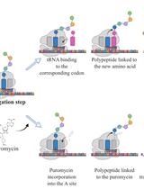
As the most energy- and metabolite-consuming process, protein synthesis is under the control of several intrinsic and extrinsic factors that determine its fine-tuning to the cellular microenvironment. Consequently, variations in protein synthesis rates occur under various physiological and pathological conditions, enabling an adaptive response by the ce•ll. For example, global protein synthesis increases upon mitogenic factors to support biomass generation and cell proliferation, while exposure to low concentrations of oxygen or nutrients require translational repression and reprogramming to avoid energy depletion and cell death. To assess fluctuations in protein synthesis rates, radioactive isotopes or radiolabeled amino acids are often used. Although highly sensitive, these techniques involve the use of potentially toxic radioactive compounds and require specific materials and processes for the use and disposal of these molecules. The development of alternative, non-radioactive methods that can be easily and safely implemented in laboratories has therefore been encouraged to avoid handling radioactivity. In this context, the SUrface SEnsing of Translation (SUnSET) method, based on the classical western blot technique, was developed by Schmidt et al. in 2009. The SUnSET is nowadays recognized as a simple alternative to radioactive methods assessing protein synthesis rates.Key features• As a structural analogue of aminoacyl-transfer RNA, puromycin incorporates into the elongating peptide chain.• Detection of puromycin-labeled peptides by western blotting reflects translation rates without the need for radioactive isotopes.• The protocol described here for in vitro applications is derived from the SUnSET method originally published by Schmidt et al. (2009).

Biological Engineering

Streamlined Adeno-Associated Virus Production Using Suspension HEK293T Cells

Streamlined Adeno-Associated Virus Production Using Suspension HEK293T Cells

AK Aditi A. Kulkarni
AS Austin G. Seal
CS Corinne Sonnet
KO Kazuhiro Oka
2439 Views
Feb 5, 2024
Recombinant adeno-associated viruses (rAAVs) are valuable viral vectors for in vivo gene transfer, also having significant ex vivo therapeutic potential. Continued efforts have focused on various gene therapy applications, capsid engineering, and scalable manufacturing processes. Adherent cells are commonly used for virus production in most basic science laboratories because of their efficiency and cost. Although suspension cells are easier to handle and scale up compared to adherent cells, their use in virus production is hampered by poor transfection efficiency. In this protocol, we developed a simple scalable AAV production protocol using serum-free-media-adapted HEK293T suspension cells and VirusGEN transfection reagent. The established protocol allows AAV production from transfection to quality analysis of purified AAV within two weeks. Typical vector yields for the described suspension system followed by iodixanol purification range from a total of 1 × 10¹³ to 1.5 × 10¹³ vg (vector genome) using 90 mL of cell suspension vs. 1 × 10¹³ to 2 × 10¹³ vg using a regular adherent cell protocol (10 × 15 cm dishes).Key features• Adeno-associated virus (AAV) production using serum-free-media-adapted HEK293T suspension cells.• Efficient transfection with VirusGEN.• High AAV yield from small-volume cell culture.Graphical overview
A Protocol for Custom Biomineralization of Enzymes in Metal–Organic Frameworks (MOFs)

A Protocol for Custom Biomineralization of Enzymes in Metal–Organic Frameworks (MOFs)

ZA Zoe Armstrong
DJ Drew Jordahl
AM Austin MacRae
QL Qiaobi Li
ML Mary Lenertz
PS Patrick Shen
AB Anastasiia Botserovska
LF Li Feng
AU Angel Ugrinov
ZY Zhongyu Yang
1314 Views
Feb 5, 2024
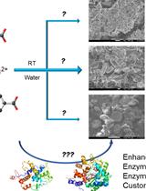
Enzyme immobilization offers a number of advantages that improve biocatalysis; however, finding a proper way to immobilize enzymes is often a challenging task. Implanting enzymes in metal–organic frameworks (MOFs) via co-crystallization, also known as biomineralization, provides enhanced reusability and stability with minimal perturbation and substrate selectivity to the enzyme. Currently, there are limited metal–ligand combinations with a proper protocol guiding the experimental procedures. We have recently explored 10 combinations that allow custom immobilization of enzymes according to enzyme stability and activity in different metals/ligands. Here, as a follow-up of that work, we present a protocol for how to carry out custom immobilization of enzymes using the available combinations of metal ions and ligands. Detailed procedures to prepare metal ions, ligands, and enzymes for their co-crystallization, together with characterization and assessment, are discussed. Precautions for each experimental step and result analysis are highlighted as well. This protocol is important for enzyme immobilization in various research and industrial fields.Key features• A wide selection of metal ions and ligands allows for the immobilization of enzymes in metal–organic frameworks (MOFs) via co-crystallization.• Step-by-step enzyme immobilization procedure via co-crystallization of metal ions, organic linkers, and enzymes.• Practical considerations and experimental conditions to synthesize the enzyme@MOF biocomposites are discussed.• The demonstrated method can be generalized to immobilize other enzymes and find other metal ion/ligand combinations to form MOFs in water and host enzymes.Graphical overview

Developmental Biology

Live Imaging Transverse Sections of Zebrafish Embryo Explants

Live Imaging Transverse Sections of Zebrafish Embryo Explants

EP Eric Paulissen
BM Benjamin L. Martin
1200 Views
Feb 5, 2024
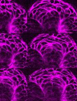
Vertebrate embryogenesis is a highly dynamic process involving coordinated cell and tissue movements that generate the final embryonic body plan. Many of these movements are difficult to image at high resolution because they occur deep within the embryo along the midline, causing light scattering and requiring longer working distances. Here, we present an explant-based method to image transverse cross sections of living zebrafish embryos. This method allows for the capture of all cell movements at high-resolution throughout the embryonic trunk, including hard-to-image deep tissues. This technique offers an alternative to expensive or computationally difficult microscopy methods.Key features• Generates intact zebrafish explants with minimal tissue disturbance.• Allows for live imaging of deep tissues normally obscured by common confocal microscopy techniques.• Immobilizes tissues for extended periods required for time-lapse imaging.• Utilizes readily available reagents and tools, which can minimize the time and cost of the procedure.Graphical overview

Immunology

Quantification of Macrophage Cellular Ferrous Iron (Fe2+) Content using a Highly Specific Fluorescent Probe in a Plate-Reader

Quantification of Macrophage Cellular Ferrous Iron (Fe2+) Content using a Highly Specific Fluorescent Probe in a Plate-Reader

PG Philipp Grubwieser
NB Natascha Brigo
MS Markus Seifert
MG Manuel Grander
IT Igor Theurl
MN Manfred Nairz
GW Günter Weiss
CP Christa Pfeifhofer-Obermair
1563 Views
Feb 5, 2024
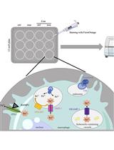
Macrophages are at the center of innate immunity and iron metabolism. In the case of an infection, macrophages adapt their cellular iron metabolism to deprive iron from invading bacteria to combat intracellular bacterial proliferation. A concise evaluation of the cellular iron content upon an infection with bacterial pathogens and diverse cellular stimuli is necessary to identify underlying mechanisms concerning iron homeostasis in macrophages. For the characterization of cellular iron levels during infection, we established an in vitro infection model where the murine macrophage cell line J774A.1 is infected with Salmonella enterica serovar Typhimurium (S.tm), the mouse counterpart to S. enterica serovar Typhi, under normal and iron-overload conditions using ferric chloride (FeCl3) treatment. To evaluate the effect of infection and iron stimulation on cellular iron levels, the macrophages are stained with FerroOrange. This fluorescent probe specifically detects Fe2+ ions and its fluorescence can be quantified photometrically in a plate reader. Importantly, FerroOrange fluorescence does not increase with chelated iron or other bivalent metal ions. In this protocol, we present a simple and reliable method to quantify cellular Fe2+ levels in cultured macrophages by applying a highly specific fluorescence probe (FerroOrange) in a TECAN Spark microplate reader. Compared to already established techniques, our protocol allows assessing cellular iron levels in innate immune cells without the use of radioactive iron isotopes or extensive sample preparation, exposing the cells to stress.Key features• Easy quantification of Fe2+ in cultured macrophages with a fluorescent probe.• Analysis of iron in living cells without the need for fixation.• Performed on a plate reader capable of 540 nm excitation and 585 nm emission by trained employees for handling biosafety level 2 bacteria.Graphical overview

Microbiology

Use of the Fluorescent Dye Thioflavin T to Track Amyloid Structures in the Pathogenic Yeast Candida albicans

Use of the Fluorescent Dye Thioflavin T to Track Amyloid Structures in the Pathogenic Yeast Candida albicans

TM Thierry Mourer
Cd Christophe d'Enfert
SB Sophie Bachellier-Bassi
1341 Views
Feb 5, 2024
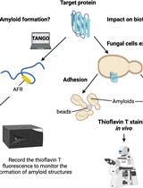
The human pathogenic yeast Candida albicans can attach to epithelial cells or indwelling medical devices to form biofilms. These microbial communities are highly problematic in the clinic as they reduce both sensitivity to antifungal drugs and detection of fungi by the immune system. Amyloid structures are highly organized quaternary structures that play a critical role in biofilm establishment by allowing fungal cells to adhere to each other. Thus, fungal amyloids are exciting targets to develop new antifungal strategies. Thioflavin T is a specific fluorescent dye widely used to study amyloid properties of target proteins in vitro (spectrophotometry) and in vivo (epifluorescence/confocal microscopy). Notably, thioflavin T has been used to demonstrate the ability of Als5, a C. albicans adhesin, to form an amyloid fiber upon adhesion. We have developed a pipeline that allows us to study amyloid properties of target proteins using thioflavin T staining in vitro and in vivo, as well as in intact fungal biofilms. In brief, we used thioflavin T to sequentially stain (i) amyloid peptides, (ii) recombinant proteins, (iii) fungal cells treated or not with amyloid peptides, (iv) fungal amyloids enriched by cell fractionation, and (v) intact biofilms of C. albicans. Contrary to other methods, our pipeline gives a complete picture of the amyloid behavior of target proteins, from in vitro analysis to intact fungal biofilms. Using this pipeline will allow an assessment of the relevance of the in vitro results in cells and the impact of amyloids on the development and/or maintenance of fungal biofilm.Key features• Study of amyloid properties of fungal proteins.• Visualization of the subcellular localization of fungal amyloid material using epifluorescence or confocal microscopy.• Unraveling of the amyloid properties of target proteins and their physiological meaning for biofilm formation.• Observation of the presence of amyloid structures with live-cell imaging on intact fungal biofilm using confocal microscopy.Graphical overview

Neuroscience

Measuring Heart Rate in Freely Moving Mice

Measuring Heart Rate in Freely Moving Mice

JS Jérémy Signoret-Genest
NS Nina Schukraft
PT Philip Tovote
1850 Views
Feb 5, 2024
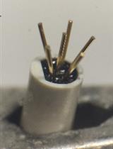
Measuring autonomic parameters like heart rate in behaving mice is not only a standard procedure in cardiovascular research but is applied in many other interdisciplinary research fields. With an electrocardiogram (ECG), the heart rate can be measured by deriving the electrical potential between subcutaneously implanted wires across the chest. This is an inexpensive and easy-to-implement technique and particularly suited for repeated recordings of up to eight weeks. This protocol describes a step-by-step guide for manufacturing the needed equipment, performing the surgical procedure of electrode implantation, and processing of acquired data, yielding accurate and reliable detection of heartbeats and calculation of heart rate (HR). We provide MATLAB graphical user interface (GUI)–based tools to extract and start processing the acquired data without a lot of coding knowledge. Finally, based on an example of a data set acquired in the context of defensive reactions, we discuss the potential and pitfalls in analyzing HR data.Key features• Next to surgical steps, the protocol provides a detailed description of manufacturing custom-made ECG connectors and a shielded, light-weight patch cable.• Suitable for recordings in which signal quality is challenged by ambient noise or noise from other recording devices.• Described for 2-channel differential recording but easily expandable to record from more channels.• Includes a summary of potential analysis methods and a discussion on the interpretation of HR dynamics in the case study of fear states.

Plant Science

Seed Collection in Temperate Trees—Clean, Fast, and Effective Extraction of Populus Seeds for Laboratory Use and Long-term Storage

Seed Collection in Temperate Trees—Clean, Fast, and Effective Extraction of Populus Seeds for Laboratory Use and Long-term Storage

NB Naima Bhutta
ON Oscar F. Nunez-Martinez
CM Carmen Mei
KB Katharina Bräutigam
1406 Views
Feb 5, 2024
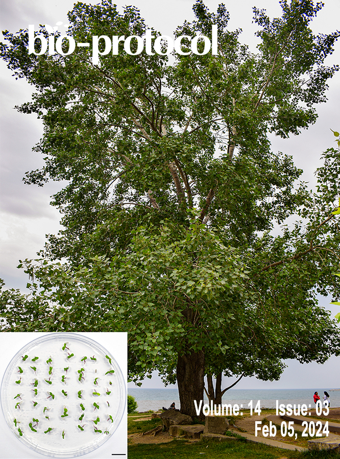
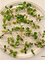
Seeds ensure the growth of a new generation of plants and are thus central to maintaining plant populations and ecosystem processes. Nevertheless, much remains to be learned about seed biology and responses of germinated seedlings to environmental challenges. Experiments aiming to close these knowledge gaps critically depend on the availability of healthy, viable seeds. Here, we report a protocol for the collection of seeds from plants in the genus Populus. This genus comprises trees with a wide distribution in temperate forests and with economic relevance, used as scientific models for perennial plants. As seed characteristics can vary drastically between taxonomic groups, protocols need to be tailored carefully. Our protocol takes the delicate nature of Populus seeds into account. It uses P. deltoides as an example and provides a template to optimize bulk seed extraction for other Populus species and plants with similar seed characteristics. The protocol is designed to only use items available in most labs and households and that can be sterilized easily. The unique characteristics of this protocol allow for the fast and effective extraction of high-quality seeds. Here, we report on seed collection, extraction, cleaning, storage, and viability tests. Moreover, extracted seeds are well suited for tissue culture and experiments under sterile conditions. Seed material obtained with this protocol can be used to further our understanding of tree seed biology, seedling performance under climate change, or diversity of forest genetic resources.Key features• Populus species produce seeds that are small, delicate, non-dormant, with plenty of seed hair. Collection of seed material needs to be timed properly.• Processing, seed extraction, seed cleaning, and storage using simple, sterilizable laboratory and household items only. Obtained seeds are pure, high quality, close to 100% viability.• Seeds work well in tissue culture and in experiments under sterile conditions.• Extractability, speed, and seed germination were studied and confirmed for Populus deltoides as an example.• Can also serve as template for bulk seed collection from other Populus species and plant groups that produce delicate seeds (with no or little modifications).Graphical overview