- Protocols

- Articles and Issues
- For Authors
- About
- Become a Reviewer

Past Issue in 2023
Volume: 13, Issue: 9
Biochemistry
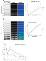
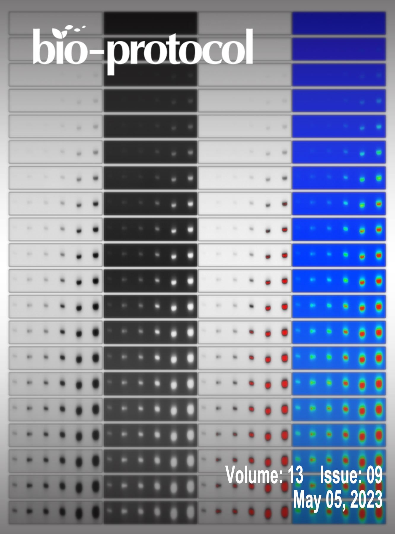
A Simple, Reproducible Procedure for Chemiluminescent Western Blot Quantification
Biological Engineering
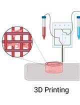
Protocol for 3D Bioprinting Mesenchymal Stem Cell–derived Neural Tissues Using a Fibrin-based Bioink
Cell Biology
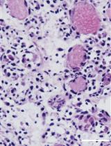
Induction of Skeletal Muscle Injury by Intramuscular Injection of Cardiotoxin in Mouse
Developmental Biology
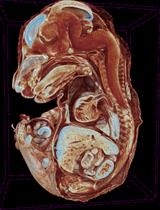
E15.5 Mouse Embryo Micro-CT Using a Bruker Skyscan 1172 Micro-CT
Drug Discovery
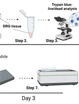
Implementation of a Drug Screening Platform to Target Gch1 Expression in Injured Mouse Dorsal Root Ganglion Neurons

Medicine
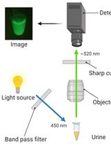
A Novel Non-invasive Qualitative Assay Using Urinary Fluorescence Imaging to Assess Kidney Disease
Microbiology
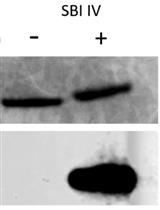
Novel Antibody-independent Method to Measure Complement Deposition on Bacteria
Neuroscience
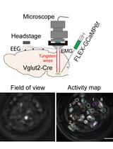
Simultaneous Microendoscopic Calcium Imaging and EEG Recording of Mouse Brain during Sleep

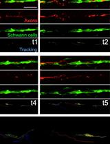
Long-term in toto Imaging of Cellular Behavior during Nerve Injury and Regeneration
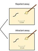
Assessment of Chemosensory Response to Volatile Compounds in Healthy, Aged, and Neurodegenerative Caenorhabditis elegans Models
Plant Science
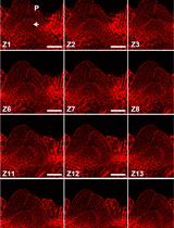
Modified Pseudo-Schiff Propidium Iodide for Staining the Shoot Apical Meristem in Arabidopsis