- Protocols

- Articles and Issues
- For Authors
- About
- Become a Reviewer

In vivo BrdU Incorporation Assay for Murine Hematopioetic Stem Cells


Published: Vol 3, Iss 21, Nov 5, 2013 DOI: 10.21769/BioProtoc.960 Views: 17707
Reviewed by: Lin FangAnonymous reviewer(s)
Original research article
The authors used this protocol in:
Jun 2013
Advertisement


Protocol Collections
Comprehensive collections of detailed, peer-reviewed protocols focusing on specific topics


Abstract
Bromodeoxyuridine (BrdU) is a thymidine analog that is incorporated into DNA during the S-phase of the cell cycle. As such, BrdU incorporation can be used to quantify the number of cells that are in S-phase in the time period during which BrdU is available. The following protocol describes an in vivo BrdU incorporation assay as a measure of cell proliferation in adult murine hematopioetic stem cells (HSCs). Specifically, BrdU incorporation was analyzed for long-term HSCs (LT-HSCs, Lin-Sca-1+c-Kit+CD34-CD135-), Short-term HSCs (ST-HSCs, Lin-Sca-1+c-Kit+CD34+CD135-) and multipotent progenitors (MPPs, Lin-Sca-1+c-Kit+CD34+CD135+) population.
Materials and Reagents
- Mouse
- BrdU
- RPMI1640
- Fetal Bovine Serum (FBS)
- Potassium bicarbonate
- Ammonium chloride
- EDTA
- BSA
- Lineage Cell Depletion Kit (including Biotin-Antibody Cocktail and Anti-Biotin MicroBeads) (Miltenyi Biotec, catalog number: 130-090-858 )
- BD Cytofix/Cytoperm Buffer (Becton, Dickinson and Company, catalog number: 554714 )
- BD Perm/Wash Buffer
- DNase (included in FITC BrdU Flow Kit) (Becton, Dickinson and Company, catalog number: 559619 )
- DPBS without calcium, magnesium (diluted from 10x DPBS) (Hyclone, catalog number: SH 30258.01 )
- Antibodies:
PE-conjugated-Sca-I (Becton, Dickinson and Company, catalog number: 553336 )
APC-conjugated-c-Kit (Becton, Dickinson and Company, catalog number: 553356 )
PerCP-eFluor 710-conjugated-CD135 (eBioscience, catalog number: 46-1351-80 )
eFluor 450-conjugated CD34 (eBioscience, catalog number: 48-0341-80 )
BrdU-FITC (included in FITC BrdU Flow Kit) (Becton, Dickinson and Company, catalog number: 559619)
- Buffer A (see Recipes)
- Red blood cell lysis buffer (see Recipes)
- Staining buffer (see Recipes)
Equipment
- Small scissors and forceps
- 60 mm tissue culture dish
- 23 G needle
- 3 cc syringe
- 15 ml centrifuge tube
- Cell strainer (Becton, Dickinson and Company, catalog number: 352340 )
- Hemacytometer
- MACS MS column (Miltenyi Biotec, catalog number: 130-042-201 )
- MiniMASC separator (Miltenyi Biotec, catalog number: 130-042-102 )
- Centrifuge
- BD LSRFortessa Analytical Flow Cytometer
Procedure
- Animal injection
i.p. Injection of BrdU (10 mg/ml) 100 μl to 8-12 weeks old mouse (50 μg/g BW, so 5 μl/g of BW), after 6 h, inject the second dose. 2 h post 2nd injection, euthanize the mice using CO2 method followed by cervical dislocation, obtain the bone marrow (BM) cells from 2 tibias and 2 femurs. Two injections ensure that BrdU can incorporate to DNA of both slow and quick turnover cells.
- Obtain total bone marrow (BM) cells
- Use small scissors and forceps, dissect out femurs and tibias from mice and place them in a 60 mm tissue culture dish containing 6 ml ice-cold RPMI1640 with 5% heat inactivated FBS. Use Kimwipe tissue to remove muscle and other tissues. Cut off both ends of each bone shaft in the dish.
- Connect the end of the bone with 23 G needle on 3 cc syringe, flush out bone marrow with RPMI1640 with 5% heat inactivated FBS into the dish. Disaggregate bone marrow tissues by repeated aspirations using the same needle. Transfer the cell suspension to 15 ml centrifuge tube.
- Spin down the cells at 350 x g for 5 min at room temperature, remove the supernatant, resuspend the cells in 1 ml of room temperature red blood cell lysis buffer and incubate at room temperature for 5 min, then add 5-10 ml of RPMI 1640 with 5% heat inactivated FBS.
- Pass the cells through a cell strainer. Collect the flow through to a new tube. Take an aliquot and count the cells in a hemacytometer. Spin down at 350 x g for 5 min at room temperature. Remove the supernatant; the cell pellet should not contain any red color. Disaggregate the cell pellet and wash the cells one time with buffer A, spin down at 350 x g for 5 min at room temperature.
- Use small scissors and forceps, dissect out femurs and tibias from mice and place them in a 60 mm tissue culture dish containing 6 ml ice-cold RPMI1640 with 5% heat inactivated FBS. Use Kimwipe tissue to remove muscle and other tissues. Cut off both ends of each bone shaft in the dish.
- Lineage cell staining (follow the mouse Lineage Cell Depletion Kit)
- Resuspend the total BM cells in buffer A (40 μl/107 cells).
- Add Biotin-Antibody Cocktail (10 μl/107 cells) to stain the lineage differentiated cells. Cocktail of biotin-conjugated monoclonal antibodies contains anti-CD5, anti-CD45R(B220), anti-CD11b, anti-Gr-1(Ly-6G/C), anti-Neutrophil (7/4) and anti-Ter-119.
- Mix well and incubate for 10 min at 4 °C.
- Add additional buffer A in media (30 μl/107 cells) then add Anti-Biotin MicroBeads (20 μl/107 cells, provided in Lineage Cell Depletion Kit).
- Mix well and incubate for 15 min at 4 °C.
- Wash cell by adding 2 ml of buffer A. Centrifuge at 300 x g for 10 min at room temperature.
- Remove the supernatant and resuspend the pellet in 0.5 ml of buffer A.
- Resuspend the total BM cells in buffer A (40 μl/107 cells).
- Lineage depleation
- Place MACS MS column in MiniMASC separator.
- Prepare column by rinsing with 0.5 ml buffer A.
- Apply cell suspension onto the column. Allow the cells to pass through and collect flow through as Lin- fraction.
- Wash column 3 times with buffer A (0.5 ml/each), wash each time once the column reservoir is empty.
- Collect all the elute (Lin-) in one tube.
- Count the Lin- cells, aliquot 1.5 x 10⁶ cells to a new tube.
- Add 2 times more staining buffer to the cell suspension.
- Centrifuge the cells at 350 x g for 5 min.
- Place MACS MS column in MiniMASC separator.
- Stain the Lin- cells with surface antigens:
Test Total (μl) Percp-eFluor 710 eFluor 450 PE APC Staining
bufferCD135 CD34 Sca-I CD117
(c-kit)1 75 1 2 0.5 0.5 71 - Stain each test sample per 1.5 x 10⁶ cells/75 μl buffer, make antibody mix as following, for more samples, increase antibody amount and staining buffer proportionally.
- Add 75 μl of antibody mix to the cell pellet. Incubate cells with antibodies for 15 minutes at room temperature (protected from light).
- Wash one time with staining buffer. Spin down for 5 minutes at 350 x g, and discard the supernatant.
- Stain each test sample per 1.5 x 10⁶ cells/75 μl buffer, make antibody mix as following, for more samples, increase antibody amount and staining buffer proportionally.
- Fix and permeabilize the cells
- Resuspend the cells in 100 μl of BD Cytofix/Cytoperm Buffer per tube.
- Incubate the cells for 15 to 30 minutes at room temperature or on ice.
- Wash the cells with 1 ml of 1x BD Perm/Wash Buffer (dilute the 10x buffer with deionized H2O). Centrifuge at 350 x g for 5 minutes at room temperature, and discard the supernatant.
- Resuspend the cells in 100 μl of BD Cytofix/Cytoperm Buffer per tube.
- Enhance the permeabilization:
- Resuspend the cells in 100 μl of BD Cytoperm Permeabilization Buffer Plus per tube. This reagent is specially formulated for the BrdU Flow kit and is used as a staining enhancer and secondary permeabilization reagent.
- Incubate the cells for 10 minutes on ice.
- Wash the cells in 1 ml of 1x BD Perm/Wash Buffer (as in step 5c).
- Resuspend the cells in 100 μl of BD Cytoperm Permeabilization Buffer Plus per tube. This reagent is specially formulated for the BrdU Flow kit and is used as a staining enhancer and secondary permeabilization reagent.
- Re-fix cells after secondary permeabilization:
- Resuspend the cells in 100 μl of BD Cytofix/Cytoperm Buffer per tube.
- Incubate the cells for 5 minutes at room temperature or on ice.
- Wash the cells in 1 ml of 1x BD Perm/Wash Buffer (as in step 5c).
- Resuspend the cells in 100 μl of BD Cytofix/Cytoperm Buffer per tube.
- Treat with DNase to expose incorporated BrdU
- Resuspend the cells in 100 μl of diluted DNase (diluted to 300 μg/ml in DPBS) per tube, (i.e. 30 μg of DNase/106 cells).
- Incubate cells for 1 hour at 37 °C.
- Wash the cells in 1 ml of 1x BD Perm/Wash Buffer (as in step 5c).
- Resuspend the cells in 100 μl of diluted DNase (diluted to 300 μg/ml in DPBS) per tube, (i.e. 30 μg of DNase/106 cells).
- BrdU intracellular antigens staining
- Make diluted BrdU antibody (1 μl to 50 μl/sample in BD Perm/Wash Buffer).
- Incubate the cells for 20 minutes at room temperature.
- Wash the cells in 1 ml of 1x BD Perm/Wash Buffer (as in step 4c).
- Make diluted BrdU antibody (1 μl to 50 μl/sample in BD Perm/Wash Buffer).
- Resuspend the cells in 0.3 ml of staining buffer and perform flow cytometry analysis. Samples can be stored overnight at 4 °C, protected from light, prior to analysis by flow cytometry.
- Flow cytometry was performed on BD LSRFortessa Analytical Flow Cytometer with the following gating strategy (Figure 1).
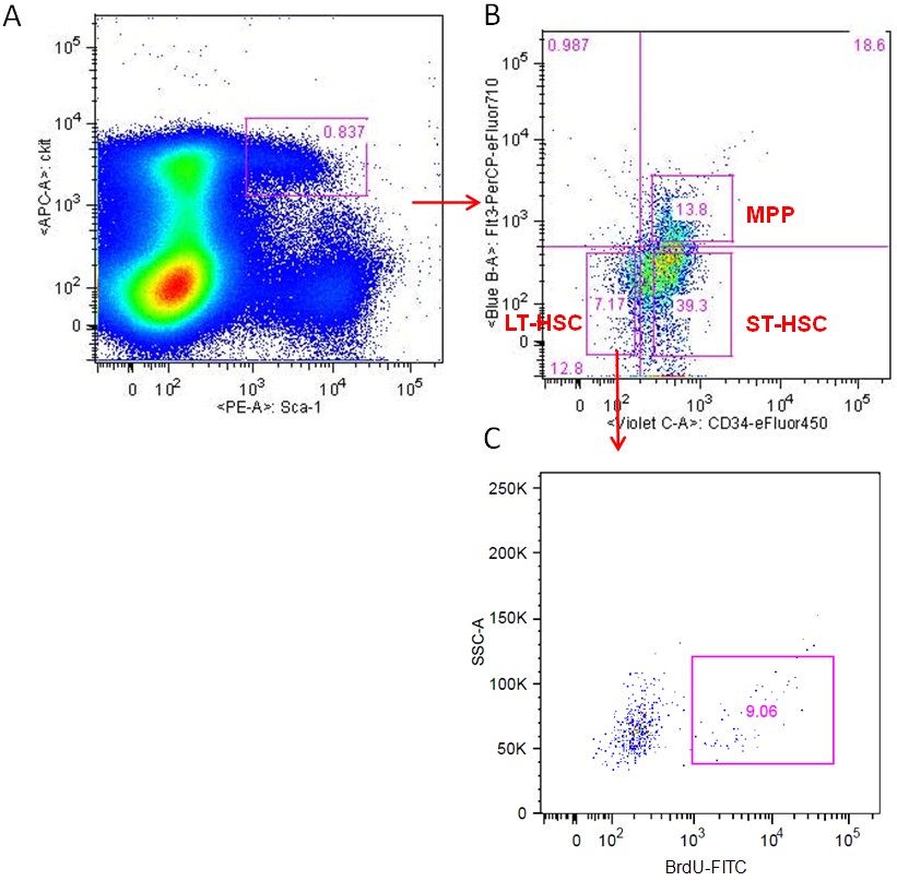
Figure 1. Gating strategy to analyze BrdU incorporation in LT-HSCs. A. BM Lin- cells were labeled with PE-Scal-I and APC-c-Kit antibodies and analyzed by flow cytometry. B. Lin-/Scal-I+/c-Kit+ (LSK) cells were gated as showed in A and the LSK cells were further analyzed with eFluor 450-CD34 and PerCP-eFluor 710-CD135 staining. Long-term HSCs (LT-HSCs, shown as Lin-Sca-1+c-Kit+CD34-CD135-), Short-term HSCs (ST-HSCs, shown as Lin-Sca-1+c-Kit+CD34+CD135-) and multipotent progenitors (MPPs, shown as Lin-Sca-1+c-Kit+CD34+CD135+) were separated as indicated. C. BrdU incorporation was further analyzed for each cell population. Data shown are LT-HSCs population analyzed for BrdU staining.
Recipes
- Buffer A
DPBS, pH 7.2 supplemented with 0.5% BSA and 2 mM EDTA
- Red blood cell lysis buffer
155 mM potassium bicarbonate
10 mM Ammonium chloride
0.1 mM of EDTA, pH = 7.4
- Staining buffer
DPBS, pH 7.2 supplememted with 0.5% BSA and 0.09% sodium azide
Acknowledgments
This protocol is adapted from An et al. (2013).
References
- An, N., Lin, Y. W., Mahajan, S., Kellner, J. N., Wang, Y., Li, Z., Kraft, A. S. and Kang, Y. (2013). Pim1 serine/threonine kinase regulates the number and functions of murine hematopoietic stem cells. Stem Cells 31(6): 1202-1212.
Article Information
Copyright
© 2013 The Authors; exclusive licensee Bio-protocol LLC.
How to cite
An, N. and Kang, Y. (2013). In vivo BrdU Incorporation Assay for Murine Hematopioetic Stem Cells. Bio-protocol 3(21): e960. DOI: 10.21769/BioProtoc.960.
Category
Stem Cell > Adult stem cell > Hematopoietic stem cell
Cell Biology > Cell viability > Cell proliferation
Molecular Biology > DNA > DNA labeling
Do you have any questions about this protocol?
Post your question to gather feedback from the community. We will also invite the authors of this article to respond.