- Protocols

- Articles and Issues
- For Authors
- About
- Become a Reviewer

In vivo Chick Chorioallantoic Membrane (CAM) Angiogenesis Assays

Published: Vol 3, Iss 18, Sep 20, 2013 DOI: 10.21769/BioProtoc.913 Views: 38772
Reviewed by: Lin FangXuecai Ge
Original research article
The authors used this protocol in:
Sep 2014
Advertisement


Protocol Collections
Comprehensive collections of detailed, peer-reviewed protocols focusing on specific topics


Related protocols
Abstract
Angiogenesis is the process of formation of new blood vessels from pre-existing vessels or endothelial cell progenitors. It plays a crucial role in tumor growth and metastasis. Tumor angiogenesis have been widely studied as an important target for suppressing tumor growth and metastasis. Here, we describe an in vivo chick embryo chorioallantoic membrane (CAM) model. The chick embryo chorioallantoic membrane is an extraembryonic and is rich of blood vessels. After exposing the vascular zone of the CAM, a sterilized filter-paper disk is employed, which is used as a carrier for being loaded with various chemicals, drugs or virus. Finally, the CAM was fixed and spread on glass slide, and the blood vessels were quantified by counting the number of blood vessel branch points. Compared with the matrigel plug angiogenesis assay, in which tumor cells are mixed with the matrigel gel (expensive) and injected into the mice, subsequently using immunohistochemistry (IHC) staining (time consuming) with the endothelial marker to indicate the presence of the newly formed capillaries, the main advantages of CAM model are its low cost, simplicity, reproducibility, and reliability. Thus, the CAM can be widely used in vivo to study both angiogenesis and anti-angiogenesis.
Keywords: Chick chorioallantoic membraneMaterials and Reagents
- Fertilized E6 chicken embryos (from Poultry Center of South China Agricultural University)
- 0.1% Benzalkonium Bromide (from Guangzhou Chemical Reagent Factory, diluted in sterile water before use)
- Methanol and acetone (1:1 in volume)
- Filter-paper disk (Whatman, catalog number: 1441150 )
- Packing film (Parafilm)
Equipment
- Incubator
- Glass slide
- Ophthalmic forceps
Procedure
- Fertilized E6 chicken embryos (48 ± 5 g) were cleaned with 0.1% Benzalkonium Bromide and preincubated at 37.5 °C in 85% humidity for 2 days.
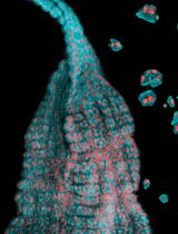
- Egg morphology appears like a meta-ellipse, with a relatively larger side and a smaller one, and the air sac is usually located on the larger side right behind the shell. After disinfection of the shell center outside the air sac with 0.1% Benzalkonium Bromide, a hole highlighted with marker pen was buffed and drilled gently over the air sac with a nipper not to break the shell, and the vascular zone was easy to be identified on the CAM (Figure 1).
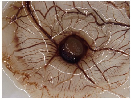
Figure 1. The vascular zone of the CAM
- Two drops of normal saline were then added to moisten the inner shell membrane adjacent to the CAM so that the membrane was easy to be separated from CAM.
- After being clamped and raised by ophthalmic forceps, the membrane and the CAM separated unforcedly, and then a 1 x 1 cm window on the membrane was sectioned to expose the vascular zone.
- A 5 mm x 5 mm sterilized filter-paper disks, which were used as a carrier for being directly loaded with indicated concentrations of chemicals or virus, were then directly applied and adhere to the vascular zone with right density of vascular.
- Upon sealing the openings with sterile flexible packing film, the eggs were further incubated for indicated periods.
- Finally, a mix of methanol and acetone (1:1 in volume) was directly added to immerse and fix the blood vessels of the experiment zone.
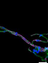
- After being clamped and raised by ophthalmic forceps, the CAM was easy to be separated from the embryo, and it was cut and spread on glass slide, and the blood vessels were viewed, photographed and quantified by counting the number of blood vessel branch points (Figure 2).
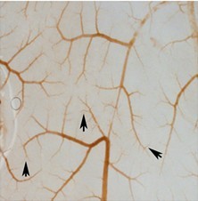
Figure 2. Blood vessel branch points on CAM. Arrow indicates new-formed blood vessel branches.
Acknowledgments
This protocol was adapted from the following published papers: Wen et al. (2013); Chen et al. (2014). This work was supported by The State Key Development Program for Basic Research of China (2009CB 918904, 2013CB945203), National Natural Sciences Foundation of China (30870955, 91029727, 30900555) and Program for New Century Excellent Talents in University (NCET-08-0646).
References
- Chen, Z., Zhang, Y., Jia, C., Wang, Y., Lai, P., Zhou, X., Wang, Y., Song, Q., Lin, J., Ren, Z., Gao, Q., Zhao, Z., Zheng, H., Wan, Z., Gao, T., Zhao, A., Dai, Y. and Bai, X. (2014). mTORC1/2 targeted by n-3 polyunsaturated fatty acids in the prevention of mammary tumorigenesis and tumor progression. Oncogene 33(37): 4548-4557.
- Wen, Z. H., Su, Y. C., Lai, P. L., Zhang, Y., Xu, Y. F., Zhao, A., Yao, G. Y., Jia, C. H., Lin, J., Xu, S., Wang, L., Wang, X. K., Liu, A. L., Jiang, Y., Dai, Y. F. and Bai, X. C. (2013). Critical role of arachidonic acid-activated mTOR signaling in breast carcinogenesis and angiogenesis. Oncogene 32(2): 160-170.
Article Information
Copyright
© 2013 The Authors; exclusive licensee Bio-protocol LLC.
How to cite
Chen, Z., Wen, Z. and Bai, X. (2013). In vivo Chick Chorioallantoic Membrane (CAM) Angiogenesis Assays. Bio-protocol 3(18): e913. DOI: 10.21769/BioProtoc.913.
Category
Cancer Biology > Angiogenesis > Drug discovery and analysis > Metabolism
Cancer Biology > Cellular energetics > Cell biology assays > Cell viability
Cell Biology > Tissue analysis > Tissue isolation
Do you have any questions about this protocol?
Post your question to gather feedback from the community. We will also invite the authors of this article to respond.