- Protocols

- Articles and Issues
- For Authors
- About
- Become a Reviewer

Transmission Electron Microscopy (TEM) Protocol: Observation Details within Cells
Published: Vol 3, Iss 13, Jul 5, 2013 DOI: 10.21769/BioProtoc.816 Views: 20286
Original research article
The authors used this protocol in:
Sep 2012
Advertisement


Protocol Collections
Comprehensive collections of detailed, peer-reviewed protocols focusing on specific topics


Related protocols
Abstract
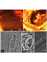
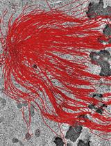
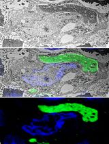
Transmission electron microscopy is a technique for observing the fine details of organelles in cells or tissues. This protocol is to be used to exam the membrane structure in cells with or without virus infection. Modifications should be made if users want to get images from tissues.
Keywords: Transmission Electron MicroscopyMaterials and Reagents
- Trypsin: working solution is 0.5% trypsin-0.2% EDTA (Life Technologies, Gibco®, catalog number: 15400-054 )
- NaH2PO4 (Kanto, catalog number: 37239-01 )
- Na2HPO4 (Chemical, catalog number: 37240-01)
- Alcohol
- Poly/Bed 812 (Electron microscopy sciences, catalog number: 14900 )
- Araldite 502 (Electron microscopy sciences, catalog number: 10900 )
- Dodecenylsuccinic anhydride (DDSA) (TAAB, catalog number: D025 )
- DMP-30 (Electron microscopy sciences, catalog number: 13600 )
- Propylene oxide (Merk, catalog number: S4912527-745 )
Note: Toxic, need to handle with care and collect after use.
- Syringe
- Dropper
- Grid (200 mesh) (TAAB)
- 0.1 M Phosphate buffer (PB) (see Recipes)
- Paraformaldehyde (4% in PB) (Bionovas, catalog number: AP0130-0500 ) (see Recipes)
- Osimium tetraoxide (1% in PB) (Electron microscopy sciences, catalog number: 51007 ) (see Recipes)
Note: Toxic, need to handle with care and collect after use.
- Epon (see Recipes)
- Toluidune Blue (Sigma-Aldrich, catalog number: NO202-146-2) (see Recipes)
- Uranyl acetate (Art, catalog number: 8473) (see Recipes)
- Lead citrate (TAAB, catalog number: L003 ) (see Recipes)
Equipment
- Eppendorf tubes
- Centrifuge
- Vacuum drying oven
- Diamond knife (Diatome)
- Ultracut (Leica)
- TEM (HITACHI, model: H-7100 )
Procedure
Note: Put all droppers and syringes into the 60 °C oven the day before this procedure to remove all the water inside all stuffs.
- After treatment, collecting cells (1 x 10⁶) with 0.5% trypsin-EDTA and washing cell with 0.5 ml 0.1 M phosphate buffer (PB) in an eppendorf tube by centrifuge (300 x g, 5 min).
- Add 0.5 ml paraformaldehyde (4% in PB) to fix cells in room temperature (RT) for at least 30 min (or store cells at 4 °C for long term storage).
- Wash cells 3 times with 0.5 ml 0.1 M PB by centrifuge (300 x g, 5 min).
- Add 0.5 ml Osimium tetraoxide (1% in PB) into the tube and incubate cells for 1 h at RT.
- Wash cells 3 times with 0.5 ml 0.1 M PB by centrifuge (300 x g, 5 min).
- Dehydrate cells with adding 0.5 ml 70% alcohol for 5 min twice→ centrifuge (300 x g, 5 min)→ remove alcohol.
- Dehydrate cells with adding 0.5 ml 85% alcohol for 5 min twice→ centrifuge (300 x g, 5 min)→ remove alcohol.
- Dehydrate cells with adding 0.5 ml 95% alcohol for 5 min for three times→centrifuge (300 x g, 5 min)→ remove alcohol.
- Dehydrate cells with adding 0.5 ml 100% alcohol for 10 min for five times→ centrifuge (300 x g, 5 min)→ remove alcohol.
Note: From step 10, all materials used should be from 60 °C oven.
- Add 0.5 ml propylene oxide into tubes and incubate for 3 min twice at RT.→ centrifuge (300 x g, 5 min)→remove propylene oxide.
- Add 1 ml (propylene oxide:Epon = 1:1) into tubes and incubate for 1 h at RT.→ centrifuge (300 x g, 5 min)→ remove Propylene oxide: Epon.
- Add 1 ml pure Epon into tubes and put the samples into the vacuum drying oven to vacuum for 1 h at RT.→ remove Epon.
- Add 1 ml pure Epon into tubes and vacuum for 1 h at RT.→ remove Epon.
- Add 1 ml pure Epon into tubes and vacuum overnight at RT.
- Put tubes into 60 °C oven for at least 48 h.
- Trimming-remove the edge of the block.
- Get thick section (1 μm) with Ultracut using diamond knife→put the thick section on a slide and stain with 0.5% toluidune blue for around 40 s to 1 min (until the edge is dried)→ wash 1 min with water → observe under microscopy to find the cells.
- Once we found the cells on the thick section, change the thickness of Ultracut to get thin sections (65-70 nm).
- Put the thin sections on the grids (3-4 sections on one grid).
- Stain sections with uranyl acetate and lead citrate for 3 min sequentially.
- Wash the grids with water for 1 min and let them dry completely.
- Get images from digital camera on TEM with identical magnificence.
Recipes
- 0.1 M Phosphate buffer (PB)
19 ml 0.2 M NaH2PO4
81 ml 0.2 M Na2HPO4
- 4% paraformaldehyde
4% paraformaldehyde in PB
- Osimium tetraoxide
1% osimium tetraoxide in PB
- Epon
Use syringe to mix all reagents below to make Epon.
Poly/Bed 812 10 ml
Araldite 502 6 ml
DDSA 18 ml
DMP-30 0.7 ml
Epon could be stored in 4 °C or -20 °C for 1-2 weeks.
- Toluidine Blue
0.5% Toluidine Blue
1% Sodium borate/in H2O
Filter with 0.22 μM filter
- Uranyl acetate
Saturated uranyl acetate in 50% Alcohol
- Lead citrate
1% lead citrate in H2O
Add 3 drops of 10 M NaOH (around 0.5 ml)
Acknowledgments
This protocol is adapted from Chau and Lu (1996) and Lee et al. (2012).
References
- Chau, Y. P. and Lu, K. S. (1996). Differential permeability of blood microvasculatures in various sympathetic ganglia of rodents. Anat Embryol (Berl) 194(3): 259-269.
- Lee, C. P., Liu, P. T., Kung, H. N., Su, M. T., Chua, H. H., Chang, Y. H., Chang, C. W., Tsai, C. H., Liu, F. T. and Chen, M. R. (2012). The ESCRT machinery is recruited by the viral BFRF1 protein to the nucleus-associated membrane for the maturation of Epstein-Barr Virus. PLoS Pathog 8(9): e1002904.
Article Information
Copyright
© 2013 The Authors; exclusive licensee Bio-protocol LLC.
How to cite
Readers should cite both the Bio-protocol article and the original research article where this protocol was used:
- Peng, W., Lu, K., Lai, S., Shy, H. and Kung, H. (2013). Transmission Electron Microscopy (TEM) Protocol: Observation Details within Cells. Bio-protocol 3(13): e816. DOI: 10.21769/BioProtoc.816.
- Lee, C. P., Liu, P. T., Kung, H. N., Su, M. T., Chua, H. H., Chang, Y. H., Chang, C. W., Tsai, C. H., Liu, F. T. and Chen, M. R. (2012). The ESCRT machinery is recruited by the viral BFRF1 protein to the nucleus-associated membrane for the maturation of Epstein-Barr Virus. PLoS Pathog 8(9): e1002904.
Category
Cell Biology > Cell imaging > Electron microscopy
Do you have any questions about this protocol?
Post your question to gather feedback from the community. We will also invite the authors of this article to respond.