- Protocols

- Articles and Issues
- For Authors
- About
- Become a Reviewer

MACS Isolation and Culture of Mouse Liver Mesothelial Cells

Published: Vol 3, Iss 13, Jul 5, 2013 DOI: 10.21769/BioProtoc.815 Views: 11696
Reviewed by: Lin Fang
Original research article
The authors used this protocol in:

Feb 2013
Advertisement


Protocol Collections
Comprehensive collections of detailed, peer-reviewed protocols focusing on specific topics


Related protocols
Abstract
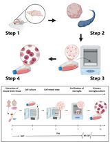
Mesothelial cells (MCs) form a single squamous epithelial cell layer and cover the surfaces of the internal organs, as well as the walls of cavities. The isolation of MCs is of great importance to study their function and characteristics for the understanding of physiology and pathophysiology of the liver. Glycoprotein M6a (GPM6A) was originally identified as a cell surface protein expressed in neurons and recently its expression was reported in epicardium and liver MCs (Wu et al., 2001; Bochmann et al., 2010; Li et al., 2012). Here we describe a method to isolate MCs from the adult mouse liver with anti-GPM6A antibodies. Under the low glucose and serum concentration, primary MCs grow and form epithelial colonies (Figure 1).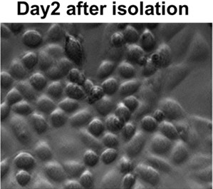
Figure 1. Liver MCs 2 days in culture (20x objective)
Materials and Reagents
- Dulbecco’s Modified Eagle’s Medium (DMEM) Low glucose with stable L-glutamine (Thermo Fisher Scientific, catalog number: SH30021.01 )
- DMEM/F-12 (Thermo Fisher Scientific, catalog number: SH30023.FS )
- Fetal Bovine Serum (FBS) (Sigma-Aldrich, catalog number: F9665 )
- PBS, pH 7.4 (Sigma-Aldrich, catalog number: P3813-10PAK )
- Bovine Collagen Solution, Type I (Advanced BioMatrix, catalog number: 5005-B )
- Pronase (Roche, catalog number: 11459643001 )
- Antibiotic-Antimycotic (100x) (Life Technologies, catalog number: 15240-062 )
- BD Falcon polypropylene conical tube 50 ml (BD Biosciences, catalog number: 352070 )
- BD Falcon cell strainer with 70 μm nylon mesh (BD Biosciences, catalog number: 352350 )
- Rat anti-mouse glycoprotein M6a (GPM6A) antibodies (MBL International, catalog number: D0553 )
- Goat anti-rat IgG microbeads (Miltenyl Biotec, catalog number: 130-048-501 )
- Hydrocortisone solution (Sigma-Aldrich, catalog number: H6909-10ML )
- Insulin-Transferrin-Selenium-X (Life Technologies, catalog number: 41400-045 )
- Ketamine (Clipper Distributing Company, catalog number: NDC57319-542-02 )
- 5% low glucose DMEM medium (see Recipes)
- MC medium (see Recipes)
- Ketamine solution (see Recipes)
Equipment
- 37 °C shaker
- Surgery Tools: Forceps, scissors and glass petri dish
- 37 °C incubator
- Centrifuge
- 24-well plate (VWR, catalog number: 29442-044 )
- G24 Environmental Incubator Shaker (New Brunswick Scientific)
- Miltenyi AutoMacs machine
Procedure
I. Before procedure
- Autoclave all surgery tools.
- Collagen-coated dishes are used for better attachment of MCs. Mix 50 μl of the collagen solution with 950 μl sterile water and coat the 24-well plate (200 μl per well).
- Put the coated dish in 37 °C incubator for at least 30 min, remove the collagen solution, and then wash with PBS 3 times. Make sure each well is completely dry before plating the cell.
- Prepare 1 mg/ml Pronase in DMEM/F-12 medium and incubate in 37 °C incubator with gentle stirring for 20 min; Filter the solution with 0.22 μm filter into 50 ml tube before using.
- Prepare 5% low glucose DMEM medium.
- Prepare MC medium.
- Prepare Ketamine solution.
- Use 3-5 C57BL/6 male or female mice (20-30 g/each) for MC isolation. Age preference at least 8 weeks old.
II. Isolation and culture of liver mesothelial cells
- Inject Ketamine solution intraperitoneally to mice with 1 ml syringe 23-25 gauge 5/8 inch needle for anesthesia, 0.1 ml per 10 mg of mouse body weight.
- Shave the hair and clean up with 70% ethanol, cut the skin, muscle and expose the abdomen cavity.
- Remove the gallbladder, cut off the ligaments connected to the liver lobes and diaphragm carefully, take out the liver lobes gently without disturbing the liver surface, and put the liver in 10 cm petri dish washing with sterile PBS.
- Transfer the livers in 50 ml tube, add 30 ml DMEM/F-12 and shake at 130 rpm in 37 °C shaker for 5-10 min washing.
- Transfer the livers to the tube with 30 ml Pronase/DMEM/F-12 medium and shake at 130 rpm in 37 °C shaker for 20 min. No need to cut the liver into small pieces.
- Filter the supernatant containing MCs by BD Falcon cell strainer.
- Collect the flow through and add 30 ml 5% low glucose DMEM.
- Spin down at 1,700 x g for 5 min at RT.
- Discard the supernatant, add 30 ml 5% low glucose DMEM, and spin down at 1,700 x g for 5 min at RT.
- Repeat step 9.
- Resuspend the pellet with 1.5 ml 5% low glucose DMEM and add 1 μl rat anti-mouse GPM6A antibodies at 1:1,500 dilutions.
- Incubate on ice for 15-30 min.
- Add 5% low glucose DMEM to 30 ml, spin down at 1,700 x g for 5 min at RT and discard supernatant.
- Add 1 ml 5% low glucose DMEM to gently suspend the pellet.
- Add 10 μl goat anti-rat IgG microbeads (1 μl/5 x 105 cells) and incubate on ice for 20 min.
- Add 5% low glucose DMEM to 30 ml, spin down at 1,700 x g for 5 min at RT and discard supernatant.
- Suspend pellet in 1 ml 5% low glucose DMEM and filter with BD Falcon cell strainer.
- Use Miltenyi AutoMACS machine to separate the MCs according to their instruction (https://www.miltenyibiotec.com). In brief, the machine will load the cell suspension to the magnetic column and the magnetically labeled GPM6A+ cells will be retained in the column under the magnetic field. After washing, GPM6A+ MCs will be eluted to a new tube.
Note: Or use Miltenyi magnetic separator to separate MC. - Add 30 ml low glucose DMEM medium and spin down at 1,700 x g for 5 min at RT, discard supernatant.
- Add 1 ml MC medium to gently suspend the pellet, count the cell number (yield: about 3 x 10⁴ MCs from 1 mouse) and plate the cells to the wells of the plate (2 x 10⁴ cells per well).
- Culture MCs in 5% CO2 at 37 °C incubator, change with MC medium every 3 days. Primary MCs form epithelial colonies and become confluent within 1 week. From 1 week after plating, some MCs start to lose epithelial cell polarity and become fibroblastic cells. Following two passages, neither epithelial nor fibroblastic MCs attach to the dish and survive.
Recipes
- 5% low glucose DMEM medium
Low glucose DMEM
5% FBS
1% Antibiotic-Antimycotic - MC medium
Low glucose DMEM
5% FBS
1% Antibiotic-Antimycotic
Hydrocortisone solution (1:1,000 dilution)
Insulin-Transferrin-Selenium-X (1:100 dilution) - Ketamine solution
4.5 ml Ketamine (conc. 100 mg/ml)
0.75 ml Xylazine (conc. 20 mg/ml)
19.5 normal saline 0.9%
References
- Wu, D. F., Koch, T., Liang, Y. J., Stumm, R., Schulz, S., Schroder, H. and Hollt, V. (2007). Membrane glycoprotein M6a interacts with the micro-opioid receptor and facilitates receptor endocytosis and recycling. J Biol Chem 282(30): 22239-22247.
- Bochmann, L., Sarathchandra, P., Mori, F., Lara-Pezzi, E., Lazzaro, D. and Rosenthal, N. (2010). Revealing new mouse epicardial cell markers through transcriptomics. PLoS One 5(6): e11429.
- Li, Y., Wang, J. and Asahina, K. (2013). Mesothelial cells give rise to hepatic stellate cells and myofibroblasts via mesothelial-mesenchymal transition in liver injury. Proc Natl Acad Sci U S A 110(6): 2324-2329.
Article Information
Copyright
© 2013 The Authors; exclusive licensee Bio-protocol LLC.
How to cite
Li, Y., Lua, I. and Asahina, K. (2013). MACS Isolation and Culture of Mouse Liver Mesothelial Cells. Bio-protocol 3(13): e815. DOI: 10.21769/BioProtoc.815.
Category
Cell Biology > Cell isolation and culture > Cell isolation
Do you have any questions about this protocol?
Post your question to gather feedback from the community. We will also invite the authors of this article to respond.