- Protocols

- Articles and Issues
- For Authors
- About
- Become a Reviewer

Immunoprecipitation for Cell Culture

Published: Vol 2, Iss 3, Feb 5, 2012 DOI: 10.21769/BioProtoc.72 Views: 17098
Advertisement


Protocol Collections
Comprehensive collections of detailed, peer-reviewed protocols focusing on specific topics


Abstract
Immunoprecipitation (IP) is a method to pull down a protein out of solution using an antibody that specifically binds to that particular protein. Immunoprecipitation is a powerful technique to isolate and concentrate a particular protein from a sample containing many thousands of different proteins, to test protein-protein interactions, and to pull multiple members of a complex out of solution by latching onto one member with an antibody. This protocol describes a general immunoprecipitation strategy using cell cultures as starting material.
Keywords: ImmunofluorscenceMaterials and Reagents
- HeLa S3 and HeLa cells were cultured in Dulbecco’s modified Eagles Medium containing 10% fetal bovine serum (Invitrogen) and antibiotics
- Protein A beads (Bio-Rad Laboratories); Protein G beads (Santa Cruz Biotechnology)
Note: For rabbit polyclonal antibodies and mouse monoclonal antibodies IgG2a, IgG2b, IgG3, we usually couple antibodies to protein A beads; For mouse monoclonal antibodies IgG1, rat monoclonal antibodies, and mouse, rat, goat, and donkey polyclonal antibodies, we usually couple antibodies to protein G beads.
- Bifunctional cross-linker dimethyl pimelimidate (DMP) (MW: 259.177) (Sigma-Aldrich, catalog number: D8388 )
- Phosphate buffered saline (PBS)
- Tween 20
- Triton X-100
- Sodium borate (pH 9.0)
- Hepes
- KCl
- NaN3
- NP-40
- Glycerol
- EGTA
- MgCl2
- DTT
- Microcystin
- Leupeptin
- Pepstatin
- Chymostatin
- β-mercaptoethanol
- Bromophenol blue
- PBST buffer (see Recipes)
- Lysis buffer (see Recipes)
- 1x SDS protein gel sample loading buffer (see Recipes)
Equipment
- Hematology/chemistry mixer (Fisher Scientific)
- Centrifuge (Eppendorf 5415D centrifuge)
- Incubator
Procedure
- Coupling antibody to Protein A beads
- Wash 30 μl beads with 20 fold volume (20 vol) PBS or PBST twice.
Notes:- For washing beads, we usually add buffer to the beads, mix in the tube several times, spin down the beads, and remove the supernatant.
- Spin down the beads at 4,000 rpm, 30 sec.
- We usually couple 1 μg antibody with 3 μl beads. If beads are stored in 1: 1 storing buffer, thus take 6 μl of the suspension of beads and buffer.
- For washing beads, we usually add buffer to the beads, mix in the tube several times, spin down the beads, and remove the supernatant.
- Dilute 10 μg antibody in 100 μl PBS.
- Add diluted antibody to beads, rotate on the Hematology/chemistry mixer equipment for 1 h at room temperature (RT).
- Spin down beads, wash and resuspend in 20 vol 0.2 M sodium borate (pH 9.0).
- Add 20 mM DMP to crosslink the antibody to the beads.
Note: We usually take dry DMP powder directly (DMP powder is stored at 4 °C) and add 5.2 mg DMP per 1 ml total sodium borate suspension.
- Incubate DMP in the sodium borate for 30 min, rotate, RT.
- Spin down the bead and wash once with 20 vol 1 M Tris-HCl (pH 7.7).
- Spin down the bead and resuspend in 20 vol 1 M Tris-HCl (pH 7.7). Incubate for 2 h, rotate, RT, to quench the activity of DMP.
- Spin down beads and wash with PBS twice.
- Spin down the beads and store antibody coupled beads in 60 μl PBS (33% beads), add 0.05% NaN3. Store the beads at 4 °C for a couple of months.
- Wash 30 μl beads with 20 fold volume (20 vol) PBS or PBST twice.
- Lysis of cells
- Lyse the cells in 7 vol lysis buffer.
- Put the lysis solution on ice for 30 min to 1 h.
- Centrifuge the lysis solution at 13,000 rpm for 10 min, 4 °C.
- Transfer supernatants. The supernatants can be stores at -80 °C for future use.
- Lyse the cells in 7 vol lysis buffer.
- Coimmunoprecipitation
- Incubate 3 μl antibody coupled beads in 250 μl cell lysis supernatants, rotate overnight at 4 °C.
- Wash beads 4 times with 20 vol lysis buffer containing 500 mM KCl and 0.5 % NP-40, and wash once with lysis buffer.
Note: For wash beads, we usually add buffer to the beads, mix the tube several times, spin down the beads, and remove the supernatant.
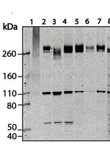
- Elute protein with 1x SDS protein gel sample loading buffer, separated by SDS-PAGE (5-15%) and analyze by western blot.
- Incubate 3 μl antibody coupled beads in 250 μl cell lysis supernatants, rotate overnight at 4 °C.
Recipes
- PBST buffer
PBS plus 0.1 % Tween 20 or 0.1 % Triton X-100
- Lysis buffer
50 mM Hepes (pH 7.4)
200 mM KCl
0.3% NP-40
10% glycerol
1 mM EGTA
1 mM MgCl2
0.5 mM DTT
0.5 μM microcystin
10 μg ml-1 each of leupeptin, pepstatin, and chymostatin
- 1x SDS protein gel sample loading buffer
50 mM Tris-HCl (pH 6.8)
2% SDS
10% glycerol
1% β-mercaptoethanol
12.5 mM EDTA
0.02 % bromophenol blue
Acknowledgments
This protocol was modified from an immunoprecipitation protocol developed in the laboratory of Dr. Guowei Fang (Department of Biology, Stanford University, Stanford, CA, USA). This work was supported by a Burroughs-Wellcome Career Award in Biomedical Research (G.F.) and by grants from National Institutes of Health (GM062852 to G.F.).
References
- Zhu, H., Coppinger, J. A., Jang, C. Y., Yates, J. R., 3rd and Fang, G. (2008). FAM29A promotes microtubule amplification via recruitment of the NEDD1-gamma-tubulin complex to the mitotic spindle. J Cell Biol 183(5): 835-848.
Article Information
Copyright
© 2012 The Authors; exclusive licensee Bio-protocol LLC.
How to cite
Zhu, H. (2012). Immunoprecipitation for Cell Culture. Bio-protocol 2(3): e72. DOI: 10.21769/BioProtoc.72.
Category
Biochemistry > Protein > Immunodetection
Do you have any questions about this protocol?
Post your question to gather feedback from the community. We will also invite the authors of this article to respond.