- Protocols

- Articles and Issues
- For Authors
- About
- Become a Reviewer

Cell Isolation of Spleen Mononuclear Cells
Published: Vol 3, Iss 9, May 5, 2013 DOI: 10.21769/BioProtoc.689 Views: 33327
Reviewed by: Lin Fang
Original research article
The authors used this protocol in:
Sep 2012
Advertisement


Protocol Collections
Comprehensive collections of detailed, peer-reviewed protocols focusing on specific topics


Related protocols
Abstract
This method allows you to isolate different subclass mononuclear cells, like B-cells, T cells, CD4+ and CD8+ T, from mouse spleen. By conjugating cells with specific antibodies and subsequently magnetic beads isolation, using the technique from Miltenyi, this allows a high purity.
Keywords: T cellMaterials and Reagents
- Antibody
- FITC-conjugated anti-CD3 antibody (BD Biosciences, catalog number: 553058 , clone 145-2C11)
- PE-conjugated anti-CD19 antibody (BD Biosciences, catalog number: 557399 , clone 1D3)
- FITC-conjugated anti-CD4 antibody (BD Biosciences, catalog number: 557667 , clone RM4-5)
- PE-conjugated anti-CD8 antibody (BD Biosciences, catalog number: 561095 , clone 53-6.7)
- FITC-conjugated anti-CD3 antibody (BD Biosciences, catalog number: 553058 , clone 145-2C11)
- Microbeads
- Anti-CD43 microbeads (Miltenyi Biotec, catalog number: 130-049-801 )
- Anti-CD90 microbeads (Miltenyi Biotec, catalog number: 130-091-376 )
- Anti-CD4 microbeads (Miltenyi Biotec, catalog number: 130-049-201 )
- Anti-CD8 microbeads (Miltenyi Biotec, catalog number: 130-091-112 )
- Anti-CD43 microbeads (Miltenyi Biotec, catalog number: 130-049-801 )
- Others
Equipment
- Scissors and forceps
- MiniMACS separation unit (Miltenyi Biotec, MiniMACS Separator, catalog number: 130-090-312 )
- Separation column (Miltenyi Biotec, separation column, Type MS, catalog number: 130-042-201 )
- Cell strainer (BD Biosciences, catalog number: 352360 )
- Flow cytometer (BD Biosciences, Coulter)
- Shaker
Procedure
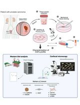
B-cells, total T cells and/or single positive CD4+ and CD8+ T cells, were purified from spleen cells by magnetic separation with the Mini-MACS system [Miltenyi, 1990] (http://www.miltenyibiotec.com). The scheme illustrates how to manage the procedure.
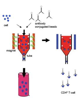
Figure 1. Schematic view of the experimental strategy using magnetic MACS beads to isolate CD4+ cells. Cells were incubated with CD4-,CD8-,CD90- and CD43-antibody conjugated magnetic beads. The cell suspensions were subjected to column selection and placed in the magnetic separator. Flow-through was discarded. Then the column was washed with separation puffer to increase purity and removed afterwards from the magnetic separator. With a plunger the magnetically labelled cells were flushed out of the column.
- Sacrifice mouse and isolate the complete spleen.
- Pass spleen through a 100-μm cell strainer to get single cells suspension by crushing with forceps and collecting the cell suspension in 5 ml PBS.
- Wash with 1x PBS & centrifuge the cells (100 x g for 5 min), then resuspend spleen cells in 80 μl ice-cold separation buffer per 107 cells. From a normal spleen you will get approximately 8 x 10⁷ cells. The overall operation temperature is room temperature. The buffers should be ice cold.
- Add a 20 μl aliquot of antibody-conjugated microbeads per 107 cells incubate for 30 min at 4-8 °C at a shaker. No prewash is needed. The number of cells is depending from the animal.
The following microbeads were used: anti-CD43 microbeads for negative isolation of resting B cells, anti-CD90 microbeads for positive isolation of total T cells, anti-CD4 and anti-CD8 microbeads for positive isolation of the respective T-cell subset.
- Wash the column by putting 5 ml 1x PBS on the top. The liquid passes the column by gravity. Then pipette the labelled cell suspension on top of a separation column, which had been washed three times with separation buffer and placed in the MiniMACS separation unit. Pass the suspension through the column.
- In case of negative selection of CD43- B cells the effluent was collected as a B-cell fraction and washed respectively centrifuged three times with 5 ml PBS.
In case of positive selection of CD90+, CD4+ or CD8+ T-cells the effluent was discarded and the columns were washed twice with 500 μl separation buffer. Subsequently, remove columns from the separator and wash magnetically labelled cells out with 1 ml separation buffer using a plunger.
- Wash the respective T-cell fraction three times with medium same as B-cells
- Assess the purity of the various cell fractions by Flow cytometry analysis using a Flow cytometer. Stain cells with fluorescein isothiocyanate (FITC)-conjugated anti-CD3 (clone 145-2C11 to detect total T-cells), phycoerythrin (PE)-conjugated anti-CD19 (clone 1D3 to detect B-cells), FITC-conjugated anti-CD4 (clone RM4-5 to detect CD4+ T-cells) and PE-conjugated anti-CD8 (clone 53-6.7 to detect CD8+ T-cells) and analyse for positive cells according to standard procedures.
Recipes
- Separation buffer
1x PBS with 5 mM EDTA and 0.5% BSA
Acknowledgments
The authors thank the German Research Foundation (DFG) and the University Erlangen-Nuremberg for funding. We thank A. von Berg and L. Sologub for excellent technical assistance.
References
- Miltenyi, S., Muller, W., Weichel, W. and Radbruch, A. (1990). High gradient magnetic cell separation with MACS. Cytometry 11(2): 231-238.
Article Information
Copyright
© 2013 The Authors; exclusive licensee Bio-protocol LLC.
How to cite
Weigmann, B. (2013). Cell Isolation of Spleen Mononuclear Cells. Bio-protocol 3(9): e689. DOI: 10.21769/BioProtoc.689.
Category
Immunology > Immune cell isolation > Lymphocyte
Cell Biology > Cell isolation and culture > Cell isolation
Do you have any questions about this protocol?
Post your question to gather feedback from the community. We will also invite the authors of this article to respond.