


- Protocols

- Articles and Issues
- For Authors
- About
- Become a Reviewer


Monitoring Protein Secretion in Living Cells With a HiBiT-Integrated RUSH System

(*contributed equally to this work, § Technical contact) Published: Vol 16, Iss 21, Nov 5, 2026 DOI: 10.21769/BioProtoc.5871 Views: 23
Reviewed by: Medhat S ElhalawanyAnonymous reviewer(s)
 PDF
 Ask a question

Original research article
The authors used this protocol in:

Sep 2025

Protocol Collections
Comprehensive collections of detailed, peer-reviewed protocols focusing on specific topics






Abstract
The cellular secretory pathway is a fundamental physiological process; however, precise, efficient, and quantitative methods for monitoring this process remain rarely explored. Although several classic approaches have been developed to investigate secretory trafficking, the RUSH (Retention Using Selective Hooks) system currently represents the most advanced technique. Here, we introduce a method termed the HiBiT-integrated RUSH system and provide a detailed protocol for its application. This approach enables real-time monitoring of the secretion of cell membrane receptors such as ACE2, PDL1, NTCP, and CD209. By converting the cellular secretion process into a quantitative curve, our method offers an objective means for result interpretation. The HiBiT-integrated RUSH system is particularly effective for studying the transport dynamics of various cell membrane receptors, thereby facilitating the identification of both common and specific regulators of the cellular secretory pathway.
Key features
• This protocol converts cellular secretion into quantitative curves, enabling objective, real-time assessment of secretory protein transport.
• The protocol is validated for monitoring ACE2 and PDL1 secretion and is applicable to diverse cell membrane receptors.
• The protocol retains the spatiotemporal control of the classic RUSH system while adding HiBiT-based sensitive, live-cell detection.
• The protocol facilitates the discovery of both common and specific regulators of the cellular secretory pathway across different cargo proteins.
Keywords: HiBiT tag Cellular secretory pathway
RUSH (Retention Using Selective Hooks)
Real-time monitoring
Quantitative secretion assay
Live-cell detection
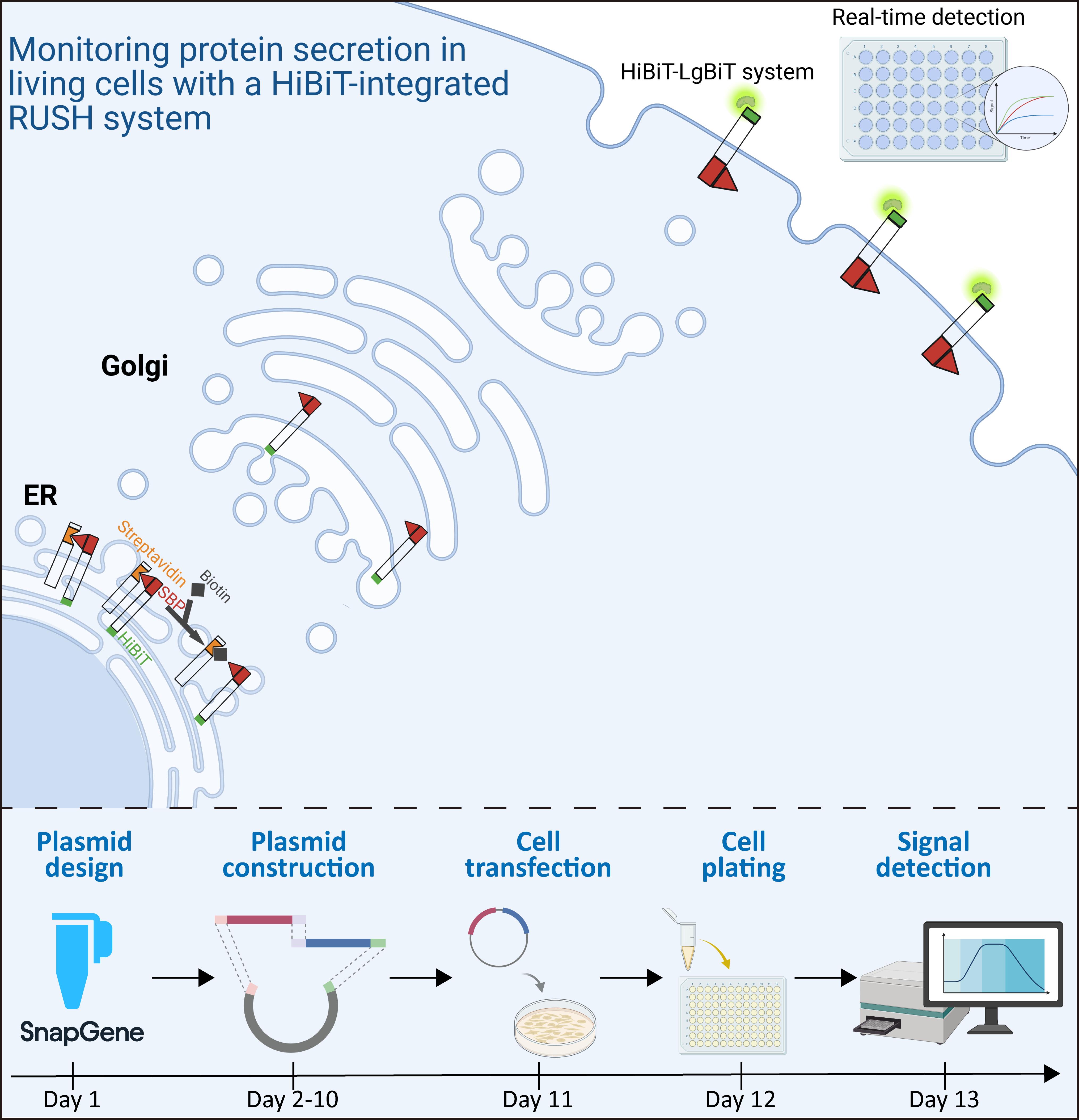
Graphical overview
Background
The cellular secretory pathway is a fundamental physiological process that refers to the transport of newly synthesized proteins to their target compartments. In eukaryotes, conventional protein secretion is primarily mediated by secretory vesicles, which transport cargo through the endoplasmic reticulum (ER) and the Golgi apparatus to the plasma membrane (PM) or extracellular space. This process is essential because it directs proteins, such as cytokines, chemokines, hormones, and receptors, to their sites of action, where they perform crucial physiological and pathological functions [1].
Several classic methods have been developed to investigate secretory trafficking, with the RUSH (Retention Using Selective Hooks) system being the most advanced technique [2–5]. The RUSH system exploits the high-affinity interaction between streptavidin (Str) and a streptavidin-binding peptide (SBP) to reversibly retain an enhanced GFP (EGFP)-tagged reporter within the ER. Subsequent addition of biotin, which competitively displaces SBP from Str, triggers reporter release. This controlled release enables quantification of secretion kinetics and status by monitoring EGFP localization dynamics via fluorescence microscopy [3].
Researchers have conducted impressive studies using the RUSH system, driving significant progress in the field of cellular trafficking [6–8]. However, the classic RUSH assay requires the acquisition and quantification of numerous fluorescence images, which poses challenges for achieving precision and efficiency, particularly constraining its use in high-throughput screening applications. Boncompain et al. [9] utilized the classic RUSH system to screen and validate three molecules from over 4,000 compounds that inhibit HIV infection by targeting CC chemokine receptor 5 (CCR5) secretion. To our knowledge, this is the only reported study to apply the RUSH technique to high-throughput screening. HiBiT is an 11-amino-acid peptide tag developed by Promega in recent years. When HiBiT-tagged proteins on the cell surface or in the extracellular space bind to the exogenously added LgBiT, a NanoBiT luciferase is generated, enabling the detection and quantification of HiBiT-tagged proteins in living cells [10,11]. Building on this advantage, we propose substituting EGFP in the RUSH reporter with HiBiT, thereby converting the fluorescent signal into a quantifiable luciferase signal, which would facilitate real-time, quantitative monitoring of reporter trafficking.
Here, we combined the HiBiT technology with the classic RUSH system to investigate the secretion process of angiotensin-converting enzyme 2 (ACE2) as a model, which is the receptor for SARS-CoV-2. Using a plate reader, we achieved real-time monitoring of the secretion dynamics, enabling a precise and efficient identification of the trafficking regulators. Notably, this strategy was also applied to monitor the secretion process of programmed death-ligand 1 (PDL1), sodium taurocholate cotransporting polypeptide (NTCP), and CD209 (also known as DC-SIGN), all of which are cell membrane receptors with important physiological functions, highlighting the significant potential of this approach in advancing the field of cellular trafficking research.
Since its introduction in 2012, RUSH has been widely used by researchers and has become a cornerstone method in the field of cellular secretion studies [3]. However, its application still faces several limitations. One of the main challenges is achieving a perfect hook, as fluorescence leakage can compromise result interpretation [12]. In addition, quantitative bioimaging remains a difficult task, especially since a single two-dimensional image cannot accurately reflect the three-dimensional nature of cells [13]. Furthermore, cellular transport is a highly dynamic process, necessitating rapid and precise data acquisition.
Denus et al. also utilized the combination of RUSH and HiBiT to quantitatively measure unconventional protein secretion, demonstrating the feasibility and compatibility of the HiBiT-integrated RUSH system [14]. However, their approach relied on endpoint measurements rather than real-time monitoring, which is essential for capturing the highly dynamic nature of cellular secretion. In this study, we employed the HiBiT-integrated RUSH system, which converts the cellular secretion process into a quantitative curve without the need for stable cell line construction or achieving a perfect hook, thereby providing an objective means for result interpretation. It is particularly effective for studying the transport dynamics of various cell membrane receptors such as ACE2, PDL1, NTCP, and CD209, facilitating the identification of both common and specific regulators of the cellular secretory process. Ongoing studies are focused on screening compound libraries using this strategy to identify potential interventions targeting the secretion of disease-associated proteins.
Materials and reagents
Biological materials
1. 293T cells (American Type Culture Collection, catalog number: CRL-3216)
2. Str–Ii_SBP-EGFP-Golgin84 plasmid (Addgene, catalog number: 65303)
3. Str-KDEL_SBP-EGFP-Ecadherin plasmid (Addgene, catalog number: 65286)
4. Str–Ii_HiBiT-ACE2-SBP-FLAG (generated in-house)
5. Str–Ii_HiBiT-PDL1-SBP-FLAG (generated in-house)
6. Str-KDEL_CD209-SBP-FLAG-HiBiT (generated in-house)
7. Str-KDEL_HiBiT-FLAG-SBP-NTCP (generated in-house)
Reagents
1. Restriction enzyme AscI (Gene-Protein Link, catalog number: G05F023)
2. Restriction enzyme XbaI (Gene-Protein Link, catalog number: G05F265)
3. Seamless Cloning kit (Beyotime, catalog number: D7010)
4. Dulbecco’s modified Eagle’s medium (DMEM) (Eallbio, catalog number: 03.1002C)
5. Fetal bovine serum (FBS) (ExCell Bio, catalog number: FSP500)
6. Penicillin–streptomycin solution (Eallbio, catalog number: 03.12001A)
7. jetPRIME transfection reagent (Polyplus, catalog number: 101000027)
8. Anti-FLAG M2 affinity gel (Merck Millipore, catalog number: A2220)
9. Nano-Glo HiBiT extracellular detection reagent (Promega, catalog number: N2421)
10. Biotin (Solarbio, catalog number: D8150)
11. Brefeldin A (BFA) (Beyotime, catalog number: S1536)
12. Trypsin-EDTA solution (Beyotime, catalog number: C0201)
13. Phosphate-buffered saline (PBS) (Beyotime, catalog number: C0221B)
Solutions
1. 100× biotin stock solution (see Recipes)
2. HiBiT detection solution (see Recipes)
Recipes
1. 100× biotin stock solution
| Reagent | Final concentration | Quantity or volume |
|---|---|---|
| Biotin (powder) | 0.96 mg/mL (100×) | 24 mg |
| DMEM | 100% | 25 mL |
Weigh 24 mg of biotin and dissolve in 25 mL of DMEM. When biotin is completely dissolved, sterilize by filtration with a 0.22 μm membrane. Store at 4 °C for 3 months.
2. HiBiT detection solution
| Reagent | Final concentration | Quantity or volume |
|---|---|---|
| LgBiT (Nano-Glo HiBiT extracellular detection reagent) | 0.5% | 0.5 μL/well |
| Furimazine (Nano-Glo HiBiT extracellular detection reagent) | 1.0% | 1 μL/well |
| DMEM | 98.5% | 98.5 μL/well |
For each well to be measured, prepare the HiBiT detection solution fresh in DMEM at the following final concentrations: 0.5% (v/v) LgBiT and 1.0% (v/v) furimazine substrate. For a single 96-well plate (100 μL per well), prepare a sufficient volume by scaling the volumes accordingly. Prepare the solution immediately before use and do not store it for later assays.
Laboratory supplies
1. Cell culture dishes, 10 cm (Servicebio, catalog number: CCD-100H)
2. 15 mL conical tubes (Servicebio, catalog number: EP-1501-J)
3. 96-well white cell culture plates (Beyotime, catalog number: FCP968)
4. 50 mL conical tubes (Servicebio, catalog number: EP-5001-J)
5. 10 μL pipette tips (Servicebio, catalog number: TP-10-C)
6. 200 μL pipette tips (Servicebio, catalog number: TP-200-C)
7. 1,000 μL pipette tips (Servicebio, catalog number: TP-1000-C)
8. 10 mL serological pipettes (Servicebio, catalog number: SP-10-SF)
Equipment
1. Plate reader capable of maintaining 37 °C (Tecan, model: Spark)
Software and datasets
1. GraphPad (GraphPad Software, Inc., Version 8.4.2)
2. SnapGene (Dotmatics, Version 4.2.4)
3. Tecan SparkControl
Note: This protocol does not contain omics data or code that need to be publicly shared. Relevant datasets will be made available upon reasonable request.
Procedure
A. Construction of HiBiT-integrated RUSH plasmids adapted for specific proteins
1. Design the HiBiT-integrated RUSH plasmid.
Note: The classical RUSH system comprises a hook (containing Streptavidin, Str) and a reporter (containing streptavidin-binding peptide, SBP). The high-affinity Str–SBP interaction anchors the EGFP-tagged reporter to the ER or Golgi apparatus; subsequent addition of biotin competitively displaces SBP and triggers reporter release. Fluorescence microscopy then monitors EGFP localization dynamics, allowing quantification of secretion kinetics and status (Figure 1A, B). In the HiBiT-integrated version, EGFP is replaced with HiBiT. When cell-surface or extracellular HiBiT-tagged proteins bind to exogenously added LgBiT, they form a functional NanoBiT luciferase detectable in living cells, enabling real-time quantitative monitoring of the secretion process. Therefore, this HiBiT-integrated RUSH system is suitable only for proteins that ultimately localize to the plasma membrane or are secreted into the extracellular space.
a. Analyze the membrane topology of the reporter protein and its signal peptide.
b. Select an appropriate backbone plasmid from the five hook constructs designed by Boncompain et al. [3] (available via Addgene, https://www.addgene.org/browse/article/10202/). For your protein of interest, choose one suitable hook and then modify the reporter part accordingly. In this study, use the Str–Ii_SBP-EGFP-Golgin84 plasmid (Addgene #65303) as the backbone for constructing the ACE2 and PDL1 reporters.
Caution: Carefully consider the membrane topology of the reporter protein to achieve successful anchoring to the hook while also ensuring that the HiBiT tag is ultimately exposed to the extracellular space.
c. Refer to the design details of the four successfully constructed HiBiT-integrated RUSH plasmids in this study (for ACE2, PDL1, NTCP, and CD209, depicted in Figures 1C–D, 3A–B, and 3D–E). Use these designs as reference; they all carefully account for membrane topology, signal peptide, successful hook anchoring, and extracellular exposure of the HiBiT tag.
2. Replace the reporter gene via molecular cloning.
a. Design primers using the SnapGene software.
b. Perform molecular cloning using your preferred method; our laboratory routinely uses a commercial seamless cloning kit.
c. Verify the construct by sequencing, then name and store the plasmid.
d. Refer to the Supplementary Information for detailed design information of the plasmids used in this protocol, including annotated plasmid maps, cloning junctions, linker sequences, positions of the HiBiT/SBP/FLAG tags, primer sequences, and the exact sequences of these reporter constructs (Figure S1–S4, Table S1).
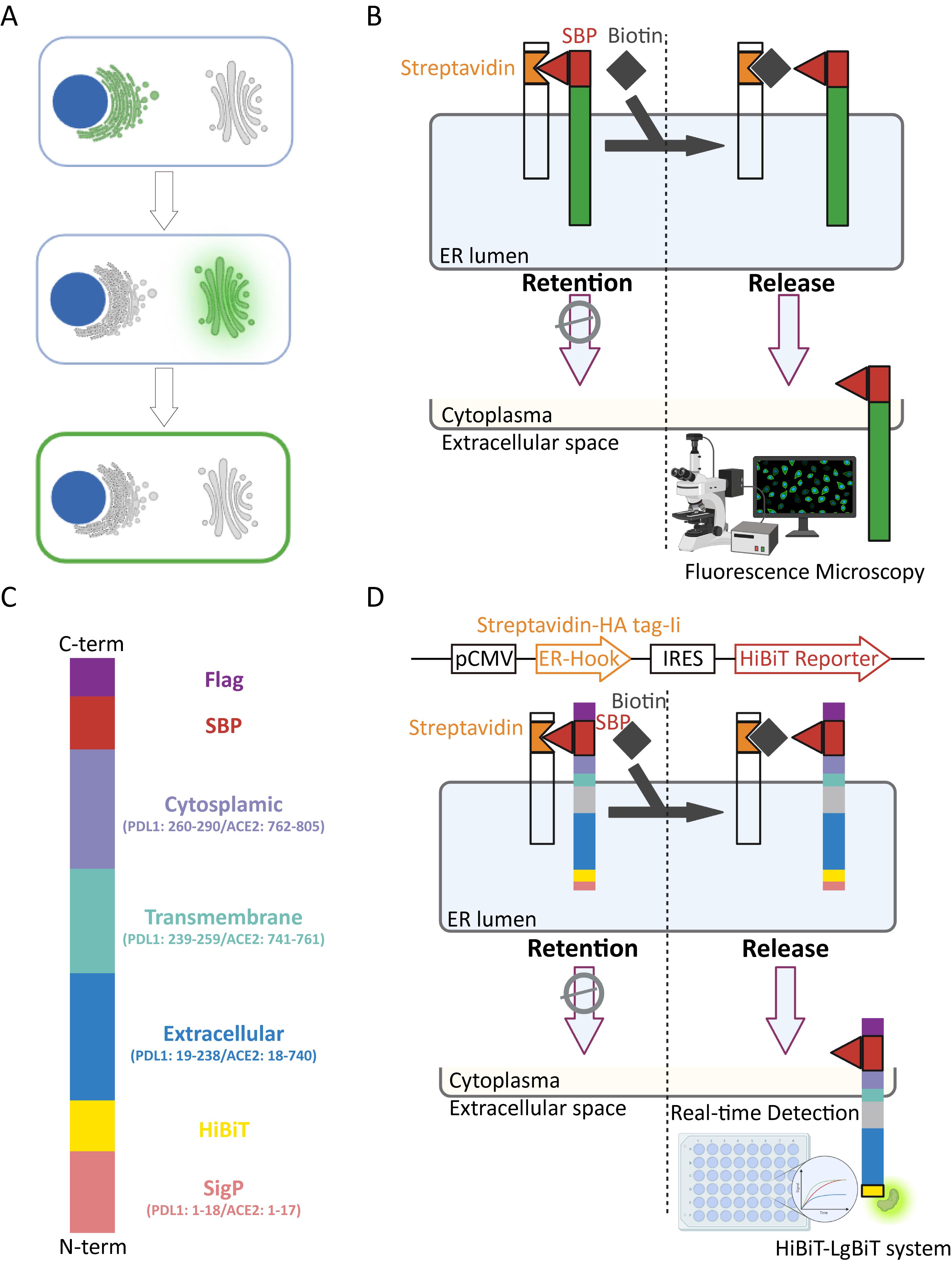
Figure 1. Rationale of HiBiT-integrated RUSH strategy. (A) The conventional RUSH system utilizes fluorescent proteins to monitor the secretion and trafficking of target proteins. (B) Biotin competes with streptavidin-binding peptide (SBP) for binding to streptavidin (Str), triggering the release of the EGFP-tagged reporter. (C) Design of reporters for ACE2 and PDL1. (D) The ER-localized hook and HiBiT reporter were engineered into a single plasmid using an internal ribosome entry site (IRES) element to construct the HiBiT-integrated RUSH plasmid, enabling real-time monitoring of protein secretion in living cells with a plate reader for detection. ACE2, angiotensin-converting enzyme 2; EGFP, enhanced GFP; ER, endoplasmic reticulum; PDL1, programmed death-ligand 1; RUSH, Retention Using Selective Hooks; SBP, streptavidin-binding peptide; Str, streptavidin.
B. Secretion dynamics assay using HiBiT-integrated RUSH strategy
1. Culture 5 × 10⁶ 293T cells in 10-cm culture dishes. The culture medium is DMEM supplemented with 10% FBS and 1% penicillin–streptomycin. Maintain the cells at 37 °C and 5% CO2. All experiments were performed using 293T cells within five passages after thawing.
2. The next day, when the cell confluence reaches 70%–80%, transfect the cells with 5 μg of the HiBiT-integrated RUSH plasmid using 10 μL of jetPRIME transfection reagent.
3. After 24 h, wash the cells with PBS, harvest by trypsinization, and seed into a 96-well white cell culture plate at a density of 20,000 cells per well in 100 μL of medium. Incubate the plate overnight to allow cell adhesion and growth to 40,000–50,000 cells per well.
Caution: We transfected the HiBiT-integrated RUSH plasmid in a 10-cm culture dish in advance, and then evenly seeded the cells into a 96-well plate the next day. This approach ensures that the total reporter expression and baseline signal remain consistent across each well, thereby eliminating the need for additional normalization. The results also demonstrated minimal well-to-well variation among replicate wells. Prewarm the plate reader to 37 °C before the measurement.
Critical: Maintain 37 °C throughout the entire process, as protein secretion is highly temperature-sensitive.
4. Detect the HiBiT signal.
a. Prepare the Nano-Glo HiBiT extracellular detection reagent by mixing LgBiT (1:200 dilution) and furimazine substrate (1:100 dilution) in complete DMEM medium (10% FBS and 1% penicillin-streptomycin) containing 40 μM biotin.
b. For experimental controls, set up two negative control groups: add 20 μM BFA (Biotin+ BFA+) to confirm that the signal is transport-specific, and include a no-biotin group (Biotin-) to measure the background signal.
Caution: Include both the BFA group and the no-biotin control group in every experiment and set up at least three replicate wells for each condition.
c. Replace the medium with the prepared reagent and immediately place the plate into the prewarmed plate reader at 37 °C.
Caution: Phenol red, serum, and penicillin–streptomycin in DMEM do not affect data acquisition or result interpretation.
d. Measure luminescence at 1-min intervals for 120–180 min.
Caution: In our experiments, luminescence was measured using a Tecan Spark multimode microplate reader equipped with a photon counting detector and controlled with Tecan SparkControl software. No gain or sensitivity adjustment was available, and automatic gain was not used. Measurements were performed in luminescence mode with an integration time of 1,000 ms per well. Plates were read from the top, which is the only reading configuration available for luminescence on this instrument, and the plate height (z-position) was detected automatically by the instrument. The settle time was set to 0 ms. No shaking was applied during measurement because the samples were mixed with the HiBiT detection reagent immediately before reading. Assays were performed in 96-well white opaque plates. Researchers should optimize the instrument settings according to their own instrument.
Caution: Achieving a perfect hook is challenging, and HiBiT signal leakage leads to a relatively high initial signal. This signal is unstable due to the LgBiT-HiBiT equilibration time and the temperature shift from the biosafety cabinet to the 37 °C plate reader. We therefore placed the plate directly into the prewarmed reader to capture the highly dynamic secretion process. Using a no-biotin control as the background effectively eliminates the influence of these condition changes.
Caution: Regarding the quantitative endpoints, our own measurements show that the majority of secretion is completed within 60 min, with the signal reaching its maximum. However, we typically measure for 120–180 min to avoid missing potential secretion processes after the peak.
e. Plot the data using GraphPad software and analyze the secretion kinetics.
Note: Refer to Figure 2A for representative results of Str–Ii_HiBiT-ACE2-SBP-FLAG and Figure 2B for representative results of Str–Ii_HiBiT-PDL1-SBP-FLAG.
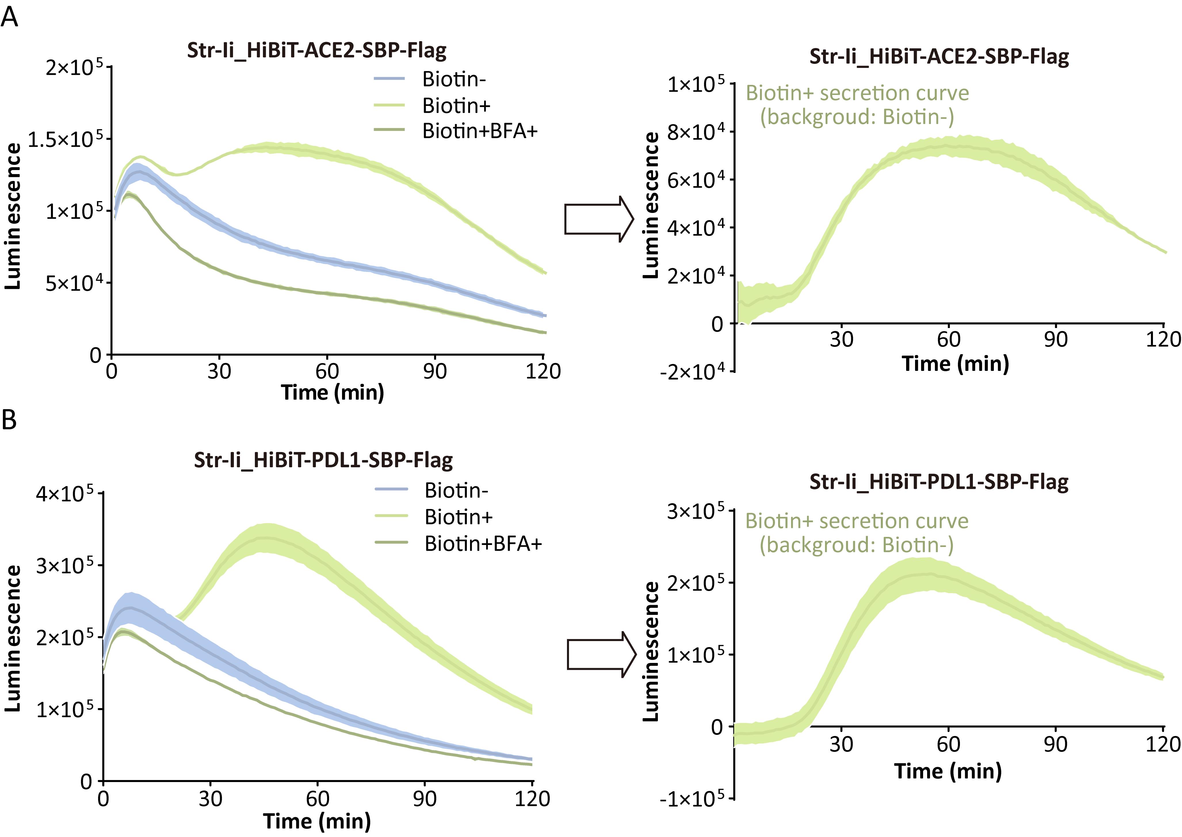
Figure 2. Secretion dynamics assay using the HiBiT-integrated RUSH strategy. (A, B) Nano-Glo HiBiT extracellular detection reagent, containing the furimazine substrate and LgBiT, was added to 293T cells transfected with Str–Ii_HiBiT-ACE2-SBP-FLAG (A) or Str–Ii_HiBiT-PDL1-SBP-FLAG (B), and luminescence was continuously monitored every minute over a 120-min period at 37 °C. Biotin-, treated without biotin; Biotin+, treated with 40 μM biotin; Biotin + BFA+, treated with 40 μM biotin and 20 μM Brefeldin A. n = 3; the shaded region around the curve represents SD. ACE2, angiotensin-converting enzyme 2; BFA, Brefeldin A; PDL1, programmed death-ligand 1; SBP, streptavidin-binding peptide; Str, streptavidin.
C. Demonstration of the general applicability of the HiBiT-integrated RUSH strategy
The HiBiT-integrated RUSH strategy was successfully used to monitor the secretion of two type-I transmembrane proteins, ACE2 and PDL1, as described in the previous sections. To demonstrate the broader applicability of this strategy across different membrane protein topologies, two additional examples are presented in Figure 3: NTCP (a multi-pass transmembrane protein) and CD209 (a type-II transmembrane protein). These examples are intended to serve as references for researchers when designing their own HiBiT-integrated RUSH plasmids. All relevant steps follow the same protocols described in sections A and B.
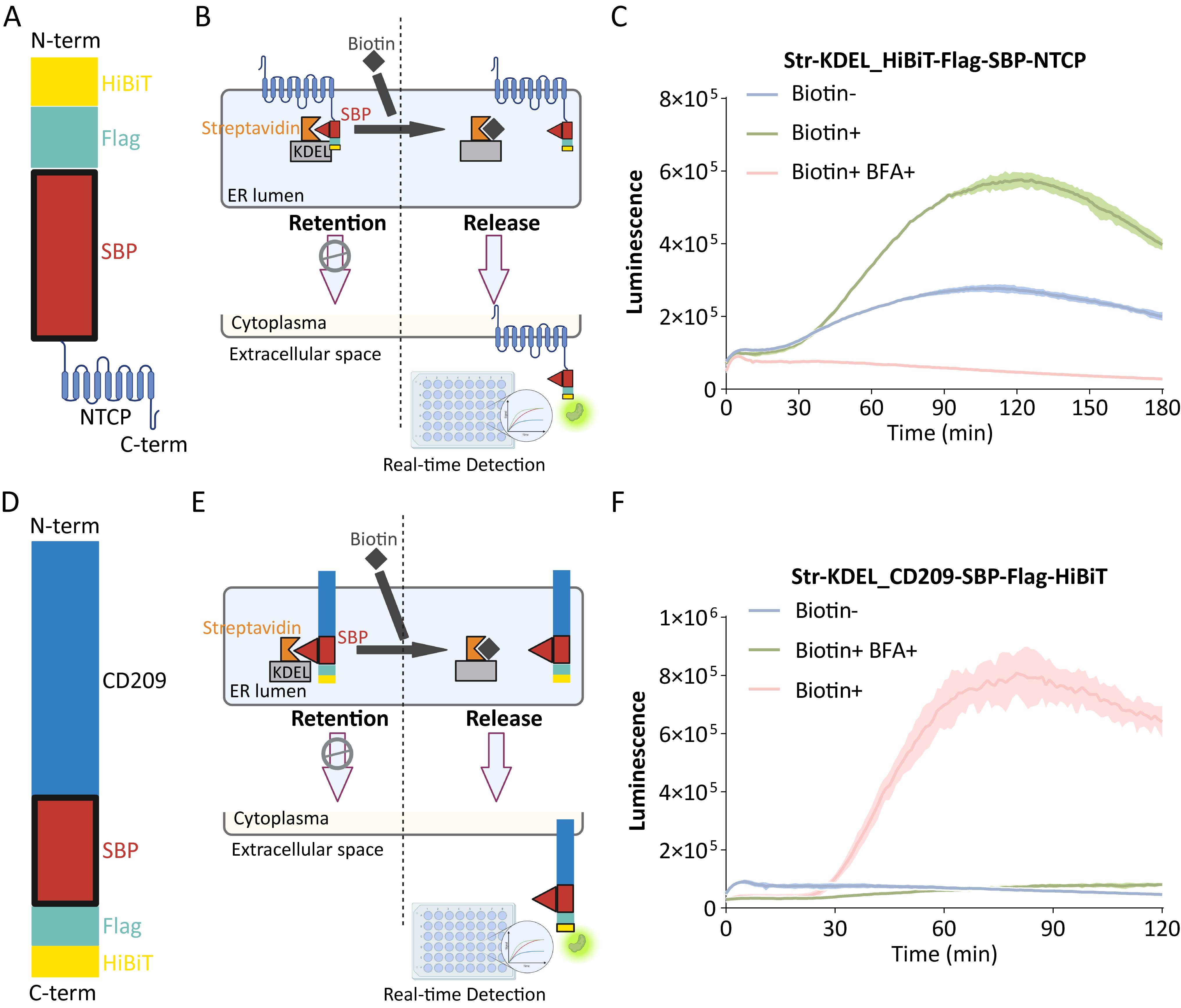
Figure 3. Design and performance of the HiBiT-integrated RUSH strategy applied to NTCP and CD209. (A) Design of the reporter for NTCP. (B) The Str-KDEL_SBP-EGFP-Ecadherin plasmid (Addgene) was used as the backbone for constructing the NTCP reporter. (C) Nano-Glo HiBiT extracellular detection reagent, containing the furimazine substrate and LgBiT, was added to 293T cells transfected with Str–KEDL_HiBiT-FLAG-SBP-NTCP, and luminescence was continuously monitored every minute over a 180-min period at 37 °C. (D) Design of the reporter for CD209. (E) The Str-KDEL_SBP-EGFP-Ecadherin plasmid (Addgene) was used as the backbone for constructing the CD209 reporter. (F) Nano-Glo HiBiT extracellular detection reagent, including the furimazine substrate and LgBiT, was added as per the manufacturer’s instructions to 293T cells transfected with Str–KEDL_CD209-SBP-FLAG-HiBiT, and luminescence was continuously monitored every minute over a 120-min period at 37 °C. Biotin-, treated without biotin; Biotin+, treated with 40 μM biotin; Biotin + BFA+, treated with 40 μM biotin and 20 μM Brefeldin A. n = 3; shaded region around the curve represents SD. NTCP, sodium taurocholate cotransporting polypeptide; CD209, cluster of differentiation 209.
Data analysis
1. Import the data recorded by the instrument into GraphPad. Select an XY table, where the X-axis represents time points starting from 0 min at 1-min intervals, and the Y-axis represents luminescence values.
2. Set up Biotin-, Biotin+, and Biotin+BFA+ groups, with at least three replicate wells per group.
3. Use the Biotin- group as the background. Subtract the paired Biotin- signal from the Biotin+ signal to obtain three values and plot the final secretion curve.
Validation of protocol
This protocol has been used and validated in the following research article:
• Zhang et al. [15]. Real-time monitoring of secretory protein traffic for investigating trafficking regulators of ACE2 in living cells. Journal of Biological Chemistry (Figures 1 and 2).
General notes and troubleshooting
General notes
1. The topology of the reporter must be carefully considered to ensure successful anchoring with the hook, while allowing the HiBiT tag to be exposed to the extracellular space for detection following its release.
2. The signal peptide of the target protein should not be overlooked, as it could lead to the removal of the HiBiT tag during protein processing.
3. Achieving a perfect hook is not strictly necessary. By including a biotin-free control as a background, accurate quantitative curves can be generated without the need to establish stable cell lines or utilize inducible expression systems.
Troubleshooting
Problem 1: Weak HiBiT signal.
Possible cause: Inappropriate temperature during the assay.
Solution: Prewarm the plate reader to 37 °C before starting the experiment.
Problem 2: No secretion-specific signal is detected.
Possible cause: Improper design of the HiBiT-integrated RUSH plasmid, resulting in failed anchoring to the hook or failure of the HiBiT tag to be exposed to the extracellular space.
Solution: Carefully consider the topology of both the hook and the reporter, as well as the signal peptide of the reporter, during the plasmid design phase.
Supplementary information
The following supporting information can be downloaded here:
1. Figure S1. Plasmid map and cloning strategy of Str-Ii_HiBiT-ACE2-SBP-FLAG.
2. Figure S2. Plasmid map and cloning strategy of Str-Ii_HiBiT-PDL1-SBP-FLAG.
3. Figure S3. Plasmid map and cloning strategy of Str-KDEL_HiBiT-FLAG-SBP-NTCP.
4. Figure S4. Plasmid map and cloning strategy of Str-KDEL_CD209-SBP-FLAG-HiBiT.
5. Table S1. Primers used for molecular cloning.
Acknowledgments
This protocol is adapted from Zhang et al. [15], which has been modified from Boncompain et al. [3,12,16].
Some components of the Graphical overview and Figures 1A, B, D and 3A, B, D, E were created and exported with BioRender.com under a paid subscription (Graphical overview: https://app.biorender.com/illustrations/6abdd619b0e09717c679070a?slideId=085320be-525a-491b-9cb5-a680c6c308e8; Figure 1A: https://app.biorender.com/illustrations/670e39a2973c45fe17fd93d8?slideId=c71907f0-adb8-48ac-aad7-2209df8a76d1; Figure 1B, D: https://app.biorender.com/illustrations/67162b847198ca31fd634c63?slideId=e6452dc0-ca56-40df-bca7-d449576bf547; Figure 3A, B, D, E: https://app.biorender.com/illustrations/6a5de85ae08e2678d9959e8d?slideId=e6452dc0-ca56-40df-bca7-d449576bf547).
Author contributions
Conceptualization, C.W. and L.W.; Investigation, M.Y., M.W., M.L., C.Z., Y.A.; Writing—Original Draft, L.W.; Writing—Review & Editing, H.Z., Xin. Y., C.W., and L.W.; Funding acquisition, L.W.; Supervision, R.P. and H.Z.
Competing interests
The authors declare that they have no competing interests.
Ethical considerations
This study did not involve human or animal subjects, and therefore ethical approval was not required.
References
- Viotti, C. (2016). ER to Golgi-Dependent Protein Secretion: The Conventional Pathway. Methods Mol Biol. 1459: 3–29. https://doi.org/10.1007/978-1-4939-3804-9_1
- Starkuviene, V., Seitz, A., Erfle, H. and Pepperkok, R. (2008). Measuring Secretory Membrane Traffic. Methods Mol Biol. 457: 193–201. https://doi.org/10.1007/978-1-59745-261-8_14
- Boncompain, G., Divoux, S., Gareil, N., de Forges, H., Lescure, A., Latreche, L., Mercanti, V., Jollivet, F., Raposo, G., Perez, F., et al. (2012). Synchronization of secretory protein traffic in populations of cells. Nat Methods. 9(5): 493–498. https://doi.org/10.1038/nmeth.1928
- Saraste, J. and Kuismanen, E. (1984). Pre- and post-Golgi vacuoles operate in the transport of semliki forest virus membrane glycoproteins to the cell surface. Cell. 38(2): 535–549. https://doi.org/10.1016/0092-8674(84)90508-7
- Arnheiter, H., Dubois-Dalcq, M. and Lazzarini, R. A. (1984). Direct visualization of protein transport and processing in the living cell by microinjection of specific antibodies. Cell. 39(1): 99–109. https://doi.org/10.1016/0092-8674(84)90195-8
- He, W., Gea-Mallorquí, E., Colin-York, H., Fritzsche, M., Gillespie, G. M., Brackenridge, S., Borrow, P. and McMichael, A. J. (2023). Intracellular trafficking of HLA-E and its regulation. J Exp Med. 220(8): e20221941. https://doi.org/10.1084/jem.20221941
- Wu, H., Wang, L. C., Sow, B. M., Leow, D., Zhu, J., Gallo, K. M., Wilsbach, K., Gupta, R., Ostrow, L. W., Yeo, C. J. J., et al. (2024). TDP43 aggregation at ER-exit sites impairs ER-to-Golgi transport. Nat Commun. 15(1): 9026. https://doi.org/10.1038/s41467-024-52706-7
- Capaci, V., Bascetta, L., Fantuz, M., Beznoussenko, G. V., Sommaggio, R., Cancila, V., Bisso, A., Campaner, E., Mironov, A. A., Wiśniewski, J. R., et al. (2020). Mutant p53 induces Golgi tubulo-vesiculation driving a prometastatic secretome. Nat Commun. 11(1): 3945. https://doi.org/10.1038/s41467-020-17596-5
- Boncompain, G., Herit, F., Tessier, S., Lescure, A., Del Nery, E., Gestraud, P., Staropoli, I., Fukata, Y., Fukata, M., Brelot, A., et al. (2019). Targeting CCR5 trafficking to inhibit HIV-1 infection. Sci Adv. 5(10): eaax0821. https://doi.org/10.1126/sciadv.aax0821
- Dixon, A. S., Schwinn, M. K., Hall, M. P., Zimmerman, K., Otto, P., Lubben, T. H., Butler, B. L., Binkowski, B. F., Machleidt, T., Kirkland, T. A., et al. (2015). NanoLuc Complementation Reporter Optimized for Accurate Measurement of Protein Interactions in Cells. ACS Chem Biol. 11(2): 400–408. https://doi.org/10.1021/acschembio.5b00753
- Nagashima, S., Primadharsini, P. P., Nishiyama, T., Takahashi, M., Murata, K. and Okamoto, H. (2023). Development of a HiBiT-tagged reporter hepatitis E virus and its utility as an antiviral drug screening platform. J Virol. 97(9): e00508–23. https://doi.org/10.1128/jvi.00508-23
- Boncompain, G. and Perez, F. (2012). Synchronizing Protein Transport in the Secretory Pathway. Curr Protoc Cell Biol. 57(1): ecb1519s57. https://doi.org/10.1002/0471143030.cb1519s57
- Chen, J., Viana, M. P. and Rafelski, S. M. (2023). When seeing is not believing: application-appropriate validation matters for quantitative bioimage analysis. Nat Methods. 20(7): 968–970. https://doi.org/10.1038/s41592-023-01881-4
- Denus, M., Filaquier, A., Fargues, W., Néel, E., Stewart, S. E., Colladant, M., Curel, T., Mezghrani, A., Marin, P., Claeysen, S., et al. (2025). A Sensitive and Versatile Cell‐Based Assay Combines Luminescence and Trapping Approaches to Monitor Unconventional Protein Secretion. Traffic. 26: e70009. https://doi.org/10.1111/tra.70009
- Zhang, C., Yang, M., An, Y., Liu, M., Li, H., Zhao, R., Yang, X., Huang, L., Zhang, Y., Peng, R., et al. (2025). Real-time monitoring of secretory protein traffic for investigating trafficking regulators of ACE2 in living cells. J Biol Chem. 301(9): 110593. https://doi.org/10.1016/j.jbc.2025.110593
- Boncompain, G. and Perez, F. (2014). Synchronization of Secretory Cargos Trafficking in Populations of Cells. Methods Mol Biol. 1174: 211–223. https://doi.org/10.1007/978-1-4939-0944-5_14
Article Information
Publication history
Received: Jul 26, 2026
Accepted: Oct 14, 2026
Available online: Oct 27, 2026
Published: Nov 5, 2026
Copyright
© 2026 The Author(s); This is an open access article under the CC BY-NC license (https://creativecommons.org/licenses/by-nc/4.0/).
How to cite
Readers should cite both the Bio-protocol article and the original research article where this protocol was used:
- Yang, M., Wang, M., Liu, M., Zhang, C., Peng, R., An, Y., Zhong, H., Wei, C., Yan, X. and Wan, L. (2026). Monitoring Protein Secretion in Living Cells With a HiBiT-Integrated RUSH System. Bio-protocol 16(21): e5871. DOI: 10.21769/BioProtoc.5871.
- Zhang, C., Yang, M., An, Y., Liu, M., Li, H., Zhao, R., Yang, X., Huang, L., Zhang, Y., Peng, R., et al. (2025). Real-time monitoring of secretory protein traffic for investigating trafficking regulators of ACE2 in living cells. J Biol Chem. 301(9): 110593. https://doi.org/10.1016/j.jbc.2025.110593

 Download Citation in RIS Format
Category
Biological Engineering > Biomedical engineering > Drug Delivery
Cell Biology > Cell-based analysis > Protein secretion
Do you have any questions about this protocol?
Post your question to gather feedback from the community. We will also invite the authors of this article to respond.
 Post a Question


