


- Protocols

- Articles and Issues
- For Authors
- About
- Become a Reviewer


A Protocol for Colorectal Tumor Spheroid Culture in Tunable Stiffness Alginate-Based Hydrogels and Subsequent Immunohistochemical Analysis

(*contributed equally to this work) Published: Vol 16, Iss 15, Aug 5, 2026 DOI: 10.21769/BioProtoc.5786 Views: 258
Reviewed by: Dipak Kumar PoriaAnonymous reviewer(s)
 PDF
PDF  Ask a question

Protocol Collections
Comprehensive collections of detailed, peer-reviewed protocols focusing on specific topics




Abstract
Tumor mechanical microenvironment, particularly extracellular matrix stiffness, plays a critical role in regulating cancer cell behavior, including proliferation, quiescence, and drug resistance. Conventional 2D culture or stiff 3D scaffolds fail to recapitulate the physiological soft (normal) or pathologically stiff (tumoral) mechanical niches. Here, we present a detailed protocol for establishing a tunable 3D tumor spheroid culture system using sodium alginate–based hydrogels crosslinked with calcium ions at different concentrations to achieve soft or stiff conditions that mimic normal colon and colorectal cancer tissues, respectively. We describe the step-by-step procedures for fabricating stiffness-tunable hydrogels, culturing colorectal cancer spheroids, releasing spheroids for downstream analysis, and performing immunohistochemical staining on intact spheroids. This protocol enables the reproducible investigation of mechanosensitive pathways and drug resistance mechanisms in a physiologically relevant 3D context.
Key features
• Tunable stiffness hydrogel system based on sodium alginate and calcium carbonate crosslinking.
• 3D tumor spheroid culture that recapitulates normal and tumor mechanical microenvironments.
• Gentle spheroid release using sodium citrate chelation, preserving morphology and viability.
• Compatible with immunohistochemistry.
Keywords: Tumor spheroid Alginate hydrogel
Matrix stiffness
3D culture
Immunohistochemistry
Alginate hydrogel
Matrix stiffness
3D culture
Immunohistochemistry
Graphical overview
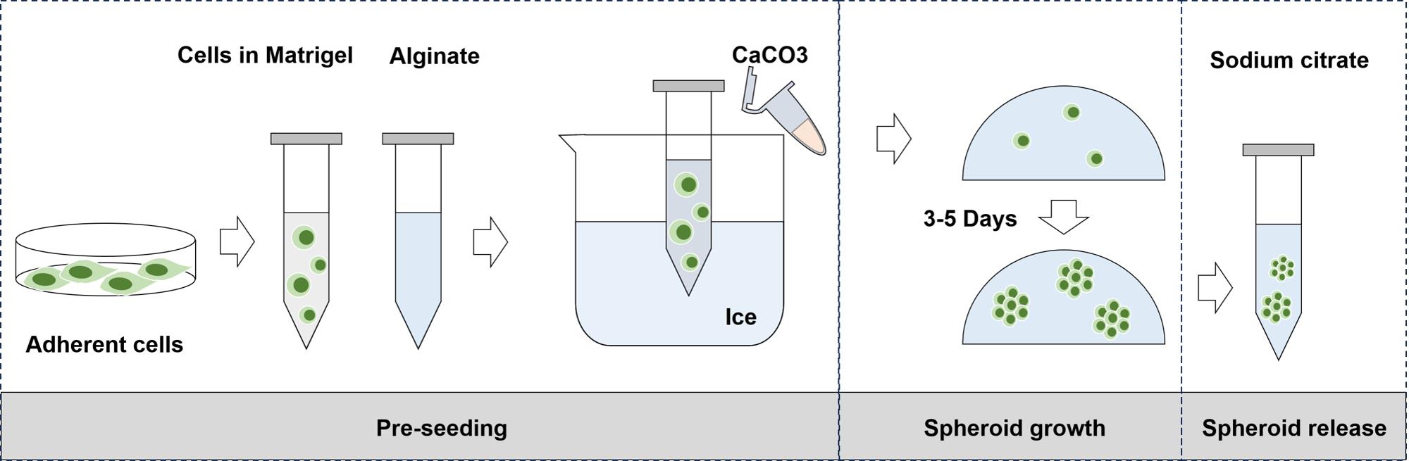
Tumor spheroid culture and release based on sodium alginate hydrogel
Background
Colorectal cancer (CRC) is the third most common cancer worldwide, with metastasis and recurrence remaining the primary causes of mortality [1,2]. The tumor microenvironment (TME) plays a crucial role in inducing poor prognosis in CRC [3]. Among various TME factors, increased matrix stiffness resulting from excessive collagen deposition and crosslinking is a fundamental physical hallmark of solid tumors [4–6]. Normal colon tissue has a stiffness of <0.5 kPa, whereas CRC tissue typically exhibits a stiffness of 1–4 kPa [7]. Elevated stiffness levels are associated with poor prognosis and drug resistance. However, most existing in vitro models fail to recapitulate this stiffness range, often using supraphysiological stiffness that does not reflect in vivo conditions. Moreover, most studies use cells seeded on the surface of hydrogels with different stiffnesses to simulate the impact of this mechanical environment on tumor behavior [8]. Yet, this two-dimensional mechanical cue differs greatly from the true in vivo environment, potentially introducing bias into the conclusions.
Sodium alginate is widely used for three-dimensional cell culture due to its excellent biocompatibility and crosslinkability [9]. To address the gap mentioned above, we employed a tunable alginate-based 3D hydrogel system that independently mimics soft (normal) and stiff (tumor) mechanical niches. Here, we provide a detailed optimized protocol for preparing stiffness-tunable hydrogels, culturing tumor spheroids, releasing them without disrupting their morphology, and performing immunohistochemical (IHC) analysis on paraffin-embedded sections. In this protocol, we use the human colorectal cancer HCT-116 and SW-620 cell lines for tumor spheroid culture and compare, via IHC, the expression of the ANXA2 protein—an annexin closely associated with the extracellular microenvironment—in tumor spheroids grown in hydrogels of different stiffnesses. This protocol offers a robust platform for investigating mechanobiology in a physiologically relevant context.
Materials and reagents
Biological materials
1. Colorectal cancer cell lines HCT-116 (ATCC, catalog number: CCL-247) and SW-620 (ATCC, catalog number: CCL-227)
Reagents
1. Sodium alginate (Sigma, catalog number: A0682)
2. Calcium carbonate (CaCO3) (Aladdin, catalog number: C100633)
3. Growth factor reduced Matrigel (Corning, catalog number: 356231)
4. Dulbecco’s modified Eagle medium (DMEM) (Gibco, catalog number: 11965092)
5. Fetal bovine serum (FBS) (Gibco, catalog number: 10099141)
6. Penicillin-streptomycin (PS), 100× (Gibco, catalog number: 15140122)
7. Phosphate-buffered saline (PBS) (Cytiva, catalog number: SH30028.FS)
8. Sodium citrate (Sigma, catalog number: S1804)
9. Paraformaldehyde (PFA), 16% EM grade (Electron Microscopy Sciences, catalog number: 15710-S)
10. Low-melting-point agarose (Sigma, catalog number: A4018)
11. Ethanol, absolute (Sinopharm, catalog number: 10009218)
12. Xylene (Sinopharm, catalog number: 10023418)
13. Citrate antigen retrieval buffer (Beyotime, catalog number: P0083)
14. Hydrogen peroxide (H2O2), 30% (Sigma, catalog number: H1009)
15. Normal goat serum (Thermo Fisher, catalog number: 16210064)
16. ANXA2 primary antibody (Cell Signaling Technology, catalog number: 8235)
17. HRP-conjugated secondary antibody (Cell Signaling Technology, catalog number: 7074)
18. DAB Substrate kit (Vector Laboratories, catalog number: SK-4100)
19. Hematoxylin (Sigma, catalog number: MHS32-1L)
20. Cytoseal 60 (Fisher Scientific, catalog number: 23-244256)
21. Tween-20 (Sigma, catalog number: P1379)
22. HCl (Bolinda, catalog number: 7647-01-0)
23. Paraffin (CITOTEST, catalog number: 80200-0015)
Solutions
1. Sodium alginate stock solution (see Recipes)
2. CaCO3 suspension (see Recipes)
3. Cell–alginate–Matrigel mixture (see Recipes)
4. Crosslinking CaCO3 volumes for stiffness tuning (see Recipes)
5. Sodium citrate release solution (see Recipes)
6. 4% PFA fixative solution (see Recipes)
7. Low-melting-point agarose for spheroid embedding (see Recipes)
8. Citrate antigen retrieval buffer (see Recipes)
9. IHC blocking buffer (see Recipes)
10. DAB substrate working solution (see Recipes)
11. Graded ethanol series for dehydration (see Recipes)
Recipes
1. Sodium alginate stock solution (2% w/v, 50 mL)
| Reagent | Final concentration | Quantity or volume |
|---|---|---|
| Sodium alginate powder | 2% (w/v) | 1 g |
| Ultrapure water | n/a | to 50 mL |
Note: Dissolve by stirring overnight at 4 °C. Adjust pH to 7.4 with 1 M NaOH. Autoclave or filter sterilize through a 0.22 μm PES membrane. Store at 4 °C for up to 3 months.
2. CaCO3 suspension (0.5 M, 10 mL)
| Reagent | Final concentration | Quantity or volume |
|---|---|---|
| CaCO3 powder | 0.5 M | 0.5 g |
| Ultrapure water | n/a | to 10 mL |
Note: Autoclave the suspension. Vortex vigorously for 1 min immediately before each use to ensure a uniform particle suspension. Do not store for more than one week.
3. Cell–alginate–Matrigel mixture (for 2 mL gel)
| Reagent | Volume per 2 mL gel |
|---|---|
| Cells in DMEM (2.5 × 10⁵ cells/mL) | 500 μL |
| Growth factor reduced Matrigel | 500 μL |
| 2% sodium alginate solution | 1 mL |
4. Crosslinking CaCO3 volumes for stiffness tuning (per 2 mL gel)
| Stiffness | Final Ca2+ concentration | Volume |
|---|---|---|
| Soft | 10 mM | 40 μL |
| Stiff | 20 mM | 80 μL |
5. Sodium citrate release solution (100 mM, 50 mL)
| Reagent | Final concentration | Quantity or volume |
|---|---|---|
| Sodium citrate | 100 mM | 1.47 g |
| 1× PBS | n/a | to 50 mL |
Note: Adjust pH to 7.4. Sterilize by filtration through a 0.22 μm PES membrane. Store at 4 °C for up to one month.
6. 4% PFA fixative solution (100 mL)
| Reagent | Final concentration | Volume |
|---|---|---|
| 16% PFA, EM grade | 4% | 25 mL |
| 1× PBS | n/a | 75 mL |
Note: Prepare fresh or store at 4 °C for up to one week, protected from light.
7. Low-melting-point agarose for spheroid embedding (2%, 50 mL)
| Reagent | Final concentration | Quantity or volume |
|---|---|---|
| Low-melting-point agarose | 2% (w/v) | 1 g |
| 1× PBS | n/a | to 50 mL |
Note: Heat in a microwave or on a hot plate until completely dissolved. Cool and maintain at 40–42 °C in a heat block before use.
8. Citrate antigen retrieval buffer (10 mM, pH 6.0, 1 L)
| Reagent | Final concentration | Quantity or volume |
|---|---|---|
| Sodium citrate tribasic dihydrate | 10 mM | 2.94 g |
| Tween-20 | 0.05% | 500 μL |
| Distilled water | to 1 L |
Note: Adjust pH to 6.0 with 1 M HCl. Store at room temperature for up to 3 months.
9. IHC blocking buffer (for peroxidase and protein blocking, 50 mL)
| Reagent | Final concentration | Volume |
|---|---|---|
| 30% H2O2 | 3% | 5 mL |
| Normal goat serum | 5% | 2.5 mL |
| 1× PBS | to 50 mL |
Note: Prepare fresh. The H2O2 blocks endogenous peroxidase, and the goat serum blocks nonspecific protein binding.
10. DAB substrate working solution (1 mL)
| Reagent | Volume |
|---|---|
| DAB chromogen concentrate | 1 drop (approximately 30 μL) |
| DAB buffer | 1 mL |
Note: Prepare fresh. Mix immediately before use. Protect from light. Discard after 30 min.
11. Graded ethanol series for dehydration (each 100 mL)
| Solution | Volume |
|---|---|
| 70% ethanol | 70 mL absolute ethanol + 30 mL distilled water |
| 80% ethanol | 80 mL absolute ethanol + 20 mL distilled water |
| 95% ethanol | 95 mL absolute ethanol + 5 mL distilled water |
| 100% ethanol | 100 mL absolute ethanol |
Laboratory supplies
1. 50 mL centrifuge tube, sterile (Celltreat, catalog number: 229106)
2. 15 mL centrifuge tube, sterile (Celltreat, catalog number: 229411)
3. 1.5 mL microcentrifuge tube, sterile (Eppendorf, catalog number: 0030120086)
4. 25 mL serological pipette, individually wrapped (Thermo Fisher, catalog number: 170357N)
5. 10 mL serological pipette, individually wrapped (Thermo Fisher, catalog number: 170356N)
6. 5 mL serological pipette, individually wrapped (Thermo Fisher, catalog number: 170355N)
7. 1,000 μL pipette tip, with filter (VWR, catalog number: 89082-350)
8. 200 μL pipette tip, with filter (VWR, catalog number: 89082-366)
9. 20 μL pipette tip, with filter (VWR, catalog number: 89082-338)
10. Wide-bore 200 μL pipette tip (VWR, catalog number: 89082-366, cut with sterile scissors)
11. Low-attachment 35 mm culture dish (NoninBio, catalog number: NBH2035)
12. Low-attachment 24-well plate (NoninBio, catalog number: NBH1124)
13. 0.22 μm PES syringe filter, 33 mm diameter (Millipore, catalog number: SLGP033RB)
14. 10 mL sterile syringe (Fisher Scientific, catalog number: 14-823-435)
15. Sterile cell scraper (Fisher Scientific, catalog number: 08-100-241)
16. Plastic embedding mold for histology (VWR, catalog number: 25608-922)
17. Tissue cassette for paraffin embedding (VWR, catalog number: 18000-134)
18. Superfrost Plus microscope slide (Thermo Fisher, catalog number: 12-550-15)
19. Coverslip, 24 × 60 mm (Fisher Scientific, catalog number: 12-543-5)
20. Microtome blade, disposable, high profile (Leica, catalog number: 819)
21. Pap pen or hydrophobic barrier pen (Vector Labs, catalog number: H-4000)
22. Humidified staining chamber (Thermo Fisher, catalog number: 50-195-9078)
23. Glass staining dish with slide rack (Thermo Fisher, catalog number: 900200)
24. Cytoseal 60 mounting medium (Fisher Scientific, catalog number: 23-244256)
25. Sterile spatula (Fisher Scientific, catalog number: 21-401-10)
26. Forceps, fine tip (Fisher Scientific, catalog number: 08-953G)
Equipment
1. Laminar flow hood (Baker, model: SterilGARD)
2. CO2 incubator (Thermo, model: Forma SteriCycle i160) (for cell culture at 37 °C, 5% CO2)
3. Benchtop centrifuge for 15/50 mL tubes (Thermo, model: Sorvall Legend X1)
4. Microcentrifuge for 1.5 mL tubes (Eppendorf, model: 5425)
5. Horizontal shaker with temperature control (Thermo Fisher, model: 88880022) (for gel dissolution and washes)
6. Heat block, capable of 40–42 °C and 65 °C (VWR, model: 13259-036)
7. Water bath, capable of 37 °C and 40 °C (Thermo Fisher, model: ISOTEMP 215)
8. Pressure cooker or electric pressure steamer for antigen retrieval (Instant Pot, model: Duo Plus) (achieves ~120 °C for 2 min)
9. Microtome (Leica, model: RM2255)
10. Tissue floatation water bath for section mounting (Leica, model: HI1210)
11. Slide drying oven (Thermo Fisher, model: 131481)
12. Light microscope for routine inspection (Olympus, model: CK2)
13. Digital light microscope for IHC imaging (Olympus, model: BX43)
14. Magnetic stirrer with heating function (Thermo Fisher, model: SP131830)
15. Analytical balance (Mettler Toledo, model: ML204T)
16. pH meter (Mettler Toledo, model: FE28)
17. Autoclave (Tuttnauer, model: 3870E)
18. Vacuum filtration system (Millipore, model: Stericup Quick Release)
19. Refrigerator (4 °C) and freezer (-20 and -80 °C) (for reagent storage)
20. Liquid nitrogen dewar for long-term storage (Taylor-Wharton, model: LS750)
Procedure
A. Fabrication of stiffness-tunable alginate hydrogels and tumor spheroid culture
1. Prepare 2% sodium alginate stock (see Recipes). Store at 4 °C.
2. Prepare a 0.5 M CaCO3 suspension freshly (see Recipes). Vortex vigorously for 30 s immediately before use to ensure uniform particle distribution.
3. Harvest cells by trypsinization, count, and resuspend in DMEM at 2.5 × 10⁵ cells/mL.
4. To prepare a final gel volume of 2 mL on ice, prepare cell–alginate–Matrigel mixture (see Recipes). Gently mix the mixture by pipetting up and down 5–6 times using a wide-bore tip.
5. Initiate gelation by adding the appropriate volume of 0.5 M CaCO3 suspension based on the desired stiffness: 40 μL for soft gels (10 mM final Ca2+) or 80 μL for stiff gels (20 mM final Ca2+). After adding the CaCO3 suspension, mix gently but thoroughly.
6. Dispense 40 μL of the mixture into a low-attachment culture dish or plate (Figure 1).
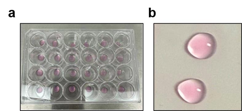
Figure 1. Crosslinking of sodium alginate solution into a hydrogel. (a) The uncrosslinked gel dispersion is shown in a 24-well low-adhesion culture plate. (b) After crosslinking, it becomes a transparent matrix, and the dome-shaped structure facilitates tumor spheroid growth.
7. Induce gelation by placing the dish in a 37 °C incubator for 30 min.
8. Overlay with 2 mL of prewarmed DMEM to promote complete ion chelation. Incubate for 1 h at 37 °C.
9. Replace the overlay DMEM with 2 mL of complete culture medium (DMEM + 10% FBS + 1% PS).
10. Culture for 3–5 days without disturbing the gel. Change medium every 2–3 days. Spheroid formation is typically observed within 3–5 days (Figure 2).
Critical: Keep all alginate–Matrigel–cell mixtures on ice before gelation to prevent premature crosslinking.
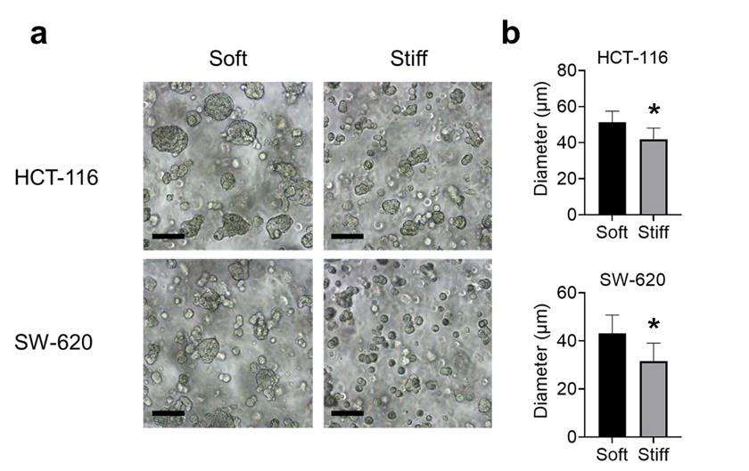
Figure 2. Tumor spheroid cultures in alginate gels with tunable stiffness. (a) HCT-116 and SW-620 spheroids grown in soft or stiff matrix after 5 days of embedded culture. Scale bars, 50 μm. (b) Quantification of tumor spheroid diameters. Data represent mean ± SD of three biological replicates, each consisting of two technical replicates. Statistical significance was determined by Student’s t-test. *p ≤ 0.05.
B. Gentle release of tumor spheroids from Matrigel–alginate hydrogel
Note: Perform all steps at room temperature unless specified.
1. Calculate the volume of sodium citrate required based on a molar ratio of sodium citrate to total CaCO3 of 1.5:1. For a 2 mL gel, the calculation is as follows:
| Gel stiffness | 0.5 M CaCO3 | Total Ca2+ (μmol) | 100 mM sodium citrate (μL) |
| Soft | 40 μL | 20 μmol | 300 μL |
| Stiff | 80 μL | 40 μmol | 600 μL |
2. Transfer the gel into a 15 mL sterile centrifuge tube using a sterile spatula or wide-bore pipette.
3. Add the calculated sodium citrate release solution (see Recipes) and bring the total volume to 10 mL with PBS.
4. Incubate on a horizontal shaker at 50 rpm for 5–10 min until the gel is completely dissolved.
5. Centrifuge at 200× g for 5 min at room temperature.
6. Carefully remove the supernatant, leaving ~100 μL to avoid losing spheroids.
7. Add 10 mL of PBS and gently resuspend by rolling the tube horizontally between the palms. Do not pipette up and down.
8. Centrifuge again at 200× g for 5 min.
9. Fix spheroids in 4% PFA for 30 min at room temperature with gentle agitation.
10. Wash once with PBS and proceed to embedding (section C).
Note: Spheroids are now ready for agarose embedding and IHC.
C. Low-melting-point agarose embedding of tumor spheroids
1. Prepare 2% low-melting-point agarose in PBS. Heat to dissolve completely, then cool and maintain at 40–42 °C in a heat block.
2. Pellet fixed spheroids by centrifugation (200× g, 5 min). Carefully remove supernatant, leaving ~50–100 μL.
3. Resuspend spheroids in 100 μL of PBS using a wide-bore tip.
4. Add 100 μL of prewarmed (40–42 °C) 2% low-melting-point agarose to the spheroid suspension. Mix gently by stirring with the pipette tip; do not pipette up and down to avoid mechanical shearing.
5. Quickly transfer the mixture to a pre-chilled plastic embedding mold.
6. Solidify at 4 °C for 30 min.
7. Extract the agarose block from the mold. Trim excess agarose around the spheroids if needed.
D. Paraffin embedding and sectioning
1. Dehydrate the agarose block through a graded ethanol series as follows: 70% ethanol for 30 min, 80% ethanol for 30 min, 95% ethanol for 30 min, and 100% ethanol for two 30-min changes.
2. Clear with xylene twice for 30 min each time.
3. Embed in paraffin using a standard histology protocol. Orient the block to ensure spheroids are positioned for sectioning.
4. Section at 4 μm thickness using a microtome.
5. Float sections in a 40 °C water bath and mount onto Superfrost Plus slides.
6. Dry slides overnight at 37 °C.
E. IHC staining
1. Deparaffinize slides by incubating them sequentially as follows: xylene for two 10-min changes, 100% ethanol for two 5-min changes, 95% ethanol for 5 min, 80% ethanol for 5 min, 70% ethanol for 5 min, and distilled water for 5 min.
2. Perform antigen retrieval by filling a pressure cooker with citrate antigen retrieval buffer (pH 6.0), immersing the slides in the buffer, heating under pressure for 2 min at 120 °C, and then allowing to cool to room temperature (approximately 20 min).
3. Block endogenous peroxidase: Incubate slides in 3% H2O2 in PBS for 10 min at room temperature, then wash 3 × 5 min with PBS.
4. Block nonspecific binding: Incubate with 5% normal goat serum in PBS for 1 h at room temperature.
5. Incubate with primary antibody: Dilute primary antibody (anti-ANXA2 1:200) in IHC blocking buffer, apply to sections, and incubate overnight at 4 °C in a humidified chamber.
6. Wash 3 × 5 min with PBS.
7. Incubate with HRP-conjugated secondary antibody: Dilute secondary antibody 1:500 in IHC blocking buffer, then incubate sections for 1 h at room temperature.
8. Wash 3 × 5 min with PBS.
9. Develop with DAB: Prepare DAB substrate according to the manufacturer's instructions, apply to sections, monitor under the microscope (typically 1–5 min), and stop the reaction by rinsing with distilled water.
10. Counterstain with hematoxylin: Apply hematoxylin for 30 s to 1 min, then rinse with running tap water for 5 min.
11. Dehydrate and mount: Dehydrate sections through a graded ethanol series: 70% ethanol for 1 minute, 80% ethanol for 1 minute, 95% ethanol for 1 min, 100% ethanol for two 1-min changes, followed by xylene for two 1-min changes. Finally, mount with Cytoseal 60 and a coverslip.
12. Image using a light microscope (Figure 3).
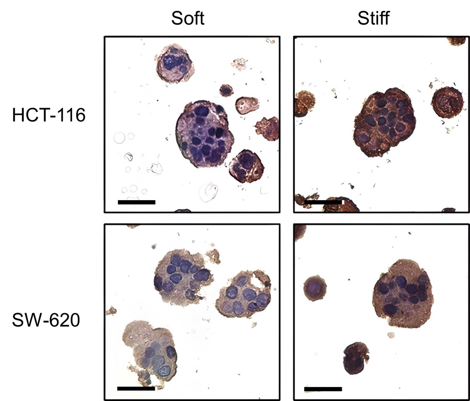
Figure 3. Immunohistochemical (IHC) staining of tumor spheroids after release from the gels. The ANXA2 protein expression was detected in HCT-116 and SW-620 spheroids grown in soft or stiff matrices. Scale bars, 50 μm.
Validation of protocol
The Matrigel–alginate matrix used in this protocol supported the growth of HCT-116 and SW-620 cells under both soft and stiff conditions. After 5 days of culture, tumor spheroids with a diameter of approximately 30–50 μm were obtained (Figure 2). These tumor spheroids were collected and sectioned, exhibiting intact morphology. Moreover, due to growth in matrices with different stiffnesses, these spheroids showed distinct ANXA2 expression levels (Figure 3). These results sufficiently validate this protocol.
This protocol is an optimized version based on a previous calcium alginate gel system and has been validated in the following research article:
Lemarie et al. [10]. Human Induced Pluripotent Spheroids’ Growth Is Driven by Viscoelastic Properties and Macrostructure of 3D Hydrogel Environment. Bioengineering (Basel). https://doi.org/10.3390/bioengineering10121418
General notes and troubleshooting
General notes
1. Always prepare the CaCO3 suspension fresh and vortex immediately before use.
2. Matrigel must be thawed overnight at 4 °C and kept on ice at all times before use. Always use pre-chilled pipette tips when handling Matrigel to avoid premature gelation at room temperature.
3. Keep alginate–Matrigel–cell mixture on ice to prevent premature gelation.
4. Spheroid release using sodium citrate is gentle but must be monitored; over-incubation (>15 min) may cause spheroid dissociation.
5. Low-melting-point agarose must be maintained at 40–42 °C.
6. Due to the small size of the spheroids (approximately 50 μm), to avoid obtaining blank sections during microtomy, we recommend embedding at least 50 spheroids per agarose block and trimming the block to expose the spheroids before collecting ribbons.
7. For rare spheroids, minimize washes and use wide-bore tips throughout.
Troubleshooting
Problem 1: Gel does not form or is too soft.
Possible causes: Insufficient CaCO3 or uneven mixing.
Solutions: Ensure CaCO3 is vortexed thoroughly; increase CaCO3 volume by 10%–20%.
Problem 2: Spheroids do not form or remain as single cells.
Possible causes: Low cell density or insufficient Matrigel.
Solutions: Use at least 2.5 × 10⁵ cells/mL; ensure Matrigel is not frozen and thawed repeatedly.
Problem 3: Spheroids dissociate during release.
Possible cause: Over-incubation in sodium citrate.
Solution: Reduce incubation time to 5–7 min; check every 2 min.
Problem 4: Spheroids are lost during agarose embedding.
Possible causes: Centrifugation too fast or pipetting too vigorous.
Solutions: Use 200× g maximum; always use wide-bore tips; roll the tube instead of pipetting.
Problem 5: Weak or no IHC signal.
Possible causes: Incomplete antigen retrieval or antibody concentration too low.
Solutions: Extend pressure cooker time to 3 min; perform titration for primary antibody.
Problem 6: High background in IHC.
Possible causes: Endogenous peroxidase not fully blocked, or secondary antibody concentration too high.
Solutions: Ensure 3% H2O2 incubation for a full 10 min; reduce secondary antibody dilution to 1:1,000.
Acknowledgments
Conceptualization, J.Y.Y.; Investigation, L.T., W.Q.; Writing—Original Draft, G.Y.T.; Writing—Review & Editing, L.T., J.Y.Y.; Funding acquisition, J.Y.Y.; Supervision, J.Y.Y.
This research was supported by the National Natural Science Foundation of China (Grant No. 82103575, Grant No.82403814), the Natural Science Foundation of Jilin Province (Grant No. YDZJ202501ZYTS045), NSFC regional innovation and development fund (U20A20360), Jilin Province Health Research Talent Special Program (2022SCZ06), the Budding Talent Support Program of China-Japan Union Hospital of Jilin University (2024CL15), and the Jilin Tobacco Industry Co., Ltd Program (KJXM-2024-10).
Competing interests
The authors declare no competing interests.
References
- Biondo, S., Frago, R. and Kreisler, E. (2024). Primary tumor resection for asymptomatic colorectal cancer with unresectable metastasis: the end of the dilemma. Ann Oncol. 35(9): 757–759. https://doi.org/10.1016/j.annonc.2024.06.017
- Siegel, R. L., Giaquinto, A. N. and Jemal, A. (2024). Cancer statistics, 2024. CA Cancer J Clin. 74(1): 12–49. https://doi.org/10.3322/caac.21820
- Zhong, X., He, X., Wang, Y., Hu, Z., Huang, H., Zhao, S., Wei, P. and Li, D. (2022). Warburg effect in colorectal cancer: the emerging roles in tumor microenvironment and therapeutic implications. J Hematol Oncol. 15(1): 160. https://doi.org/10.1186/s13045-022-01358-5
- Li, X., Pan, J., Liu, T., Yin, W., Miao, Q., Zhao, Z., Gao, Y., Zheng, W., Li, H., Deng, R., et al. (2023). Novel TCF21(high) pericyte subpopulation promotes colorectal cancer metastasis by remodelling perivascular matrix. Gut. 72(4): 710-721. https://doi.org/10.1136/gutjnl-2022-327913
- Wang, K., Ning, S., Zhang, S., Jiang, M., Huang, Y., Pei, H., Li, M. and Tan, F. (2025). Extracellular matrix stiffness regulates colorectal cancer progression via HSF4. J Exp Clin Cancer Res. 44(1): 30. https://doi.org/10.1186/s13046-025-03297-8
- Zheng, Y., Zhou, R., Cai, J., Yang, N., Wen, Z., Zhang, Z., Sun, H., Huang, G., Guan, Y., Huang, N., et al. (2023). Matrix Stiffness Triggers Lipid Metabolic Cross-talk between Tumor and Stromal Cells to Mediate Bevacizumab Resistance in Colorectal Cancer Liver Metastases. Cancer Res. 83(21): 3577–3592. https://doi.org/10.1158/0008-5472.CAN-23-0025
- Liu, C., Pei, H. and Tan, F. (2020). Matrix Stiffness and Colorectal Cancer. Onco Targets Ther. 13: 2747–2755. https://doi.org/10.2147/OTT.S231010
- Ye, Y., Jia, Y. and Li, W. (2025). TAGLN: a stiffness-driven prognostic biomarker for metastatic colorectal cancer. Cancer Cell Int. 26(1): 54. https://doi.org/10.1186/s12935-025-04157-3
- Jiang, T., Munguia-Lopez, J., Flores-Torres, S., Grant, J., Vijayakumar, S., De Leon-Rodriguez, A. and Kinsella, J. M. (2018). Bioprintable Alginate/Gelatin Hydrogel 3D In Vitro Model Systems Induce Cell Spheroid Formation. J Vis Exp. (137). https://doi.org/10.3791/57826
- Lemarie, L., Dargar, T., Grosjean, I., Gache, V., Courtial, E. J. and Sohier, J. (2023). Human Induced Pluripotent Spheroids' Growth Is Driven by Viscoelastic Properties and Macrostructure of 3D Hydrogel Environment. Bioengineering (Basel). 10(12). https://doi.org/10.3390/bioengineering10121418
Article Information
Publication history
Received: Apr 18, 2026
Accepted: Jun 28, 2026
Available online: Jul 21, 2026
Published: Aug 5, 2026
Copyright
© 2026 The Author(s); This is an open access article under the CC BY-NC license (https://creativecommons.org/licenses/by-nc/4.0/).
How to cite
Liu, T., Guo, Y., Wang, Q. and Jia, Y. (2026). A Protocol for Colorectal Tumor Spheroid Culture in Tunable Stiffness Alginate-Based Hydrogels and Subsequent Immunohistochemical Analysis. Bio-protocol 16(15): e5786. DOI: 10.21769/BioProtoc.5786.

 Download Citation in RIS Format
Category
Cancer Biology > Microenvironment
Cell Biology > Cell isolation and culture > 3D cell culture
Do you have any questions about this protocol?
Post your question to gather feedback from the community. We will also invite the authors of this article to respond.
 Post a Question


