


- Protocols

- Articles and Issues
- For Authors
- About
- Become a Reviewer


Gene Editing in Chlamydomonas Using the SCREAM Technique

(§Technical contact: i.ross@imb.uq.edu.au) Published: Vol 16, Iss 14, Jul 20, 2026 DOI: 10.21769/BioProtoc.5759 Views: 411
Reviewed by: Diarmuid Seosamh Ó’MaoiléidighAnonymous reviewer(s)
 PDF
PDF  Ask a question
Original research article
The authors used this protocol in:
Aug 2024

Protocol Collections
Comprehensive collections of detailed, peer-reviewed protocols focusing on specific topics




Abstract
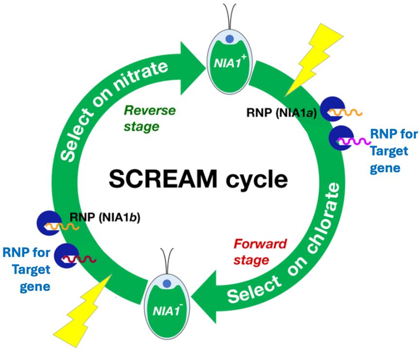
In the model alga Chlamydomonas reinhardtii, CRISPR (clustered regularly interspaced short palindromic repeat)-based gene editing using Cas (CRISPR-associated) enzymes enables both (a) insertion of large gene cassettes and (b) the creation of knockouts based on the introduction of indels, and specific mutations via mutation-directing oligonucleotides. Owing to the relatively low efficiency of this process, selection markers are frequently used to enrich the candidate pool prior to screening, which typically employs PCR. Unfortunately, few selection markers are available for Chlamydomonas. Furthermore, each marker requires different selection media, and deletion of the selectable marker can be difficult. When multiple successive gene editing steps are required, the use of these markers becomes onerous. The SCREAM (sequential CRISPR via recycling endogenous auxotrophic markers) technique employs an endogenous gene as a marker, the mutation of which can be selected both in the forward (loss of function) and reverse (gain of function) directions. During the first gene editing step, crRNA and mutation-directing oligonucleotides are provided for both the marker and the first target gene (Target 1). Candidates with edited marker genes are selected by loss of marker function, prior to screening for the desired modification of the first target gene. Using a successful candidate, a subsequent gene editing step directs reversion of the mutant marker gene to wild-type status, with candidates being selected on auxotrophic media to detect the regain of function of the auxotrophic marker to wild type (i.e., reversion). Simultaneously, a second target gene modification is produced using Target 2–specific crRNA and oligonucleotides. Revertants, now with a wild-type auxotrophic marker, are then screened for the specific mutation of Target 2. This reversion strategy enables a single selectable marker to be reused indefinitely, facilitating the creation of many successive mutations in a single cell line. As the marker can be completely reconstituted, strains can be created in which only the target gene is altered. Employment of homology-directed repair, using single-stranded oligonucleotides for mutation creation, enables the creation of site-directed mutants, tag insertion, and gene knockouts or reversion, rather than the insertion of large gene cassettes. In this implementation, nitrate reductase is used as the endogenous auxotrophic marker, and the adenine phosphoribosyltransferase gene is used as an example of a target gene.
Key features
• A single endogenous selectable marker gene is used for all successive mutational steps, employing only two standardized selection media.
• An indefinite series of gene edits created in a single cell line enables functional analysis of redundant gene families.
• Suitable applications include gene tagging, gene knockout, restoration of mutated genes to wild type, and site-directed mutagenesis to study gene function.
• Widely applicable to existing cell lines.
Keywords: CRISPR Microalgae
Chlamydomonas
SCREAM
Selection markers
NIA1
Nit1
Nitrate reductase
Microalgae
Chlamydomonas
SCREAM
Selection markers
NIA1
Nit1
Nitrate reductase
Graphical overview
Background
The genetic manipulation of Chlamydomonas reinhardtii using CRISPR (clustered regularly interspaced short palindromic repeat) gene editing typically employs Cas (CRISPR-associated) enzymes, such as the widely employed Cas9, together with CRISPR RNAs (crRNA). CRISPR gene editing involves the creation of a double-strand break, either to facilitate disruption of the gene (e.g., via creation of an indel that leads to a frameshift) or to insert a transgene at a desired location to create a new function (e.g., an enzyme for a new metabolic pathway). Indel formation is typically produced via non-homologous end joining (NHEJ) DNA repair mechanisms [1]. However, CRISPR/Cas9 can also be used to create precise alterations to an existing gene element (e.g., promoter, coding region). This is achieved by exploiting homology-directed repair (HDR) by the endogenous polymerase theta [2]. This repair enzyme mediates microhomology-mediated end joining (MMEJ), also known as theta-mediated end joining (TMEJ), with the repair templates typically being small homologous regions on either side of the double-strand break. In CRISPR approaches, exogenously supplied single-stranded oligonucleotides act as templates to engineer the sequence at the repaired site [3,4]. This allows precise gene tagging, site-directed mutagenesis, and promoter engineering. Transgene insertion occurs at a very low frequency (~1 in 105), which requires the use of a marker on the transgene itself. Oligonucleotide-directed HDR in Chlamydomonas is also intrinsically inefficient and highly locus-dependent, typically ~0.1%–1% [5]. Although precise editing efficiencies up to ~10% have been reported for specific genes under optimized conditions [3], HDR remains generally too inefficient and variable to reliably identify edited cells without selection, reflecting a persistent bias toward non-homologous repair pathways. The use of parallel crRNAs against both an unselectable target gene and a selectable marker gene enables the selection of cells in which the marker is modified, i.e., an enrichment of cells that are competent to carry out the CRISPR/Cas9 reaction, even if not all of these will also have the target gene modified. This greatly simplifies the screening task. For a single CRISPR-mediated editing task, there are several markers to choose from, including both auxotrophic (e.g., ARG7) and antibiotic resistance markers (e.g., for paromomycin, hygromycin). Here, a target gene is typically disrupted by insertion of the exogenously supplied marker DNA (with or without additional functional DNA being supplied). But when multiple successive steps are required, the number of available markers is rapidly depleted. Removing a resistance marker at this stage, assuming it is located remote from the target gene, requires back-crossing with extensive validation of the crosses and also introduces a new genetic background. To solve this problem, the sequential CRISPR via recycling endogenous auxotrophic markers (SCREAM) protocol employs an endogenous marker gene that can be selected both for and against disruption, the archetypal example being the nitrate reductase gene NIA1 (previously known as Nit1) which, when functional, enables growth on nitrate (auxotrophic selection), but when knocked out confers resistance to the herbicide chlorate (antibiotic resistance) [5]. NIA1 is used in this protocol to illustrate the method, but other cyclical marker genes could be used instead. For example, mutations in the nucleotide salvage pathway enzyme adenine phosphoribosyltransferase (APRT) confer resistance to 2-fluoroadenine. Some Chlamydomonas cell lines lack a functional NIA1 gene, due to point mutations (which should, in principle, be repairable) or transposable element insertions (which may preclude efficient restoration by this approach). This limits the use of NIA1 as a marker strategy. Alternative selectable markers are therefore under evaluation for use with these cell lines.
SCREAM is a two-stage process in which the marker gene is first mutated, then reverted to the wild-type genotype. In the first stage, the population of NIA1-mutated cells is enriched for cells in which an accompanying target gene (Target 1) has been edited. These can then be screened for the alteration of the target gene (e.g., by PCR, for deletions that change amplicon size, or for the presence of an edited oligonucleotide insertion). Subsequently, Cas9 and HDR can be used to revert the mutated NIA1 gene back to the wild-type state, with auxotrophic selection (survival on nitrate) to identify NIA1 wild-type cells, and thereby enrich for cells in which a second target gene (Target 2) has been edited.
Because the marker is returned to the wild-type state, the marker system is regenerated and ready to use for an indefinite series of future modifications. A standard set of NIA1 gRNAs, which will be the same for each future experiment, is typically used. Target gene sequencing is used to confirm that the desired mutation has been introduced. This method is thus applicable to any gene editing that falls within the scope of oligonucleotide-directed HDR. The example here uses the cw15 (lacking a cell wall) strain CC-1883, which has a wild-type NIA1 gene. Strains with a cell wall are transfected at lower efficiency, but apart from this, it has been shown that they are also amenable to CRISPR modification. The example used here is the introduction of a specific mutation in the APRT gene, which can be used as a positive control by testing for the resulting resistance to 2-fluoroadenine. Notes on adapting this protocol for other target genes and cell lines are provided at the end of the protocol.
Materials and reagents
Biological materials
1. Chlamydomonas reinhardtii strain CC-1883, from The Chlamydomonas Resource Center, St Paul, Minneapolis (www.chlamycollection.org)
Reagents
1. Guide RNAs, supplied by Integrated DNA Technologies; AltRTM (see Table 1)
Table 1. Guide RNAs used in this protocol
| Name | Target gene | Gene ID† | Sequence (5′-3′) |
| NIA1_Disrupt | NIA1 (Nit1) | Cre09.g410950 | CCACCUACUGGACGGGCGUG |
| NIA1_Revert | NIA1 | Cre09.g410950 | CCACCUACUGAUCAGGCGUG* |
| APRT_Ex1 | APRT | Cre17.g704850 | UGCUGUACUGGAACGCCUGG |
| APRT_Ex3 | APRT | Cre17.g704850 | GCACAACGCGUUGCCCGGGC |
| †Phytozome database, phytozome-next.jgi.doe.gov *Bold residues show mutated residues targeted during the reversion cycle | |||
2. Single-stranded oligonucleotides (ssODN) used for HDR; supplied by Integrated DNA Technologies (see Table 2)
Table 2. Single-stranded DNA HDR oligonucleotides used in this protocol
| ssODN name | Gene | Function | Sequence (5´-3´) |
| NIA1_STOP_BclI | NIA1 | Insert STOP codon and insert BclI site | CTGAAGAAGAGCATTGGCTTCAACTGGGGCCCTTGTGCCACCAGCACCACCTACTGATCAGGCGTGCGGCTGCGCGACCTGTTGCAGCACGCCG |
| NIA1_Revert-to-WT | NIA1 | Repair mutated gene | CTGAAGAAGAGCATTGGCTTCAACTGGGGCCCTTGTGCCACGAGCACCACCTACTGGACGGGCGTGCGGCTCCGCGACCT |
| APRT_STOP_BclI | APRT | Insert STOP codon and insert BclI site | G*G*GTATTCTGTTCTGGGATGTCACCACCATCATGCTGAACCACCAGTGAT CAGTACAGCATTGACCTGTTCGCTGAGCAGTACAAGGACAAGAA*G*A |
| *Phosphorothioate linkages | |||
3. PCR primers (Integrated DNA Technologies) (see Table 3)
Table 3. Single-stranded DNA PCR primers used in this protocol
| Name | Gene | Amplicon length | Sequence (5´-3´) |
|---|---|---|---|
| NIA1_F | NIA1 | 547 | CAACAAGCCGTTGACTTTGA |
| NIA1_R | NIA1 | GGCATACATGCACTCACACC | |
| APRT_F | APRT | 920 | ATGGCTGACGTTGAGGC |
| APRT_R | APRT | TGCTGCCGTCTTCACAAA |
4. tracrRNA (20 nmoles) (IDT, catalog number: 1072533)
5. IDT duplex buffer (30 mM HEPES, pH 7.5, 100 mM potassium acetate; catalog number 11-01-03-01); duplex buffer is normally provided with tracrRNA and gRNAs ordered from IDT
6. Cas9 enzyme (IDT Alt-RTM S.p. Cas9 Nuclease V3, 500 µg, catalog number: 1081059)
7. PCR polymerase typically used for amplicons >1 kb (Q5 Hot Start High-Fidelity DNA Polymerase) (New England Biolabs, catalog number: M0493S/L; includes polymerase, 5× reaction buffer, and optional 5× high GC enhancer)
8. PCR polymerase typically used for amplicons <1 kb (Platinum II Hot-Start PCR Master Mix 2×, 200 reactions) (Thermo Fisher, catalog number: 14000013; includes optional 5× high GC enhancer).
9. Proteinase K, ~20 mg/mL (Thermo Fisher, catalog number: EO00491)
10. Hutner’s trace elements (available from the Chlamydomonas Resource Center, www.chlamycollection.org)
11. Potassium chlorate (KClO3) (Merck, catalog number: 255572-100G, CAS: 3811-04-9)
12. Sucrose (Chem-Supply, catalog number: SA030-5KG, CAS: 57-50-1)
13. Urea [CO(NH2)2] (Merck, catalog number: U0631-1KG, CAS: 57-13-6)
14. Dipotassium hydrogen phosphate (K2HPO4) (Sigma, catalog number: P8281-500G, CAS: 7758-11-4)
15. Potassium dihydrogen phosphate (KH2PO4) (Sigma, catalog number: P5655-500G, CAS 7778-77-0)
16. Potassium nitrate (KNO3) (Sigma, catalog number: P8291-500G, CAS: 7757-79-1)
17. Potassium chloride (KCl) (Chem-Supply, catalog number: PA054-500G, CAS: 7447-40-7)
18. Magnesium sulfate heptahydrate (MgSO4·7H2O) (Sigma, catalog number: M2773-1KG, CAS: 10034-99-8)
19. Magnesium chloride hexahydrate (MgCl2·6H2O) (Chem-Supply, catalog number: MA029-500G, CAS: 7791-18-6)
20. Calcium chloride dihydrate (CaCl2·2H2O) (Sigma, catalog number: 223506-500G, CAS: 10035-04-8)
21. Tris(hydroxymethyl)aminomethane (Tris), ultrapure grade (Astral Scientific, catalog number: BIO3094T-5X1KG, CAS: 77-86-1)
22. Glacial acetic acid (CH3COOH) (Merck, catalog number: 1000632511, CAS: 64-19-7)
23. Sodium dodecyl sulfate (SDS) (Sigma, catalog number: L4390-1KG, CAS: 151-21-3)
24. Agar (LabChem, Thermo Fisher, catalog number: AJA863)
25. Corn starch (Coles Cornflour, Coles supermarkets)
26. Hydrochloric acid (HCl) (Sigma-Aldrich, catalog number: 258148, CAS: 7647-01-0)
27. Ethanol (CH3CH2OH) (Chem-Supply, catalog number: EA043, CAS: 64-17-5)
Solutions
1. Phosphate stock for nitrate media (see Recipes)
2. Potassium nitrate 2 M stock (see Recipes)
3. Nitrate medium salts stock (see Recipes)
4. Nitrate medium (NM) pH 7.0 (see Recipes)
5. Nitrate medium Tris with sucrose (NM-ToS) (see Recipes)
6. Urea 2 M stock (see Recipes)
7. Potassium chlorate stock 40× (see Recipes)
8. Duplex buffer (supplied by IDT with gRNAs and tracrRNA) (see Recipes)
9. DNA isolation buffer pH 8.3 (see Recipes)
10. Sterile corn starch stock for spreading on plates (see Recipes)
Recipes
1. Phosphate stock for nitrate media
| Reagent | Final concentration | Quantity or volume |
|---|---|---|
| K2HPO4 | 1.653 M | 28.8 g |
| KH2PO4 | 1.05 M | 14.4 g |
| MilliQ water | To 100 mL | |
| Total phosphate | 2.711 M |
Autoclave.
2. Potassium nitrate 2 M stock
| Reagent | Final concentration | Quantity or volume |
|---|---|---|
| KNO3 | 2.0 M | 20.22 g |
| MilliQ water | n/a | To 100 mL |
Autoclave.
3. Nitrate medium salts stock
| Reagent | Final concentration | Quantity or volume |
|---|---|---|
| MgSO4·7H2O | 16.22 mM | 4.0 g |
| CaCl2·2H2O | 13.6 g/L | 2.0 g |
| MilliQ water | n/a | To 1 L |
Autoclave.
4. Nitrate medium (NM) pH 7.0
| Reagent | Final concentration | Quantity or volume |
|---|---|---|
| Tris-HCl | 20 mM | 2.42 g |
| Nitrate medium salts (Recipe 3) | 13.6 g/L | 25 mL |
| Potassium nitrate 2 M stock (Recipe 2) | 5 mM | 2.5 mL |
| Hutner’s trace elements | n/a | 1.0 mL |
| Glacial acetic acid | Total acetate 17.5 mM | 1.0 mL |
| MilliQ water | n/a | to 1 L |
Autoclave.
5. Nitrate medium Tris with sucrose (NM-ToS)
| Reagent | Final concentration | Quantity or volume |
|---|---|---|
| Sucrose | 40 mM | 1.369 g |
| Nitrate medium | n/a | To 100 mL |
Filter sterilize, do not autoclave.
6. Urea 2 M stock
| Reagent | Final concentration | Quantity or volume |
|---|---|---|
| CO(NH2)2 | 2.0 M | 17.0 g |
| MilliQ water | n/a | To 100 mL |
Filter sterilize, do not autoclave.
7. Potassium chlorate stock 40×
| Reagent | Final concentration | Quantity or volume |
|---|---|---|
| KClO3 | 0.4 M | 4.902 g |
| MilliQ water | n/a | To 100 mL |
Filter sterilize, do not autoclave.
8. Duplex buffer (supplied by IDT with gRNAs and tracrRNA)
| Reagent | Final concentration | Quantity or volume |
|---|---|---|
| HEPES pH 7.5 | 30 mM | 0.7149 g |
| NaOH 1 M stock | As required to bring pH to 7.5 | |
| Potassium acetate | 100 mM | 0.9815 g |
| MilliQ water | n/a | To 100 mL |
9. DNA isolation buffer pH 8.3
| Reagent | Final concentration | Quantity or volume |
|---|---|---|
| Tris-HCl pH 8.3 1 M stock | 50 mM | 1.21 g |
| KCl 1 M stock | 50 mM | 5 mL |
| MgCl2·6H2O 0.1 M stock | 2.5 mM | 2.5 mL |
| SDS 20% stock | 0.5% | 2.5 mL |
| HCl 32% | n/a | As required to bring pH to 8.3 |
| MilliQ water | n/a | To 100 mL |
10. Sterile corn starch stock for spreading on plates
| Reagent | Final concentration | Quantity or volume |
|---|---|---|
| Corn starch | 30% w/v | 15 g |
| Ethanol | n/a | To 50 mL |
Ethanol sterilized, do not autoclave.
Laboratory supplies
1. 1.5 mL safe-lock tubes (Fisher Scientific, Eppendorf, catalog number: 15625367)
2. Screw cap tube, 15 mL (Merck, catalog number: CLS430791)
3. Screw cap tube, 50 mL (Merck, catalog number: CLS430829)
4. No. 10 scalpel blade, 100 (LabCo, catalog number: BI0161-01)
5. Microscope slides (Menzel-Glaser, catalog number: EPBRSF21201)
6. 250 mL narrow-mouth Erlenmeyer flask (Merck, Corning, catalog number: Z232947)
7. 0.1 cm gap Gene Pulser electroporation cuvettes (Bio-Rad, catalog number: 1652089)
8. 200 μL filter tips, sterile, RNase/DNase free (LABCON, catalog number: 1059-965-008)
9. 10 μL filter tips, sterile, RNase/DNase free (LABCON, catalog number: 1036-260-000)
10. 100 mm × 100 mm × 20 mm square plates, sterile (Sarstedt Australia, catalog number: 82.9923.422)
11. Benchtop cooler (ThermoFisher, catalog number: 5115-0032)
12. Polypropylene micro tube rack, 96 well (LabCo Scientific, catalog number: 650.100.260)
13. Test tube racks for 15 and 50 mL tubes (Thermo Fisher, catalog number: 99019)
14. 1.8 mL cryotubes (Merck, catalog number: CLS430488)
15. 5 mL serological pipettes (Merck, catalog number: CLS4487)
16. 10 mL serological pipettes (Merck, catalog number: CLS4488)
17. 25 mL serological pipettes (Merck, catalog number: CLS4489)
18. PCR tubes 0.2 mL (Axygen Inc., catalog number: PCR-02-C)
19. PCR tubes 0.2 mL SnapStrip II (Scientific Specialities Inc., catalog number: 324500)
20. 200 μL tips (Greiner, catalog number: 775350)
21. 1000 μL tips (Axygen, catalog number: T-1000-B)
22. 90 mm sterile plates (Sarstedt Australia, catalog number: 82.1473.001)
23. Inoculation loop (Sarstedt Australia, catalog number: 6.1562.050)
24. Reagent reservoirs (LabCo, catalog number: 650.500.152)
25. 96-well deep-well plate (Merck, catalog number: CLS3596)
26. PCR microplate (Bio-Rad, catalog number: MLP9601)
27. Microplate sealing film, Rayon (Fisher Scientific, Axygen, catalog number: 11326254)
28. Syringe filter 0.22 micron (sterile) (Merck Millipore, catalog number: SLGP033RS)
29. Parafilm (Merck, catalog number: HS234526B)
Equipment
1. Mline 8-channel pipettor 30–300 μL (Sartorius, catalog number: 725140)
2. Single channel pipettors 0–10 μL, 10–100 μL, 100–1,000 μL (Eppendorf, catalog number: 3123000900)
3. S1 pipette filler (Thermo Fisher, catalog number: 9511)
4. Benchtop centrifuge 5810 R (Eppendorf, catalog number: 2230000080)
5. Pico 17 microcentrifuge (Thermo Fisher Scientific, catalog number: 75002410)
6. Bio-Rad T100 thermal cycler (Bio-Rad, catalog number: 1861096)
7. BR-2000 vortexer (Bio-Rad, catalog number: 1660611)
8. Hotblock (Major Science Elite Dry Bath incubator EL02, catalog number: EL-02-110/220)
9. Light meter (Walz, model: ULM-500)
10. C10 Benchtop Platform Shaker (New Brunswick Scientific, catalog number: M1245-0001)
11. Gene Pulser XCell electroporation apparatus, comprising main unit, capacitance extender module, pulse controller module, and ShockPod cuvette chamber (Bio-Rad, catalog number: 1652660)
12. ChemiDoc MP imaging system (Bio-Rad, catalog number: 1708280)
13. Mini-sub Cell GT electrophoresis tank (Bio-Rad, catalog number: 1704466)
14. Electrophoresis PowerPac HC (Bio-Rad, catalog number: 164-5052)
15. Counting chamber Neubauer pattern (Merck, catalog number: BR718605)
16. Horizontal laminar flow cabinet (Gelaire, catalog number: HLAF-1800; Email Westinghouse AirPure Laminar Flow Cabinet, catalog/model number: 1687-1400)
17. Nanodrop 2000c (Thermo Fisher, catalog number: ND-2000C)
18. Inverted microscope (Nikon Ts2) equipped with Kopa camera and software
Software and datasets
1. CRISPR RGEN tools: http://www.rgenome.net “Cas-OFFinder: A fast and versatile algorithm that searches for potential off-target sites of Cas9 RNA-guided endonucleases” [6] and “Microhomology-based choice of Cas9 nuclease target sites” [7], free
2. Snapgene v8.2.1 (Dotmatics, www.snapgene.com), requires license
3. Benchling (Biology software), 2024, https://www.benchling.com/, free alternative to Snapgene
4. Fiji (ImageJ; National Institutes of Health. https://imagej.net/software/fiji/downloads), free
5. Image Lab Software, Bio-Rad (Version 4.1) (SOFT-LIT-170-9690-ILSPC-V-4-1), free (current version requires a license)
6. Microsoft Excel (Microsoft.com), requires a license but is widely available. Free alternative: Apache OpenOffice https://openoffice.apache.org
Procedure
This protocol describes the use of NIA1 as a marker to knock out a single target gene (APRT) by using two APRT gRNAs to create a deletion. Additional strategies for target gene modification, including site-directed mutagenesis, oligonucleotide insertion (e.g., protein tagging), multiplexing, and efficient pool screening, are provided in the General notes section. The medium used here is based on the traditional tris acetate phosphate (TAP) recipe of Gorman and Levine [8], which contains Hutner’s trace elements. An alternative formulation with defined mineral salts is provided by Kropat et al. [9], which has been increasingly adopted in recent years.
A. Preparation of CC-1883 cells
1. Inoculate a 250 mL glass Erlenmeyer flask of nitrate medium (NM; see Recipe 5) with a small loop of stock plate culture of CC-1883 about a week before the experiment.
2. Grow the culture at 20–30 rpm under 100–200 μE/m2/s light (light source details above) at room temperature (22–27 °C). The culture is grown in NM to ensure the NIA1 (Nit1) gene is induced. Monitor the cell density and split the culture if necessary to maintain at ~1–2 million cells/mL.
3. Cells should be maintained in logarithmic growth phase with viability greater than 99%.
4. Grow between 2–3 million cells/mL so the culture is not light-limited. Cultures should be maintained above 1 × 10⁶ cells/mL, as lower densities complicate quantitative harvesting due to adhesion to centrifuge tube walls.
5. Cell synchronization is not routinely employed. Synchronization has been reported to improve efficiency [10]; however, reported efficiency gains are relatively modest and introduce additional experimental complexity.
6. As 5 million cells are needed per electroporation (~2 mL of culture), grow sufficient culture to cover this requirement. A large batch of culture is not needed; 30–50 mL is typically used unless many reactions are required.
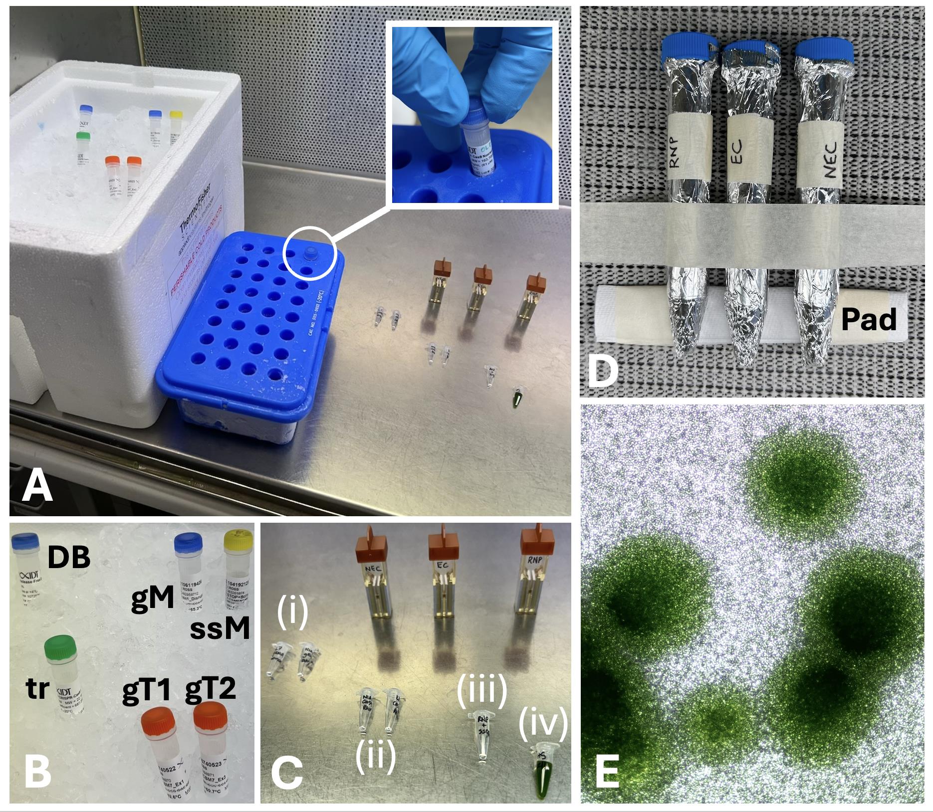
Figure 1. Layout of a minimal experiment with a single target gene. (A) CRISPR reagents in ice box, Cas9 enzyme in cooler (see also inset), with gRNAs, reagent premixes, heat-shocked cells (each in PCR tubes), and electroporation cuvettes ready for controls (NEC, non-electroporated control; EC, electroporated control) and the Cas9 ribonuclear protein (RNP) reaction. (B) Reagents include duplex buffer (DB), marker gRNA (gM), single-stranded oligonucleotide for the marker HDR (ssM), tracerRNA (tr), and two guide RNAs for the target gene (gT1, gT2) designed to create a deletion mutant. (C) (i) Annealed tracrRNA-gRNA for the marker (NIA1 gRNA) and the target gene (gRNAs 1 and 2), (ii) annealed gRNAs with buffer and Cas9 added to form RNPs, (iii) ssODN aliquoted, ready for mixing with Cas9 RNPs, and (iv) heat-shocked cells ready for addition to electroporation cuvettes. (D) After electroporation, cells are transferred to 10 mL of recovery medium in 15 mL Falcon tubes, wrapped in foil to protect from light, and laid on a shaking platform with a paper pad to hold the tube level, for effective mixing. (E) Photomicrograph of Chlamydomonas colonies in starch layer on chlorate selection plates. The starch layer maintains hydration, enhancing the survival of single-cell-wall-deficient mutant cells prior to their expansion into colonies [12].
B. Preparation of annealed guide RNA stocks (day 1)
1. In this section, equimolar amounts of guide RNA and tracrRNA are annealed.
2. While maintaining sterile conditions in a laminar flow cabinet at all times, aliquot 2 μL of NIA1_Disrupt gRNA stock (200 µM in duplex buffer) into a sterile 0.2 mL PCR tube.
3. Aliquot 1 μL each of the target gene (e.g., APRT_Ex1 and APRT_Ex3) gRNA stocks (200 µM in duplex buffer) into a second PCR tube.
4. Add 2 μL of the tracrRNA stock (200 µM) to each tube.
5. Close the tubes and heat to 95 °C for 5 min in a thermal cycler, ramping down to 25 °C at 0.1 °C/s. Allow to cool to room temperature for 5 min.
Critical: Set the hot lid of the thermal cycler to 100 °C to prevent condensation of the liquid under the lid and subsequent loss from the tube. Inspect after the reaction has cooled to ensure that the reaction mix has not dried out. As an alternative, use a 95 °C water bath with the tube sealed with Parafilm and immersed, which provides even heating, or a hot block with water in the well of the block and the tube sealed with Parafilm and taped down so that it is fully immersed in the water.
6. Centrifuge the tubes briefly to bring any condensed liquid on the walls or lid back to the bottom of the tubes.
7. These tubes are designated as the “annealed gRNA stocks.” Excess stock can be stored at -20 °C for future reactions.
C. Preparation of Cas9-gRNA binding reactions (RNPs) (day 1)
Note: In this section, the different gRNAs are mixed with Cas9 separately before being combined in the reaction mix. This was designed to ensure that the Cas9 binds quantitatively to each gRNA in case some gRNAs bind preferentially to Cas9. This separate Cas9 binding step may not be essential; however, empirical validation has not been performed.
1. Set up an ice bath containing gRNAs, oligonucleotide stocks, and IDT duplex buffer (Figure 1A–B).
2. Use a -20 °C benchtop cooler to maintain Cas9 stock at -20 °C.
3. Prepare a new sterile 0.2 mL PCR tube for the Cas9 binding reaction (Figure 1C).
4. Transfer an aliquot of IDT duplex buffer (1.76 μL) to the tube.
5. Add 0.4 μL of annealed NIA1_Disrupt gRNA stock to the tube (the “NIA1” reaction).
6. Add 0.34 μL of IDT Cas9 enzyme stock to the NIA1 reaction tube for a final volume of 2.5 μL and leave at room temperature for 20 min to allow binding. Then, if not ready for immediate use, transfer to an ice bath until electroporation is carried out. The use of ice may be conservative, as the vendor suggests that Cas9 RNPs are stable at room temperature for up to three days; however, the stability of RNPs has not yet been tested with this protocol.
7. Repeat for the target gene gRNAs (APRT_Ex1 and APRT_Ex3) to prepare the target gene–Cas9 binding mix. If two gRNAs are being used for the target gene, as in this example, 0.2 μL of each is typically used. These reactions are referred to as the “Cas9 RNPs.” As there is a 2-fold molar excess of RNA over Cas9, the precision of the RNA pipetting is not critical, but the pipettor should be designed to pipette sub-microliter amounts, and the tips should be carefully observed during the procedure to check that the amount delivered is approximately correct.
Notes:
1. According to IDT, Cas9 RNPs are quite stable and can be prepared well before the cells are ready, with no lack of activity. However, to date, we have timed the procedure so that the 20-min Cas9 binding step concludes just as the 20-min heat shock procedure finishes. This requires sections C and D to be conducted more or less concurrently. Depending on the number of electroporation experiments to be conducted, the amount of premix solution can be scaled.
2. An alternative strategy is to edit the target gene by HDR. In this case, a single gRNA (e.g., APRT_Ex1) is combined with an HDR-directing ssODN (e.g., APRT_STOP_BclI; Table 2). In this case, in Table 4, the “APRT tube” would be composed of 0.51 μL of IDT duplex buffer, 0.4 μL of the single gRNA (pre-hybridized with tracrRNA), and 1.25 μL of the stock ssODN (made in IDT duplex buffer), yielding the same final volume of 2.5 μL, and a similar final composition to the NIA1 Tube.
Table 4. Formation of Cas9 ribonucleoprotein (RNP) complexes
| Reagent | NIA1 tube | APRT tube | Final concentration | |
| IDT duplex buffer | 1.76 μL | 1.76 μL | 0.7× | |
| RNP mix (100 μM; section B) | 0.4 μL | 0.4 μL* | 16 μM | |
| Cas9 stock (62 μM) | 0.34 μL | 0.34 μL | 8.4 μM | |
| Total volume: | 2.5 μL | 2.5 μL | ||
| *comprises 0.2 μL each of APRT_Ex1 and APRT_Ex3; see Table 1 | ||||
D. Harvesting and heat shock of CC-1883 cells (day 1)
Note: Counting the cells prior to concentrating them is important for consistency. Including a heat shock step has been shown to improve the rate of oligonucleotide-mediated homology-directed repair.
1. Sterile culture conditions must be maintained throughout while handling cells.
2. Time the procedure so that the heat shock step concludes within 30 min of the cells being used for electroporation.
3. Remove a drop of culture and, using a hemocytometer, estimate the cell density of the culture. Then, transfer 30–50 mL of culture to a weighed 50 mL Falcon tube.
4. Centrifuge at 800× g for 10 min at room temperature (22–25 °C) to pellet the cells.
5. Remove all but ~0.5 mL of the supernatant using a cell culture pipettor, then briefly re-centrifuge (800× g for 1 min) to bring residual liquid to the bottom of the tube.
6. Remove the remaining liquid from the pellet using a sterile 1 mL pipettor. Estimate the volume of the cell pellet by re-weighing the pre-weighed tube, or by visual comparison to a tube containing known volumes.
7. Taking the pellet volume into account, add sufficient NM-ToS (see Recipe 5) to create a cell suspension at 250 million cells/mL. Using a sterile pipettor, mix gently but thoroughly so there are no lumps.
8. Transfer 200 μL of the cell suspension into a sterile 0.2 mL PCR tube for the heat shock step. If more cells are required, use more 0.2 mL tubes.
9. Using a thermal cycler, heat shock the cell suspension at 42 °C for 20 min. Then, remove the cell suspension and return to room temperature in a sterile laminar flow hood.
E. Electroporation of cells with Cas9-binding mixes and HDR oligonucleotides (day 1)
Note: Electroporation transfers the RNPs into the cell and involves a trade-off between efficiency and cell destruction. Under the conditions described below, electroporation yields acceptable transformation efficiency with minimal cell damage to the fragile CC-1883 cells. However, electroporation conditions should be optimized for each cell line. For example, for the 21gr wild-type cell line CC-1690 (which has wild-type cell walls), we have employed the conditions described by Kelterborn et al. [11] with good results. An electroporation mixture is made for each set of gRNAs to be used.
1. Set aside three sterile 1 mm gap electroporation cuvettes in a sterile laminar flow hood (Figure 1C). The first will contain 20 μL of heat-shocked cells and 5 μL of duplex buffer, but will not be electroporated (non-electroporated control, NEC). The second will contain 20 μL of cells and a 5 μL dummy reaction mixture and will be electroporated (electroporated control, EC). The third will contain 20 μL of cells and 5 μL of the actual RNP complex (RNP reaction).
2. The final RNP electroporation mixture is made in a separate 0.2 mL PCR tube under sterile conditions (Table 5). In this protocol example, a single target gene is employed, requiring only one tube. Start by combining in order: (i) 1.25 μL of NIA1 RNP (i.e., annealed NIA1_Disrupt gRNA–Cas9 reaction mix), (ii) 2.5 μL of the APRT RNP, and (iii) 1.25 μL of NIA1_STOP_BclI single-stranded HDR oligonucleotide (“ssODN”; 400 μM stock) for a final volume of 5 μL. Mix gently and store on ice. This is designated as the “RNP electroporation mix.” Although the APRT (target gene) RNP load is twice that of the NIA1 RNP load, sufficient NIA1 gRNA is present to generate more colonies than can be readily screened.
Note: A 0–2 mL pipettor is used for dispensing the small volumes required. Duplicate or triplicate reactions can be employed if a quantitative estimate is to be made of the efficiency of the reaction. However, this is unnecessary if the aim is to create mutants, since each electroporation reaction will generate a large number of independent mutants. If the target gene employs a single gRNA and an ssODN instead of dual gRNAs as described here, the mix is adjusted as for the NIA1 RNP mix.
Table 5. Preparation of electroporation mix
| Reagent | Volume |
| APRT RNP mix* | 1.25 μL |
| NIA1_Disrupt RNP mix | 1.25 μL |
| NIA1_STOP_BclI ssODN (400 μM) | 2.5 μL |
| Total volume | 5.0 μL |
| *Add NIA1 mix and ssODN directly to the APRT tube to maximize the addition of the target gene gRNAs | |
3. Using long fine pipette tips and a 20 μL pipettor, add 20 μL of cells (5 million cells) to the NEC cuvette, followed by 5 μL of NM-ToS buffer (for routine reactions) or dummy electroporation mix (for quantitative experiments). Mix gently but thoroughly, ensuring the mixture is well distributed in the well of the cuvette and on the electrodes and avoiding bubble formation.
Critical: The sucrose in the NM-ToS protects the cells from lysis [12] and is consequently important during the actual electroporation step. However, it is not required for the recovery medium.
4. Once mixed, transfer the contents to a 15 mL Falcon tube containing 10 mL of recovery medium (NM+U, i.e., nitrate medium + 2 mM urea). Wrap in aluminum foil and place on a flask shaker platform (20–30 rpm) with a paper pad taped under the bottom of the tube so that the liquid level is horizontal (Figure 1D). This ensures good mixing of the cell suspension.
Note: The presence of urea in the recovery medium ensures that cells that have undergone NIA1 gene editing still have a source of nitrogen, which does not inhibit the expression of the NIA1 locus (as ammonium would do).
5. Prepare the EC cuvette in the same way. For the RNP cuvette, add 20 μL of heat-shocked cells directly to the PCR tube containing the 5 μL of RNP plus ssODN reagents (RNP electroporation mix) to maximize the RNP transfer to the cell aliquot, and then transfer the whole mixture to the RNP cuvette. We employ a Bio-Rad Gene Pulser equipped with a capacitance extender and an exponential decay function. For CC-1883, set the voltage to 220 V, the capacitance to 25 μF, and the resistance to “infinite resistance.”
Note: Cell lines are typically optimized using a plasmid encoding AphVIII (which confers resistance to paromomycin), since the optimum conditions for plasmid transfection seem to compare well with optimal conditions for RNP transfection.
5. Immediately after electroporation of each cuvette, transfer the contents of the cuvette to a 15 mL Falcon tube containing 10 mL of NM+U recovery medium. Rinse the cuvette at least twice with the recovery medium from the Falcon tube to ensure good recovery of the cells. Wrap the tubes with aluminum foil and place on a shaker as for the NEC tube.
Note: The cells in recovery medium are placed in the dark for at least 1 h, and then at low light (~20–50 μE/m2/s). This provides an initial dark time for the cells to repair membranes damaged during electroporation (the presence of acetate in the medium provides a carbon source and mitigates the loss of light). The low light conditions inhibit cell division, which would result in dilution of the RNP and oligonucleotides.
6. Leave the recovery tubes shaking at least overnight. This gives time for the gene editing reaction to proceed. For quantitative experiments, cells that are modified by Cas9 will eventually divide, giving rise to some clonal colonies. Therefore, for quantitative experiments, we plate out the next day. But for routine generation of gene-edited cells (this protocol), we leave the recovery tubes for 48 h prior to plating, to increase the likelihood of obtaining successive gene editing events when a longer recovery time is employed.
F. Preparing selective media (day 2)
1. On day 2, prepare 24 plates of nitrate medium agar with additional 2 mM urea and 10 mM potassium chlorate, in a sterile laminar flow hood, as follows:
a. Because urea is heat-sensitive, place 25 μL of the 2 M urea stock and 0.4 mL of potassium chlorate stock in each empty plate and add 15 mL of liquid NM agar, held at ~50 °C in a water bath. Mix well to uniformly suspend the urea and chlorate and then allow the plate to cool.
Caution: Chlorate, a herbicide, is toxic. Use gloves and eye protection and label stocks appropriately.
b. Prepare 20 plates for the RNP reaction and 4 plates for the NEC and EC control plates.
c. Store plates in their plastic sleeve, overnight at room temperature.
2. Using a 15 g aliquot of the sterile starch in ethanol (see Recipe 10), wash three times using sterile MilliQ water, centrifuging for 30 s each time in a benchtop centrifuge to pellet the starch. Finally, resuspend in sterile nitrate medium. Store at room temperature until day 3.
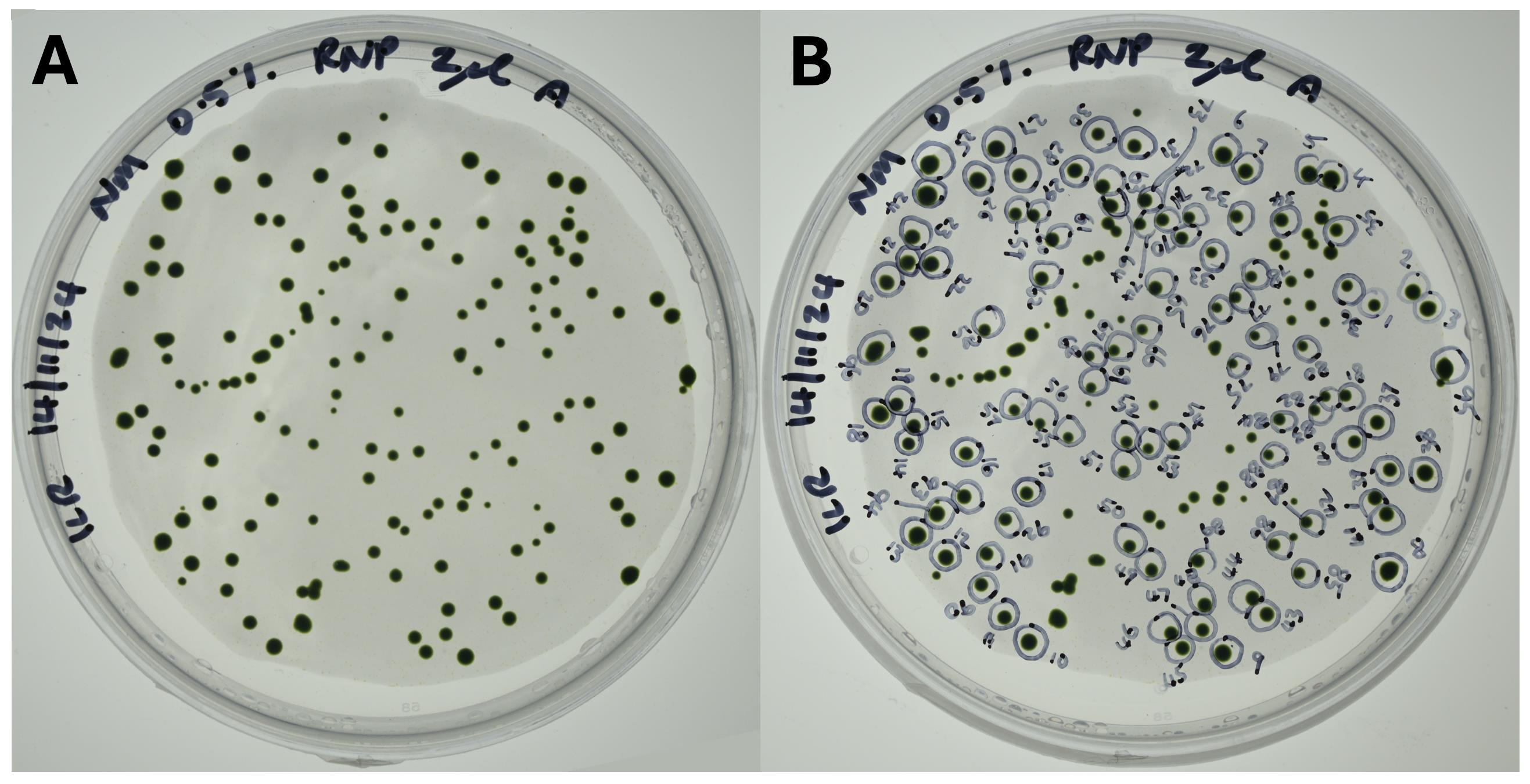
Figure 2. Selection plates with starch overlay. (A) A 2 μL cell resuspension plated out on chlorate selection plates with a starch overlay (pale grey zone over most of the plate). (B) The same plate after colonies are picked into 96-well plates prior to PCR analysis. Colonies are numbered for future reference. This enables re-picking if necessary.
G. Plating transformed cell suspension on selective media (day 3)
Note: In this step, knockouts of the NIA1 gene are detected via resistance of NIA1 knockouts to the herbicide potassium chlorate (sodium chlorate is also effective). Not all chlorate-resistant colonies are due to specific editing of the NIA1 gene, and not all clones edited at the NIA1 locus have precise insertion of the single-stranded HDR oligonucleotide. Finally, not all clones with NIA1 gene editing will also carry an edited target gene. Therefore, chlorate-resistant colonies are designated “candidates” until their correctness is checked by PCR screening and sequencing.
1. Take a 7 μL aliquot of each tube and count cells using the hemocytometer. This will allow calibration of efficiency with the cell numbers spread on the selection plates. Harvest the tubes of recovery medium by centrifugation at 800× g for 10 min at room temperature in a benchtop centrifuge. Remove the supernatant with a cell culture pipettor.
Note: We typically find that the NEC control has a greater cell density than the EC control due to the cell damage caused by electroporation of the EC control. The NEC control, therefore, allows assessment of the losses in cell density due to the electroporation itself, while still controlling for losses due to handling and washing of the cuvettes.
2. Re-centrifuge for 1 min to bring down the remaining liquid from the sides of the tube and remove from the cell pellet using a 200 μL pipettor and a slightly bent tip.
3. Resuspend the cell pellet gently but thoroughly in ~150–400 μL of NM+U recovery medium. The volume required is determined by summing the volume of cell suspension required for the number of screening plates employed. In this example protocol, Table 6 illustrates the process. The aim is that all cell suspension will be used up (unless a few microliters are required for counting or other purposes). The dilution series ensures that at least some plates will have an optimal number of colonies for counting, picking, and statistical analysis.
4. Lay out the plates in a sterile laminar flow hood, pre-labeled with their designated volume of cell suspension.
5. For each plate, pour 0.4 mL of starch suspension onto the surface and immediately add the aliquot of cell suspension. Using a loop or spreader, mix the cells well into the starch solution, and then spread evenly over the surface of the plate. The aim is to have a uniform, smooth starch suspension, with the cells evenly distributed within it.
6. After spreading the starch solution, put the lid on the plate and seal with parafilm. Place right side up under 100–200 μE/m-2/s light at room temperature.
7. (Optional but recommended until experience is gained) Similarly, plate one or more aliquots of cell suspension onto TAP plates (TAP check plates), at the same cell concentration as the most diluted selection plate. This will act as a CFU assay for viable cells and allows for (i) confirmation that the cells are inherently viable and (ii) estimation of the viable cell density, so that the efficiency of candidate clones per unit of viable cells can be calculated.
8. The next day, assuming the starch layer remains solid and excess moisture has been absorbed into the agar, turn the plates upside down to maintain a moist but not overly wet starch layer and to minimize condensation on the lid.
9. Leave the plates until small colonies appear, typically 4–10 days. Wild-type cells initially form tiny colonies and may take several days to start to die off. Check daily under an inverted microscope to confirm that colonies are healthy Chlamydomonas colonies and that the plates are free from fungal or bacterial contamination.
Table 6. Plan for selection plate requirements and total cell suspension volume
| A. Plates plan | B. Volume per plate | # Plates | Cumulative volume* | |||
| Total plates available | 24 | 2 μL | 4 | 8 μL | ||
| NEC control plates | 2 | 4 μL | 4 | 16 μL | ||
| EC control plates | 2 | 8 μL | 4 | 32 μL | ||
| Dilution series plates | 20 | 16 μL | 3 | 48 μL | ||
| 32 μL | 3 | 96 μL | ||||
| 64 μL | 2 | 128 μL | ||||
| Total plates: | 20 | 328 μL | ||||
| *Cumulative volume of cell suspension required for plating: here, transformed cells should be resuspended in 328 μL of NM+U recovery medium, and aliquots plated out as shown in column B. This scheme can be adapted for different numbers of available selection plates, reaction mixes, and desired replicates. | ||||||
H. Colony picking in preparation for PCR screening
Note: Isolation of quality genomic DNA for PCR is a common problem with Chlamydomonas. The procedure used here is convenient and rapid.
1. When colonies are of a suitable size, but have not yet become overgrown (Figures 1E and 2A), pick them into 96-well plates containing 200 μL of NM + U (nitrate medium + 2 mM urea) per well. Using a sterile 10 μL pipettor, pick individual colonies, preferably those that are well separated from other colonies.
Critical: Due to potential chlorate genotoxicity, we prefer not to select for long periods on chlorate-containing media. However, be aware that on chlorate selection, wild-type cells underlying the resistant colony (Figure 1E) may remain viable, so plating into the conventional Chlamydomonas medium (TAP; tris acetate phosphate) without chlorate will typically select against the desired clones and favor wild-type cells. Similarly, if a detrimental physiological phenotype is suspected for knockout clones, it may be advantageous to retain chlorate selection while handling and screening candidates until a clonal population is assured.
2. Start with the most dilute plates and pick colonies (Figure 2B), working up to the more concentrated plates until two plates of clones have been isolated. We generally find that this is sufficient to obtain a set of unique clones, but fewer or more plates can be collected if desired. Seal the plates with Parafilm or tape to prevent accidental dislodgment of the lids.
3. Grow the plates under moderate light without shaking until the cells have expanded to form a green layer at the base.
4. (Optional) Using a multi-well pipettor, plate out arrays of clones onto large square agar plates containing nitrate medium + 2 mM urea as a precaution in case of well plate contamination.
5. (Optional but recommended) Prepare similar arrays on nitrate medium without urea, to check for non-NIA1 mutations. Clones should grow poorly or not at all on this medium. Colonies that can grow well may be contaminated with wild-type cells, which typically grow out as small microspots within the droplet. Alternatively, if a clone thrives as a uniform spot, it may have mutations elsewhere in the nitrate assimilation pathway (e.g., nitrate transporters) that do not prevent survival on nitrate as an N-source. Under efficient CRISPR conditions, such clones are infrequently observed.
6. Isolate genomic DNA for PCR screening as follows: Resuspend cells in the 96-well selection plates and transfer 20 mL aliquots into 200 mL PCR tubes. To each tube, add 20 mL of lysis buffer (10 mM Tris pH 8.3, 50 mM KCl, 2.5 mM MgCl2, 0.5% SDS) and 1× proteinase K (freshly prepared from the 200× main stock). Incubate tubes in a hot block at 65 °C for 1 h, then inactivate proteinase K at 95 °C for 15 min. Centrifuge for 5 min in a Pico 17 microcentrifuge (or equivalent) to pellet the cell debris. Use 1 mL of the supernatant per PCR reaction. If cell-based contaminants interfere with PCR (or if using Taq polymerase), the supernatant should be diluted up to 100-fold.
Pause point: Genomic DNA can be readily stored at -80 °C if necessary, but do not repeatedly freeze-thaw.
I. PCR screening
Note: Because PCR is a routine tool, and because the PCR primers, polymerase type, and conditions need to be adapted for the desired target gene, we do not include detailed PCR notes as part of the protocol. However, because efficient screening is critical to deal with large numbers of clones, some broad descriptions of the screening procedure are provided. Table 7 provides typical PCR conditions employed for the NIA1 and APRT primers listed in Table 3. Platinum II is less expensive and works well for smaller amplicons. For amplicons over ~600 bp, Platinum II may be less effective, and it may be necessary to use a proofreading polymerase like Q5 polymerase. Both options are illustrated here. Table 8 provides typical thermocycler settings. We typically use a touchdown protocol for amplification from genomic DNA.
1. Using a suitable PCR protocol, test the pools for deletion between the gRNAs in the target gene (here, the APRT gene). For example, the primers in Table 3 provide an amplicon of 920 nt for the native APRT sequence and 228 nt for the deletion mutant.
2. If, instead of a deletion, the targeting strategy has employed the insertion of a unique oligonucleotide sequence by ssODN-directed HDR (e.g., for protein tagging or gene knockout), then the unique inserted oligonucleotide sequence can be used in conjunction with a gene-specific primer to amplify only those clones containing the inserted oligonucleotide sequence. This allows pooling of clones during initial candidate screening, which is most efficient when the percentage of insertion mutants is low (e.g., less than ~15%). When precise insertion of a donor oligonucleotide at the target locus is required, the amplicon size can be predicted, so variant amplicon sizes can flag most cases where additional indels have occurred. When gene knockout is all that is required, these additional indels are of little consequence. Once pools that contain mutant target genes have been identified, individual wells are then screened to identify likely candidates.
3. If, instead of oligonucleotide insertion, site-directed mutation has been employed (e.g., point mutation), then the donor oligonucleotide will not provide a detectable change in amplicon size and may only yield minimal sequence changes, making the design of a specific mutant primer more challenging. In this case, the use of the donor oligonucleotide to introduce silent third base changes around the point mutation site can provide enough specificity to amplify mutants over wild-type clones. Another screening strategy is to design the mutation-directing oligonucleotide so that it alters a restriction site at the site of mutation. Restriction digestion of the amplicon can then be used to test whether the amplicon contains the altered sequence [5].
4. Once identified, the set of likely target gene mutant candidates then undergoes sequence verification. Amplicons are produced for both the NIA1 gene and the target gene for sequence verification. The desired clones are those in which the target gene contains a suitable mutation, and in which the NIA1 gene has also been precisely edited (enabling later reversal to the wild type using the standard reversion gRNA “NIA1_Revert” Table 1). If precise modification of the target gene is needed (e.g., for side-directed mutagenesis, tag addition), then more clones may need to be checked.
5. (Optional) If quantitative real-time PCR is available, which allows for high resolution melting (HRM) analysis, then qPCR primers can be designed to screen a whole 96-well plate of clones at the same time, especially in the case of site-directed mutagenesis, where minimal sequence changes have been made.
Table 7. PCR setup for NIA1 and APRT primers (master mix composition for 20 reactions, each 14 µL + 1 µL template).
| Q5 polymerase | Platinum II polymerase | |||
| Reagent | µL | Reagent | µL | |
| 5× HiGC reagent | 60 | 5× HiGC reagent | 60 | |
| Nuclease-free water | 131 | Nuclease-free water | 50 | |
| Forward primer (10 µM) | 10 | Forward primer (10 µM) | 10 | |
| Reverse primer (10 µM) | 10 | Reverse primer (10 µM) | 10 | |
| 5× reaction buffer | 60 | Platinum II MM 2× | 150 | |
| 10 mM dNTPs | 6 | |||
| Q5 polymerase | 3 | |||
| Master mix total volume | 280 | Master mix total volume | 280 | |
Table 8. Thermocycler touchdown (TD) PCR protocols for NIA1 and APRT
| NIA1 | APRT | ||||
| Step | Temperature (°C) | Time (s) | Temperature (°C) | Time (s) | |
| Initial denaturation | 95 | 300 | 95 | 300 | |
| Cycle denaturation | 95 | 30 | 95 | 30 | |
| TD annealing | 68–61 (-1 °C per cycle, 8 cycles) | 30 | 63–58 (-1 °C per cycle, 5 cycles) | 30 | |
| Post-TD annealing | 61 | 30 | 58 | 30 | |
| Extension | 72 | 45 | 72 | 45 | |
| Cycle denaturation | 95 | 30 | 95 | 30 | |
| Final extension | 72 | 300 | 72 | 300 | |
| Termination | 12 | Hold | 12 | Hold | |
| Reaction volume 15 µL; hot lid temperature 105 °C. If touchdown is not used, annealing temperatures for NIA1 and APRT primer sets are 61 °C and 58 °C, respectively. | |||||
J. Reversion of the NIA1 gene to the wild-type sequence
Note: The key advantage of SCREAM for repeated sequential gene editing is the ability to revert the selectable marker to the wild-type sequence and to select for this event. In this case, the NIA1 guide RNA is directed against the modified gene in the NIA1 clone isolated from the above procedure. Instead of chlorate selection to detect a NIA1-negative genotype, the restoration of the wild-type sequence is detected by selection on nitrate medium without urea, so that in dilute plate culture where no organic nitrogen is available, only cells able to process nitrate to nitrite can survive and become colonies. The procedure is essentially the same as that above, with differences described below.
1. Grow cells in nitrate medium with 2 mM urea, instead of in nitrate medium alone, because they lack NIA1 and so require urea for growth and survival. Unlike ammonium, urea does not suppress nitrate reductase gene expression.
2. The gRNA used is directed against the modified genotype of the NIA1- cells. This gRNA only targets cells in which the NIA1 gene has been modified as planned.
3. The HDR oligonucleotide used is one that restores the wild-type sequence. The ssODN used here has two silent non-wild-type “check nucleotides,” which maintain efficient codon usage and can be used during sequencing to confirm that the HDR process has occurred, and that apparent “revertants” are not in fact due to wild-type cells that have carried through the clonal mutant selection. These check nucleotides are not normally required.
4. The selective method is an auxotrophic selection, which consists of nitrate medium alone. If the parent culture consists of pure mutants, only cells in which nitrate reductase function has been restored are capable of surviving and forming colonies on nitrate medium.
5. A second target gene can be deployed so that each half-cycle yields a new gene editing event. Because auxotrophic selection requires restoration of the function of the NIA1 gene, we find that ~9 out of 10 candidates have correct HDR insertion, resulting in a clean wild-type NIA1+ genotype. This step can also be employed to simply reverse the NIA1 mutation to wild type if desired.
Data analysis
Independent clones. The creation of a successful mutant is verified by sequencing at both the marker (NIA1) and target locus. As noted, multiple independent clones can be isolated from a single electroporation, so that biological replicates of the RNP reaction are not required except if the experimenter is interested in acquiring quantitative data about the efficiency of the process. However, phenotypic examination of a single mutant is insufficient for robust conclusions about the effect of the edited site, because random DNA integration events are known to occur in Chlamydomonas, and there is no way to simply identify the presence of such events apart from whole genome sequencing. Consequently, we typically isolate a set of independent “correct” mutants to act as controls for phenotypes, which are attributed to the introduced gene edits, even if, for practical reasons, only a single clone can typically be carried forward for subsequent gene editing steps. These are cryopreserved for future recovery if the generation of mutants from a different clonal lineage is required.
CRISPR experiment efficiency. The efficiency of the CRISPR experiment can be divided into candidate generation (assessed by the number of colonies emerging from a given electroporation cell) and target gene editing frequency, assessed by the percentage of desired changes (e.g., target gene disruption) among the pool of candidates examined. For the cell wall–deficient CC-1883 cells and the NIA1 gRNA used here, we obtain around ~5,000 colonies, substantially exceeding practical screening capacity and representing NIA1 editing in ~0.5%–1% of the original cell population [5]. For cell wall–containing cells, the electroporation efficiency is the major barrier, but electroporation of cell wall–containing cells can still be successful, for example, using the protocol described by Yamano et al. [13]. In contrast, the target gene editing frequency varies widely from gene to gene, depending on the efficiency of gRNA design, the accessibility of the target locus, and the constraints on gRNA design. Gene disruption via oligonucleotide insertion or regional deletion enables great flexibility with gRNA design. In contrast, precise nucleotide changes at a specific site (for example, for single amino acid replacement) severely constrain the gRNAs that are possible near that site and may result in lower efficiency. Using APRT, we have obtained a 45% gene disruption rate using oligonucleotide insertion. For other genes (e.g., Chlamydomonas light-harvesting genes), we have obtained gene disruption rates from 2% to 15%.
Assessing the efficiency of candidate generation. Using SCREAM, the overall efficiency of experiments with different target genes can be compared to each other. This is because a consistent selectable marker (e.g., NIA1) is used, so the number of candidate colonies obtained is dependent on the rate of NIA1 mutation, and not dependent on the mutation of the target gene, unless there is an overriding negative effect on viability of the engineered target mutation. Because the cell density is measured in the recovery medium, and because all cells in the recovery medium are concentrated, resuspended in a known volume, and plated out in a dilution series, colony counting provides the means to assess the number of colonies per million cells plated, and colonies per million cells electroporated, a measure of overall efficiency.
To assess this, one or more dilution plates are chosen, in which colonies are well separated, to enable accurate colony counting. For the highest accuracy, several independent dilution plates are used, and the mean colonies per unit resuspended cell mix is calculated. Note that this procedure assumes a high viability of colony-forming units. For cell wall–deficient strains, the use of the starch method is important because the efficiency of recovery of viable cells on bare plates is otherwise low. Wild-type strains are much more robust, and starch overlays are not required, but the use of 0.6% agar and prevention of excessive plate drying are recommended to ensure a high viability of plated cells. The use and accuracy of statistical tests will depend on the existence of primary data (e.g., number of independent plates) and pipetting accuracy. We typically use the standard deviation of colony estimates from several independent plates, which controls for the multiple sources of error within a given electroporation; but for true gRNA efficiency estimation, the use of replicate electroporation reactions, and ideally separate experiments on different days, is desirable. Calculations of efficiency provide reassurance that the protocol is working as intended. If many NIA1 mutants are obtained but not target mutations, then there is a problem with the target gRNA design or the HDR-directing ssODN, or there is a strongly negative effect on viability of the introduced mutation.
Image processing and data analysis. A high resolution RGB image of the plate is acquired using a digital camera. ImageJ is then used to process the image using standard approaches. The blue channel (in which the colonies show the most contrast against the background) is used. The colony area, free of artifacts such as plate edges, is thresholded and converted to a binary image, and closely joined colonies are separated using a watershed algorithm. Particle analysis is checked visually for accuracy, and the number of colonies over a particular size is quantified. Sizing is used because small plate defects can be mistaken for colonies.
Assessed by colony counting, the efficiency of a given gRNA is not dependent solely on the gRNA sequence but also on the chromosomal accessibility of the locus, so we typically use conditions in which both the marker (here, NIA1) gene and the desired target gene are expected to be induced. Note that the gRNA design for a target gene is often constrained by what sites are available around the point of mutation. Consequently, some target gRNAs will necessarily have low efficiency scores when assessed using tools like RGEN Cas Designer (http://www.rgenome.net/cas-designer/) or CRISPOR (https://crispor.gi.ucsc.edu). As such, the guide RNA targeting the marker gene (here, NIA1) should ideally be less efficient than the target gene gRNA. On the other hand, a marker guide RNA designed to have a very low efficiency will yield few candidate colonies. As the marker gRNA cannot be designed anew for each target gene, a compromise is needed. The NIA1_Disrupt gRNA employed here has a moderately low score in CRISPOR (25; Doench-RuleSet3) but still yields many colonies in a typical experiment. It is expected that for target genes with limited gRNA design flexibility, the use of marker gRNAs with lower scores will boost target gene mutation rates; this is under investigation in our laboratory. The current NIA1 gRNA typically has a CRISPR transformation efficiency of 0.5%, i.e., it provides around one colony per 200 cells electroporated.
Validation of protocol
This protocol has been used and validated in the following research article:
Ross et al. [5]. A cyclical marker system enables indefinite series of oligonucleotide-directed gene editing in Chlamydomonas reinhardtii. Plant Physiology 196(4): 2330–2345 (see, particularly, Figures 6 and 7).
General notes and troubleshooting
General notes
1. Sterile conditions must be maintained throughout because any contamination of the gRNA premix will result in contamination of the screening plates.
2. Time the annealing and Cas9 reaction so that the 20-min Cas9 binding step concludes just as the 20-min heat shock procedure is concluding. This requires sections C and D to be conducted more or less concurrently.
3. Single-stranded ODNs with and without phosphorothioate linkages have both been used successfully. Although we have not systematically examined this variable, we have no data to suggest that phosphorothioate linkages improve HDR rates.
4. The specific ssODN used can also vary in efficiency. Although an extensive consideration of ssODN design is not possible here, the ssODN should span the Cas9 cut site, and the homology arms should each be 30–60 nt. As one possible contributor to low efficiency is secondary structure formation, the design of ssODNs should be conducted to minimize the secondary structure, such as hairpin formation. It may be necessary to trial different ssODN designs if low target gene frequencies are experienced.
5. Depending on the number of electroporations desired, the amount of premix can be scaled. This protocol describes a single reaction aimed at one target gene (APRT), but typically, several reactions would be undertaken in parallel. Although scaling to multiple electroporations is technically straightforward, the plating and screening are the most labor-intensive steps, so numerous CRISPR-mediated gene editing reactions should only be employed if the resulting screening workload is feasible, for example, via qPCR.
6. A dummy electroporation mix can be used for quantitative experiments, in which the HDR oligonucleotides are replaced by irrelevant oligonucleotides (with no homology to the Chlamydomonas genome), and the gRNA-Cas9 mixtures are replaced by irrelevant gRNAs (for example, a gRNA with no homology to the Chlamydomonas genome). However, the critical factor for electroporation efficiency is the salt content of the electroporation mixture, so any alternative oligonucleotides should have comparable salt contents (e.g., be desalted and resuspended in the same buffer).
7. Multiplexing has been used successfully to simultaneously create gene editing in more than one gene, but care must be taken so that multiplexed experiments do not incur more screening effort than achieving the same result by sequential steps. The use of the oligo insertion method coupled with a generic primer, which binds to multiple members of a gene family, achieves this aim, since initial screening for insertion mutants does not need to be gene-specific.
Adaptations for cell wall–containing strains
Strains with a wild-type cell wall have lower efficiencies of electroporation [14]. Three approaches can be undertaken to surmount this barrier. First, and most simply, the reaction can be scaled up to yield more colonies. Our initial protocol [5] used five-fold greater quantities and a 4 mm cuvette and was only scaled down to save on reagent costs. Second, the electroporation conditions should be varied. For the CC-1690, we employ a square wave pulse (rather than an exponential pulse) with an 800 V, 5 ms initial pulse (“poring pulse”) and 5 pulses at 40 V (“transfer pulses”) with a 10 ms pulse interval. Finally, numerous other authors have used the Chlamydomonas gamete lysin to degrade the cell wall prior to electroporation, enabling a simple exponential pulse to be successful [15–18]. In all cases, the parameters should be trialled for each strain using plasmid electroporation or, ideally, Cas9 RNP electroporation with an easily scorable marker like APRT or NIA1.
Target gene modification strategy
The advantage of SCREAM is that the marker gene (e.g., NIA1) can remain constant for a long series of modifications. In contrast, each experimenter will need to make unique, specific changes to the target gene(s). This means that no prescribed protocol can be given for target gene gRNA design or site modification. However, desired changes typically fall into three categories:
a. Knockout of the target gene, either by insertion of stop codons or by deletion of a region.
b. Site-directed mutagenesis of the target gene (e.g., changing one amino acid).
c. Knock-in of a new sequence to the target gene (e.g., adding a peptide tag to a protein).
Each of these requires a tailored approach in terms of gRNA design, and some general principles are provided below, along with some pros and cons of the different approaches.
Gene knockout. This is typically accomplished by the generation of small indels (e.g., those classically produced by NHEJ and a single gRNA), by insertion of one or more deliberate stop codons (as used for the NIA1 gene) and/or a unique primer sequence, using a single-stranded oligonucleotide for HDR, or finally, by deletion of a gene region using two or more gRNAs. Gene disruption usually allows considerable choice of the gRNA site so that efficient gRNAs can be used. Each approach has pros and cons.
(i) Single gRNAs: We have found the use of single gRNAs without an HDR template to be inefficient, likely because NHEJ is usually an efficient repair process, and the gRNA target site is simply recreated when NHEJ is successful in double-strand break repair. It is likely to be used mainly when a selectable marker is not required (e.g., when an independently selectable phenotype results from the target gene modification).
(ii) Single gRNA with a ssODN template for HDR: The use of HDR to create a precise mutation (e.g., stop codon insertion) is not only readily reversible (for example, to prove that a phenotype is in fact due to the introduced mutation) but can be used to remove or create a restriction enzyme site that can be used to interrogate PCR amplicons for the desired change. On the other hand, the change in size of the PCR amplicon across the break site is usually too small to see directly from PCR. (iii) Introduction of a unique PCR primer: Another way to use HDR is to insert a larger, unique sequence that can be probed using a specific primer that will only appear when a successful oligonucleotide insertion has occurred. This has the advantage that candidate DNA can be pooled and the presence of a positive clone can be readily identified in a large pool (e.g., a whole 96-well plate), a key strategy if the target gene is inefficiently modified relative to the marker gene.
(iv) Using dual gRNAs to create deletions: Finally, the use of dual gRNAs to produce a deletion has three potential advantages. First, the loss of a large region of the gene removes the possibility of residual function (or gain of function) of the non-deleted region. Second, it eliminates the gene sequence from the genome (for example, if a modified gene homolog is to be inserted into the genome and its presence screened by PCR). Third, the resulting large change in PCR amplicon size makes positive candidates easy to identify during PCR screening, including from (limited) pools of clones, as the smaller amplicon is usually favoured during amplification. It should also be kept in mind that this is an efficient strategy for removal of large regions of regulatory elements such as promoters and enhancers, for example, during the search for transcription factor binding sites.
Site-directed mutagenesis (SDM). The process of precisely modifying one or more amino acids at a specific site in a protein usually mandates the use of HDR unless an indel is expected that will accomplish the desired mutation, or unless a random set of mutants at the SDM site is desired (in which case an ssODN including random sequence at the SDM site is still probably a more efficient approach). The need for a precise site severely restricts the choice of gRNA, as the ssODN used for HDR needs to accommodate not only the desired sequence changes but also the 45 nt homology arms on either side. This means that the CRISPR cleavage site should be reasonably close to the desired SDM site, and the use of an inefficient gRNA may be necessary. Consequently, target gene modification efficiency relative to the marker needs to be prioritized along with pool screening. The omission of the NIA1 marker ssODN can boost target gene efficiency, as TMEJ with a ssODN template is more efficient than NHEJ, but at the cost of random indel formation at the NIA1 site (necessitating a new gRNA and ssODN to be designed for the NIA1 reversion to wild-type step for each resulting clone). Pool screening is difficult when a single, precise one or two nucleotide change is made, because the ability to screen for amplicon size changes, restriction site introduction, or unique PCR primer creation are all limited. To deal with this situation, we make additional third base codon changes to the surrounding codons, so that a unique primer site is created but without changes to the amino acid sequence. Due to restricted codon usage by Chlamydomonas, this introduces the possibility of poorer mRNA translation and subsequent alteration of the protein abundance, a situation that would need to be controlled for in phenotypic screening.
New sequence knock-in. The knock-in of a new amino acid sequence in a protein (or perhaps a transcription factor binding site in a promoter) can be accomplished using HDR, but it should be kept in mind that the larger the sequence to be knocked in, the less efficient the HDR process is. In practical terms, the use of unlinked markers like NIA1 is likely to be successful for knocking in up to 60 nt, but as the insert size increases, it becomes inefficient. Larger sequences (for example, whole genes) therefore require a linked marker such as the AphVIII gene to screen for successful knock-in candidates. Consequently, SCREAM is useful for tagging proteins, for alteration of intron-exon structure, for insertion of transcription factor binding site, and similar changes requiring up to 50–60 nucleotides, but it is not designed for gene knock-in.
Troubleshooting
Problem 1: Bacterial colonies on screening plates.
Possible cause: Contamination of one or more reagents and/or introduction of contamination during handling.
Solution: Test reagents using LB agar plates and TAP plates to confirm a lack of contamination. Check the primary cell line for contamination using TAP plates with 4 g/L yeast extract (TAPY media). Clean pipettes, especially inside the barrels and the pistons. If no contamination is found, repeat the experiment.
Problem 2: Few or no colonies on TAP check plates for EC and RNP, but many for NEC.
Possible cause: Electroporation problems. This may be caused by excess salt in the mixture, for example, from the ssODN or from a PCR product that contains salt from DNA amplicon purification; check the time constant, which should be between 3 and 6 ms. An additional possible cause is excessive voltage, leading to elevated cell mortality.
Solution: Count the cell density immediately following transfer to the recovery medium and look for excessive cell debris. Potential sources of excess salt in the electroporation mixture should be assessed. Conduct a voltage titration using AphVIII containing 1 μg of plasmid in duplex buffer (taking care to minimize salt from the plasmid preparation) instead of ssODN, and spread cells on plates containing 10 μg/mL paromomycin to identify an efficient voltage.
Problem 3: Similar low numbers of colonies (per million cells plated) on all screening plates including the control plates (forward screen-chlorate selection).
Possible cause: Since there are several genes that generate chlorate-resistant colonies when mutated, typically nitrate transporters, there is always a background of chlorate-resistant colonies on the NEC and EC plates. Unlike true NIA1 mutants, such mutants typically grow successfully on nitrate plates. If the NIA1 gRNA is working efficiently, gene-edited NIA1 candidates (which cannot grow on nitrate) will be far more prevalent, such that the presence of occasional background colonies can be neglected.
Solution: Calculate the rate of colony formation for the RNP plates vs. the EC control. If they are comparable, the Cas9-NIA1 RNP has not formed correctly. Check the annealing process (making sure Cas9 is added only after the annealed tracrRNA and gRNA have cooled). The Cas9 may have lost activity, the gRNA and/or tracrRNA may be degraded, or there may be RNase present in the reagents or released from the cells. Trial reagent replacement.
Problem 4: Some growth of NIA1-mutant cells on nitrate check plates (forward screen–nitrate selection).
Possible cause: Chlorate inhibits the growth of wild-type cells, but, in the presence of other sources of nitrogen (especially organic nitrogen from cell debris), wild-type cells can survive. Therefore, when a “mutant” colony is picked, some wild-type cells may also be picked. If the candidate is grown in normal TAP media or nitrate medium with urea, they will survive and grow alongside the mutant cells. Subsequent plating on nitrate plates without urea will reveal “spotty” droplets as the wild-type cells grow out against the non-growing mutants. Alternatively, colonies on nitrate check plates that have a diffuse “slow growing” appearance are probably due to the use of many cells, whereby dying cells provide a source of organic nitrogen for the survivors.
Solution: Validated candidates should be spread very thinly on selective media (with a starch overlay for cell wall–deficient strains), and clean single colonies picked.
Problem 5: No colonies on screening plates (reverse screen–nitrate selection).
Possible cause: Because the spontaneous recovery of an inactive nitrate reductase is unlikely, and few other mutations can compensate for NR deficiency, the background for the reverse screen is usually very clean, with very few or no colonies on the EC or NEC plates. Therefore, failure of the NIA1 reversion to wild type is the probable cause.
Solution: Apart from reagent failures of the type described in Problem 3, check that the gRNA used is the one designed against the mutated site, not the wild-type site, and check that the ssODN used is the one designed to restore the wild-type enzyme.
Problem 6: Many colonies on nitrate plates, including control plates (reverse screen nitrate selection).
Possible cause: A wild-type cell line, or a clone contaminated with wild-type cells, has been used. See Problem 4 for an explanation as to why wild-type cells may be present in mutant cell lines.
Solution: Thinly spread the starting clone on selective media and pick a clean single colony free of wild-type cells. Check that the mutant gene is still present.
Problem 7: Low rates of target gene modification relative to the NIA1 marker gene.
Possible cause: The target gene gRNA is inefficient relative to the NIA1 gRNA, one or more target gene reagents are defective, the accessibility of the target locus is low compared to NIA1, or the introduced mutation is physiologically detrimental.
Solution: Check the integrity of the target gene reagents. Test the use of HDR for the target gene but NHEJ for the NIA marker (i.e., omit the NIA1 ssODN). This will reduce the overall number of colonies due to less efficient NIA1 modification, but may improve the target gene hit rate compared to the marker. If a detrimental effect of the mutated target gene is suspected, try a gRNA/ssODN pair that creates an innocuous change to the target gene.
Acknowledgments
Conceptualization, I.L.R.; Investigation, I.LR.; Writing—Original Draft, I.L.R.; Writing—Review & Editing, B.H.; Funding acquisition, B.H.; Supervision, B.H. This work was supported by ARC Discovery project DP240101727. The method described here is developed from that published in [5] and from subsequent work by I.L.R.
Competing interests
The authors declare no conflicts of interest.
References
- Vogt, A., He, Y. and Lees-Miller, S. P. (2023). How to fix DNA breaks: new insights into the mechanism of non-homologous end joining. Biochem Soc Trans. 51(5): 1789–1800. https://doi.org/10.1042/bst20220741
- Sfeir, A., Tijsterman, M. and McVey, M. (2024). Microhomology-Mediated End-Joining Chronicles: Tracing the Evolutionary Footprints of Genome Protection. Annu Rev Cell Dev Biol. 40(1): 195–218. https://doi.org/10.1146/annurev-cellbio-111822-014426
- Ferenczi, A., Chew, Y. P., Kroll, E., von Koppenfels, C., Hudson, A. and Molnar, A. (2021). Mechanistic and genetic basis of single-strand templated repair at Cas12a-induced DNA breaks in Chlamydomonas reinhardtii. Nat Commun. 12(1): e1038/s41467–021–27004–1. https://doi.org/10.1038/s41467-021-27004-1
- Sizova, I., Kelterborn, S., Verbenko, V., Kateriya, S. and Hegemann, P. (2021). Chlamydomonas POLQ is necessary for CRISPR/Cas9-mediated gene targeting. G3 Genes|Genomes|Genetics. 11(7): e1093/g3journal/jkab114. https://doi.org/10.1093/g3journal/jkab114
- Ross, I. L., Le, H. P., Budiman, S., Xiong, D., Hemker, F., Millen, E. A., Oey, M. and Hankamer, B. (2024). A cyclical marker system enables indefinite series of oligonucleotide-directed gene editing in Chlamydomonas reinhardtii. Plant Physiol. 196(4): 2330–2345. https://doi.org/10.1093/plphys/kiae427
- Bae, S., Park, J. and Kim, J. S. (2014). Cas-OFFinder: a fast and versatile algorithm that searches for potential off-target sites of Cas9 RNA-guided endonucleases. Bioinformatics. 30(10): 1473–1475. https://doi.org/10.1093/bioinformatics/btu048
- Bae, S., Kweon, J., Kim, H. S. and Kim, J. S. (2014). Microhomology-based choice of Cas9 nuclease target sites. Nat Methods. 11(7): 705–706. https://doi.org/10.1038/nmeth.3015
- Gorman, D. S. and Levine, R. P. (1965). Cytochrome f and plastocyanin: their sequence in the photosynthetic electron transport chain of Chlamydomonas reinhardi. Proc Natl Acad Sci USA. 54(6): 1665–1669. https://doi.org/10.1073/pnas.54.6.1665
- Kropat, J., Hong‐Hermesdorf, A., Casero, D., Ent, P., Castruita, M., Pellegrini, M., Merchant, S. S. and Malasarn, D. (2011). A revised mineral nutrient supplement increases biomass and growth rate in Chlamydomonas reinhardtii. Plant J. 66(5): 770–780. https://doi.org/10.1111/j.1365-313x.2011.04537.x
- Angstenberger, M., de Signori, F., Vecchi, V., Dall’Osto, L. and Bassi, R. (2020). Cell synchronization enhances nuclear transformation and genome editing via Cas9 enabling homologous recombination in Chlamydomonas reinhardtii. ACS Synth Biol. 9(10): 2840–2850. https://doi.org/10.1021/acssynbio.0c00390
- Kelterborn, S., Boehning, F., Sizova, I., Baidukova, O., Evers, H. and Hegemann, P. (2022). Gene editing in green alga Chlamydomonas reinhardtii via CRISPR-Cas9 ribonucleoproteins. Methods Mol Biol. 2379: 45–65. https://doi.org/10.1007/978-1-0716-1791-5_3
- Shimogawara, K., Fujiwara, S., Grossman, A. and Usuda, H. (1998). High-efficiency transformation of Chlamydomonas reinhardtii by electroporation. Genetics. 148(4): 1821–1828. https://doi.org/10.1093/genetics/148.4.1821
- Yamano, T., Iguchi, H. and Fukuzawa, H. (2013). Rapid transformation of Chlamydomonas reinhardtii without cell-wall removal. J Biosci Bioeng. 115(6): 691–694. https://doi.org/10.1016/j.jbiosc.2012.12.020
- Jeon, K., Suresh, A. and Kim, Y. C. (2013). Highly efficient molecular delivery into Chlamydomonas reinhardtii by electroporation. Korean J Chem Eng. 30(8): 1626–1630. https://doi.org/10.1007/s11814-013-0098-4
- Akella, S., Ma, X., Bacova, R., Harmer, Z. P., Kolackova, M., Wen, X., Wright, D. A., Spalding, M. H., Weeks, D. P., Cerutti, H., et al. (2021). Co-targeting strategy for precise, scarless gene editing with CRISPR/Cas9 and donor ssODNs in Chlamydomonas. Plant Physiol. 187(4): 2637–2655. https://doi.org/10.1093/plphys/kiab418
- De Silvio, M. A., Sánchez-Retuerta, C., Ruiz-Sola, M. Ã., Baidukova, O. and Monte, E. (2025). Corrigendum to ‘A quick-to-implement and optimized CRISPR-Cas9 protocol to obtain insertional and small indel mutants in Chlamydomonas reinhardtii’ [MethodsX 15 (2025) 103416]. MethodsX. 15: 103501. https://doi.org/10.1016/j.mex.2025.103501
- Kindle, K. L. (1990). High-frequency nuclear transformation of Chlamydomonas reinhardtii. Proc Natl Acad Sci USA. 87(3): 1228–1232. https://doi.org/10.1073/pnas.87.3.1228
- Nievergelt, A. P., Diener, D. R., Bogdanova, A., Brown, T. and Pigino, G. (2023). Efficient precision editing of endogenous Chlamydomonas reinhardtii genes with CRISPR-Cas. Cell Rep Methods. 3(8): 100562. https://doi.org/10.1016/j.crmeth.2023.100562
Article Information
Publication history
Received: Feb 5, 2026
Accepted: May 27, 2026
Available online: Jun 29, 2026
Published: Jul 20, 2026
Copyright
© 2026 The Author(s); This is an open access article under the CC BY-NC license (https://creativecommons.org/licenses/by-nc/4.0/).
How to cite
Readers should cite both the Bio-protocol article and the original research article where this protocol was used:
- Ross, I. L. and Hankamer, B. (2026). Gene Editing in Chlamydomonas Using the SCREAM Technique. Bio-protocol 16(14): e5759. DOI: 10.21769/BioProtoc.5759.
- Ross, I. L., Le, H. P., Budiman, S., Xiong, D., Hemker, F., Millen, E. A., Oey, M. and Hankamer, B. (2024). A cyclical marker system enables indefinite series of oligonucleotide-directed gene editing in Chlamydomonas reinhardtii. Plant Physiol. 196(4): 2330–2345. https://doi.org/10.1093/plphys/kiae427

 Download Citation in RIS Format
Category
Plant Science > Plant molecular biology > DNA
Cell Biology > Cell engineering > CRISPR-cas9
Plant Science > Phycology > Nuclear transformation
Do you have any questions about this protocol?
Post your question to gather feedback from the community. We will also invite the authors of this article to respond.
 Post a Question


