


- Protocols

- Articles and Issues
- For Authors
- About
- Become a Reviewer


Simple Electroporation of Chlamydomonas reinhardtii Strains With an Intact Cell Wall


(*contributed equally to this work) Published: Vol 16, Iss 14, Jul 20, 2026 DOI: 10.21769/BioProtoc.5740 Views: 539
Reviewed by: Lucy XieAnonymous reviewer(s)
 PDF
PDF  Ask a question
Original research article
The authors used this protocol in:
Oct 2022
Advertisement


Protocol Collections
Comprehensive collections of detailed, peer-reviewed protocols focusing on specific topics




Abstract
Chlamydomonas reinhardtii is a model green alga extensively used to study photosynthesis and cilia using molecular biology and genetics. Electroporation is a very common technique to integrate DNA into the nuclear genome, which is essential to generate mutant collections and express transgenes. Here, we describe a simple, fast, and efficient protocol to transform strains with an intact cell wall. The technique achieves good transformation efficiency without cell wall digestion or the use of commercial kits and is compatible with the widely available Gene Pulser electroporation system.
Key features
• High transformation efficiency of Chlamydomonas reinhardtii strains with an intact cell wall.
• Faster than currently available electroporation protocols.
Keywords: Chlamydomonas reinhardtii Microalgae
Transformation
Electroporation
Cell wall
Microalgae
Transformation
Electroporation
Cell wall
Background
Chlamydomonas reinhardtii is a model green alga amenable to genetic engineering. Electroporation is a common technique to randomly integrate DNA into the nuclear genome through non-homologous end joining repair [1]. Several protocols have been developed, but they often require cell-wall-less strains, digestion of the cell wall, specialized electroporators (e.g., NEPA21, Nepagene) [2], or a commercial kit (i.e., MAX Efficiency Transformation Reagent for Algae kit, Invitrogen). Here, we describe a simple, fast, and efficient method to transform Chlamydomonas reinhardtii strains with an intact cell wall. Compared to previously published electroporation protocols by Crozet et al. [3,4] and Onishi and Pringle [5], we simplified the procedure by removing the cold incubation (20–30 min), allowing the electroporation part of our protocol (section C) to take approximately 30 min. We used a CHES pH 9.25 electroporation buffer [5], which has been reported to yield a high number of transformants and to be efficient with several transgenes and strains, including those with an intact cell wall [5–8]. Our protocol is suitable for the generation of randomly mutagenized mutant collections and transgenic approaches (e.g., complementation of mutants or protein localization) but not for the delivery of Cas9 ribonucleoprotein complexes.
Materials and reagents
Biological materials
1. Chlamydomonas reinhardtii, tested strains: CC-4051 (4A-), CC-5101 (T222+), CC-4425 (D66), CC-124, CC-4533, CC-5325 (available at the Chlamydomonas Resource Center, www.chlamycollection.org)
Note: This protocol does not work with true cell-wall-less strains like UVM4, for which glass bead transformation is recommended [9].
2. Expression plasmid for Chlamydomonas reinhardtii
Note: This protocol does not cover plasmid design and cloning, but we recommend using MoClo vectors [3,9] (available at the Chlamydomonas Resource Center, www.chlamycollection.org/product/moclo-toolkit).
Reagents
1. Tris base (tris(hydroxymethyl)aminomethane) (Fisher Scientific, catalog number: BP152-1)
2. Potassium phosphate dibasic (K2HPO4) (Fisher Scientific, catalog number: BP363-500)
3. Potassium phosphate monobasic (KH2PO4) (VWR, catalog number: EMD-PX1565-1)
4. Ammonium chloride (NH4Cl) (VWR, catalog number: BDH9208-500G)
5. Magnesium sulphate heptahydrate (MgSO4·7H2O) (Fisher Scientific, catalog number: M63-500)
6. Calcium chloride dihydrate (CaCl2·2H2O) (Fisher Scientific, catalog number: C79-500)
7. Ethylenediaminetetraacetic acid disodium salt dihydrate (EDTA-Na2) (Sigma-Aldrich, catalog number: E4884)
8. Potassium hydroxide (KOH) (Sigma-Aldrich, catalog number: P6310)
9. Ammonium molybdate tetrahydrate [(NH4)6Mo7O24] (Fisher Scientific, catalog number: A674-500)
10. Zinc sulfate heptahydrate (ZnSO4·7H2O) (Sigma-Aldrich, catalog number: Z4750)
11. Manganese(II) chloride tetrahydrate (MnCl2·4H2O) (Sigma-Aldrich, catalog number: M3634)
12. Iron(III) chloride hexahydrate (FeCl3·6H2O) (Sigma-Aldrich, catalog number: F2877)
13. Sodium carbonate (Na2CO3) (Sigma-Aldrich, catalog number: BP357-1)
14. Copper(II) chloride dihydrate (CuCl2·2H2O) (Sigma-Aldrich, catalog number: 307483)
15. Glacial acetic acid (CH3COOH) (VWR, catalog number: EMD-AX0073-9)
16. Hydrochloric acid (HCl) (Fisher Scientific, catalog number: A144SI-212)
17. Agar (VWR, catalog number: 90000-762)
18. Blasticidin S hydrochloride (Fisher Scientific, catalog number: 50-712-729)
19. Zeocin (Thermo Fisher Scientific, catalog number: R25001)
20. Hygromycin B (Thermo Fisher Scientific, catalog number: 10687010)
21. Kanamycin monosulfate (TCI, catalog number: K0047)
22. Paromomycin sulfate (Sigma-Aldrich, catalog number: P5057)
23. Spectinomycin dihydrochloride pentahydrate (Sigma-Aldrich, catalog number: S4014)
24. Nourseothricin sulfate (GoldBio, catalog number: N-500)
25. N-cyclohexyl-2-aminoethanesulfonic acid (CHES) (Sigma-Aldrich, catalog number: C2885)
26. Sucrose (Millipore, catalog number: 573113)
27. Sorbitol (Thermo Fisher Scientific, catalog number: 036404.36)
Solutions
1. 1 M Tris base (see Recipes)
2. Phosphate buffer II (see Recipes)
3. Solution A (see Recipes)
4. Kropat’s stock solutions (see Recipes)
a. 125 mM EDTA-Na2 stock solution
b. 285 μM (NH4)6Mo7O24
5. Kropat’s trace elements solutions (1,000×-concentrated) (see Recipes)
a. 25 mM EDTA-Na2
b. 28.5 μM (NH4)6Mo7O24
c. Zn-EDTA (2.5 mM ZnSO4, 2.75 mM EDTA-Na2)
d. Mn-EDTA (6 mM MnCl2, 6 mM EDTA-Na2)
e. Fe-EDTA (20 mL FeCl3, 22 mM EDTA-Na2, 22 mM Na2CO3)
f. Cu-EDTA (2 mM CuCl2, 2 mM EDTA-Na2)
6. Liquid TAP (see Recipes)
7. TAP plates with antibiotics (see Recipes)
8. CHES buffer (see Recipes)
9. TS40 (40 mM sucrose in TAP) (see Recipes)
Recipes
1. 1 M Tris base
a. Weigh 121.14 g of Tris.
b. Dissolve in 800 mL of distilled water.
c. Adjust the volume to 1,000 mL with distilled water.
2. Phosphate buffer II
a. Weigh 10.8 g of K2HPO4 and 5.6 g of KH2PO4.
b. Dissolve one by one in 80 mL of distilled water.
c. Adjust the volume to 100 mL with distilled water.
3. Solution A
a. Weigh 20.0 g of NH4Cl, 5.0 g of MgSO4·7H2O, and 2.5 g of CaCl2·2H2O.
b. Dissolve one by one in 400 mL of distilled water.
c. Adjust the volume to 500 mL with distilled water.
4. Kropat’s stock solutions
Kropat’s trace elements solutions preparation is based on the following protocol www.chlamycollection.org/content/uploads/2022/05/Kropats-Trace-Elements-safety-update.pdf [10]. Selenite is omitted as it is not necessary for this protocol. If sodium selenite is used, it should be handled with care, as it is toxic.
a. 125 mM EDTA-Na2 stock solution
i. Dissolve 11.63 g of EDTA-Na2 in 250 mL of distilled water.
ii. Adjust to pH 8.0 with KOH pellets.
iii. Adjust the volume to 300 mL with distilled water.
b. 285 μM (NH4)6Mo7O24
i. Dissolve 88.0 mg of (NH4)6Mo7O24 in 200 mL of distilled water.
ii. Adjust the volume to 250 mL with distilled water.
5. Kropat’s trace elements solutions (1,000×-concentrated)
a. 25 mM EDTA-Na2
Mix 50 mL of 125 mM EDTA-Na2 and 200 mL of distilled water.
b. 28.5 μM (NH4)6Mo7O24
Mix 25 mL of 285 μM (NH4)6Mo7O24 and 225 mL of distilled water.
c. Zn-EDTA (2.5 mM ZnSO4, 2.75 mM EDTA-Na2)
i. Dissolve 180.0 mg of ZnSO4.7H2O in 200 mL of distilled water.
ii. Add 5.5 mL of 125 mM EDTA-Na2.
iii. Adjust the volume to 250 mL with distilled water.
d. Mn-EDTA (6 mM MnCl2, 6 mM EDTA-Na2)
i. Dissolve 297.0 mg of MnCl2.4H2O in 200 mL of distilled water.
ii. Add 12 mL of 125 mM EDTA-Na2.
iii. Adjust the volume to 250 mL with distilled water.
e. Fe-EDTA (20 mL FeCl3, 22 mM EDTA-Na2, 22 mM Na2CO3)
i. Dissolve 2.05 g of EDTA-Na2 and 0.58 g of Na2CO3 in 200 mL of distilled water.
ii. Add and dissolve 1.35 g of FeCl3.6H2O.
iii. Adjust the volume to 250 mL with distilled water.
f. Cu-EDTA (2 mM CuCl2, 2 mM EDTA-Na2)
i. Dissolve 85.0 mg of CuCl2.2H2O in 200 mL of distilled water.
ii. Add 4 mL of 125 mM EDTA-Na2.
iii. Adjust the volume to 250 mL with distilled water.
6. Liquid TAP
a. Add the following solutions to 900 mL of distilled water: 20 mL of 1 M Tris base, 1 mL of phosphate buffer II, 10 mL of solution A, 1 mL each of Kropat’s trace elements solutions [25 mM EDTA-Na2, 28.5 μM (NH4)6Mo7O24, Zn-EDTA, Mn-EDTA, Fe-EDTA, and Cu-EDTA], and 1 mL of glacial acetic acid.
b. Adjust pH to 7.0 with concentrated HCl.
c. Adjust the volume to 1,000 mL with distilled water.
d. Autoclave for 30–40 min.
7. TAP plates with antibiotics
a. Prepare TAP as in Recipe 6 (without the autoclaving step).
b. Add 15 g of agar per liter.
c. Autoclave for 30–40 min.
d. Mix well and let the solution reach a temperature of 40–50 °C.
e. Add relevant antibiotic at the following final concentration: blasticidin S (50 mg/L), zeocin (15 mg/L), hygromycin B (20 mg/L), kanamycin (50 mg/L), paromomycin (20 mg/L), spectinomycin (100 mg/L), or nourseothricin (10 mg/L) [4,11].
f. Mix well and pour plates.
g. Store at 4–8 °C for up to 2 months.
8. CHES buffer (10 mM CHES pH 9.25, 40 mM sucrose, 10 mM sorbitol)
a. Weigh 1 g of CHES, 6.84 g of sucrose, and 0.91 g of sorbitol.
b. Dissolve one by one in 400 mL of distilled water.
c. Adjust pH to 9.25.
d. Bring to 500 mL with distilled water.
e. Filter sterilize.
9. TS40 (40 mM sucrose in TAP)
a. Dissolve 6.84 g of sucrose in 500 mL of TAP.
b. Filter sterilize.
Laboratory supplies
1. Gel extraction kit (e.g., Monarch Spin DNA, New England Biolabs, catalog number: T1120S)
2. Bottle top 0.45 μm filers (e.g., Nalgene Rapid flow, Thermo Scientific, catalog number: 565-0010)
3. 4-mm-gapped electroporation cuvettes (USA Scientific, catalog number: 9104-6050)
4. Parafilm or breathable tape (3M, catalog number: Vent Tape 394)
Equipment
1. Gene Pulser with Pulse Controller (Bio-Rad, catalog number: 1652662)
2. NanoDrop spectrophotometer (ThermoFisher Scientific, model: NanoDrop Lite Plus)
3. Hemocytometer or cell counter (e.g., Beckman Coulter, model: Multisizer 4e Coulter Counter)
4. Growth chamber with light and temperature control (e.g., Percival, Conviron, BioChamber) and a rotary shaker
Note: A simpler culture setup, such as hanging fluorescent bulbs over a shaker at room temperature, has yielded similar results.
5. Standard laboratory equipment (e.g., pipettes, centrifuge, microcentrifuge, laminar flow hood, glass beakers, 15- and 50-mL sterile tubes)
Procedure
A. Purification of the DNA cassette
1. Digest a sufficient quantity of plasmid DNA (typically 5–10 µg) with appropriate restriction enzymes (typically BbsI or BsaI for MoClo plasmids [3,9]) to isolate the desired cassette. If the size of the cassette is close to that of the vector backbone fragment, additionally cut the latter with another restriction enzyme to ensure good separation. It is also possible to linearize the plasmid without purifying the expression cassette on the gel [6].
2. Incubate the reaction for 3 h to overnight at 37 °C.
3. Separate the cassette by agarose gel electrophoresis and isolate it by gel purification.
4. Measure the DNA concentration with a NanoDrop spectrophotometer. Typical recovery yield is 60%–80%.
B. Chlamydomonas culture and harvesting
1. From this point on, all steps must be performed under sterile conditions, for example, in a laminar flow hood.
2. Inoculate Chlamydomonas reinhardtii culture in liquid TAP. Adjust the volume depending on the number of transformations; the minimum is 25 mL per transformation if cells are harvested at 2 × 10⁶ cells/mL.
3. Incubate the culture at 25 °C with shaking (~120 rpm) and constant light (50–100 μmol photons m-2·s-1) until it reaches a concentration of 2–5 × 10⁶ cells/mL, which takes 2–3 days. Cell concentration can be measured with a hemocytometer or a cell counter. Using optical density has not specifically been tested, but may be used following the methods developed in [12]. It is recommended to maintain the culture under 5 × 10⁶ cells/mL by dilution when necessary. This protocol works with stationary phase cultures as well, but efficiency might be slightly reduced.
C. Electroporation and recovery
1. In 50 mL tubes, harvest 50 × 106 cells per transformation by centrifugation (2,500× g, room temperature, 5 min). Calculate the volume to harvest as follows: V = 5 × 10⁷/concentration. For example, if the culture concentration is 2 × 10⁶ cells/mL, the volume is V = 50 × 106/2 × 106 = 25 mL per transformation.
2. Remove all the supernatant and resuspend the cell pellet in CHES buffer to a final concentration of 2 × 10⁸ cells/mL.
3. In a 4-mm-gapped electroporation cuvette, mix 250 μL of the cell suspension with 50–500 ng of the DNA cassette. 200 ng of DNA is usually sufficient, but the quantity may need to be adjusted depending on the expression cassette. Several samples can be prepared at the same time, as incubation time has little effect on the transformation efficiency.
4. Right before electroporation, gently flick the cuvette to resuspend the cells. If bubbles are present, tap the cuvette on the bench to eliminate them.
5. Proceed to the electroporation at 600 V, 25 µF, and 1,000 Ω. Electroporation also works with Bio-Rad systems that lack resistance control.
Note: The measured pulse time has been between 10 and 15 ms for successful transformations.
6. Transfer the cells to a 15 mL tube filled with approximately 10 mL of TS40 and rinse the cuvette twice with 500 μL of TS40 to recover all the cells.
7. Incubate the tubes at 25 °C and a light intensity of ~50 μmol photons m-2·s-1 horizontally on a rotary shaker at ~120 rpm (or in a tube rotator at moderate speed) for 12–16 h.
Note: Do not skip this recovery step, as it is essential for the cells to express the resistance gene.
D. Selection of transformants
1. Harvest cells by centrifugation (2,500× g, room temperature, 5 min).
2. Pour off almost all the supernatant, leaving a volume of ~300–500 μL.
3. Resuspend the cell pellet in the remaining liquid, spread on TAP agar plates supplemented with the relevant antibiotic, and dry.
4. Seal the plates with parafilm or breathable tape and incubate at 25 °C and 50–100 μmol photons m-2·s-1.
5. Colonies should appear after 4–6 days (Figure 1).
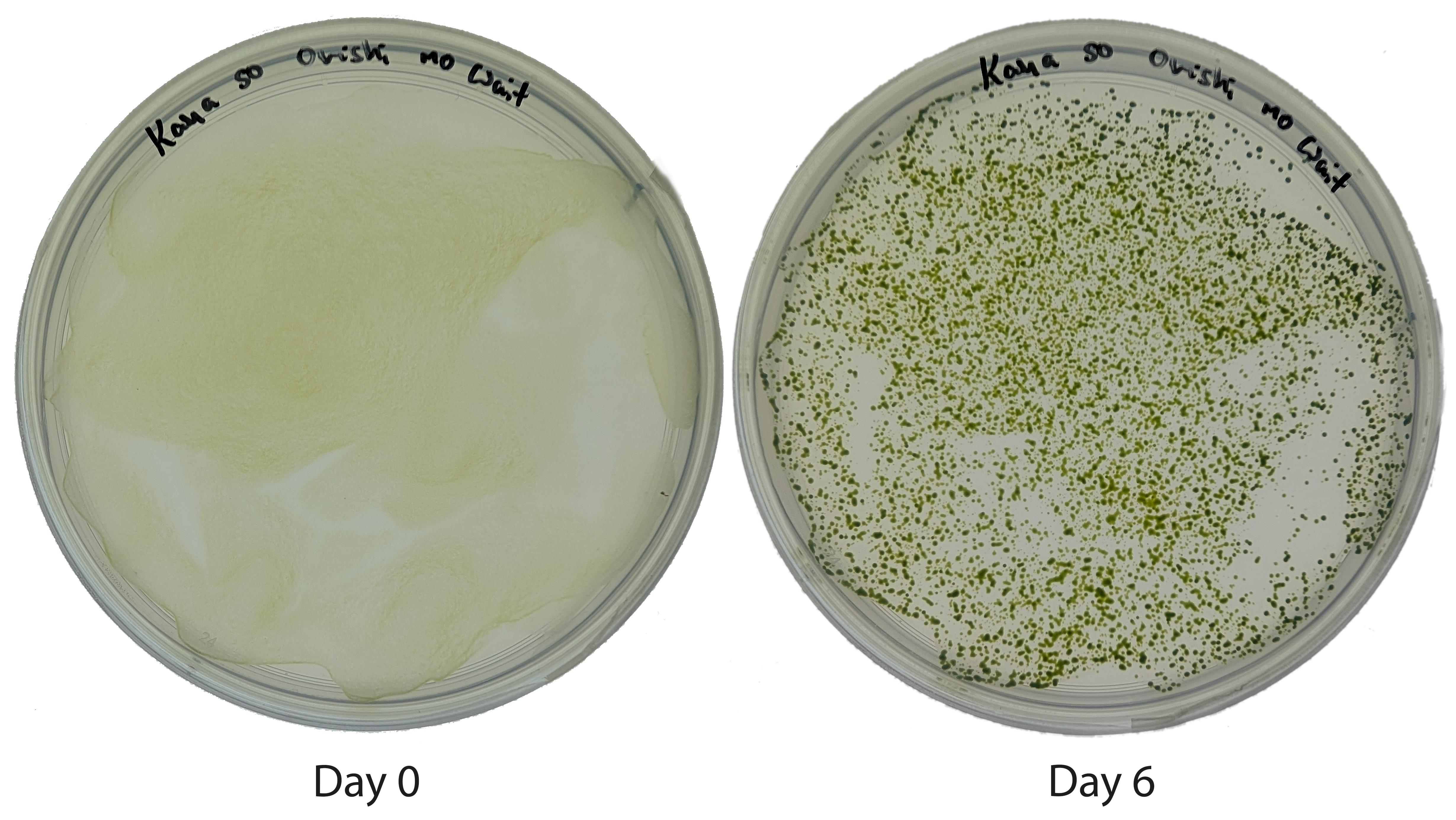
Figure 1. Typical successful electroporation. Pictures were taken 0 and 6 days after plating. See Figure 2 legend for more details.
Validation of protocol
This protocol or a very similar one was successfully used in previous publications [5–8]. Here, it was further validated by comparing it with the “Crozet” protocol [4] and the MAX Efficiency commercial kit. Our protocol produced comparable results in terms of number of transformants or transgene expression, as estimated here by mVenus fluorescence (Figure 2). Transformant characterization depends on the goal of the experiment; it may include detection or localization of the insertion locus by PCR [4,13] or detection of the produced protein [6].
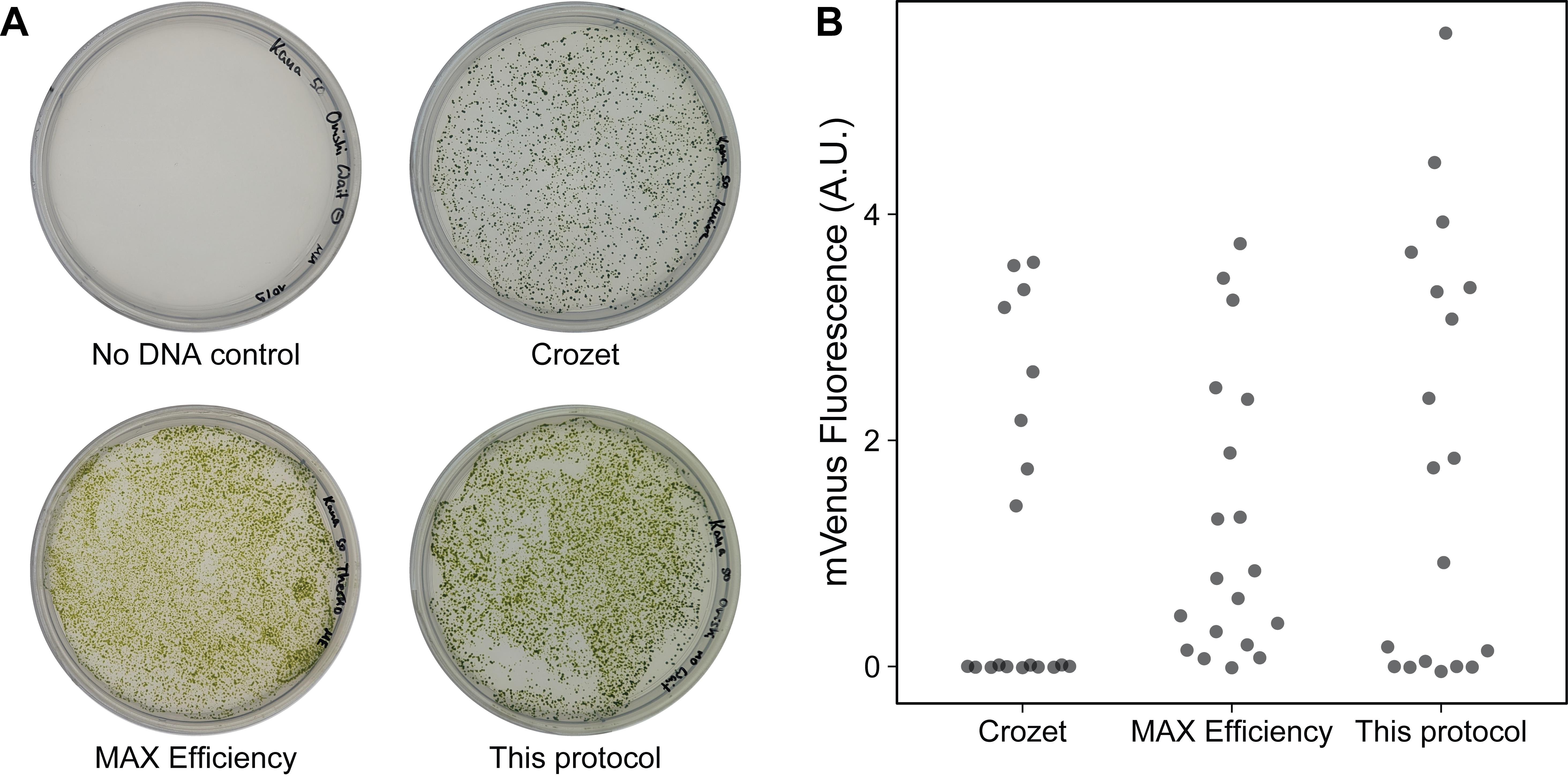
Figure 2. Validation of the electroporation protocol in a wild-type strain with an intact cell wall. (A) The plasmid pFCM-010 (pAR-NPTII-tRbcS2-pAβSAP-mVenus-tPsaD, Dataset S1) was built with the Chlamydomonas MoClo toolkit [3,14]. 500 ng of purified cassette were transformed into the 4A- wild type (with an intact cell wall) with our simplified protocol, the “Crozet” protocol [3,4], or the MAX Efficiency Transformation Reagent for Algae kit (Invitrogen) and selected on TAP+kanamycin plates. The transformation efficiency of our protocol was estimated at 13.3 × 10³ transformants/μg of DNA or 134 transformants/106 cells; that of the “Crozet” protocol was 7.0 × 10³ transformants/μg of DNA or 70 transformants/106 cells. Note that the number of transformants may depend on the expression cassette. Pictures were taken after 6 days. (B) Fluorescence of mVenus in individual transformants generated with the three protocols. 96-well plates containing 200 μL of cell culture per well were measured with a Tecan Infinite M1000 PRO plate reader (excitation: 505 nm; emission: 530 nm) and normalized with chlorophyll fluorescence (excitation: 430 nm; emission: 655 nm).
Troubleshooting
Problem: Low transformation efficiency.
Possible cause: Saturated or unhealthy culture.
Solution: Repeat the experiment and check that the preculture contains mostly single cells. If the strain is motile, cells should be swimming. Also, check that the culture is not contaminated by plating cells on Luria broth.
Supplementary information
The following supporting information can be downloaded here:
1. Dataset S1. Plasmid map of pFCM-010.
Acknowledgments
Conceptualization, M.M., F.C.; Investigation, M.M., F.C., E.L., M.H.; Data Analysis, M.M., F.C., S.W., M.S., K.K.N.; Writing—Original Draft, M.M., F.C.; Writing—Review & Editing, M.M., F.C., E.L., M.H., S.W., M.S., K.K.N.; Funding acquisition, M.S., K.K.N.; Supervision, F.C., S.W., K.K.N.
We acknowledge Dr. Masayuki Onishi for developing the original version of the protocol and Dr. Olivier D. Caspari for providing it to us. This work was supported by the Howard Hughes Medical Institute, the BioComp 4.0 program (RPTU), and UC Berkeley Sponsored Projects for Undergraduate Research (SPUR) program. K.K.N. is an investigator of the Howard Hughes Medical Institute, and S.W. is supported by the U.S. Department of Energy, Office of Science, through the Photosynthetic Systems program in the Office of Basic Energy Sciences.
Competing interests
The authors declare no conflicts of interest.
References
- Jinkerson, R. E. and Jonikas, M. C. (2015). Molecular techniques to interrogate and edit the Chlamydomonas nuclear genome. Plant J. 82(3): 393–412. https://doi.org/10.1111/tpj.12801
- Yamano, T. and Fukuzawa, H. (2020). Transformation of the Model Microalga Chlamydomonas reinhardtii Without Cell-Wall Removal. In S. Li, L. Chang, and J. Teissie (Eds.), Electroporation Protocols: Microorganism, Mammalian System, and Nanodevice (pp. 155–161). New York, NY: Springer US. https://doi.org/10.1007/978-1-4939-9740-4_16
- Crozet, P., Navarro, F. J., Willmund, F., Mehrshahi, P., Bakowski, K., Lauersen, K. J., Pérez-Pérez, M. E., Auroy, P., Gorchs Rovira, A., Sauret-Gueto, S., et al. (2018). Birth of a Photosynthetic Chassis: A MoClo Toolkit Enabling Synthetic Biology in the Microalga Chlamydomonas reinhardtii. ACS Synth Biol. 7(9): 2074–2086. https://doi.org/10.1021/acssynbio.8b00251
- de Carpentier, F., Le Peillet, J., Boisset, N. D., Crozet, P., Lemaire, S. D. and Danon, A. (2020). Blasticidin S Deaminase: A New Efficient Selectable Marker for Chlamydomonas reinhardtii. Front Plant Sci. 11: e00242. https://doi.org/10.3389/fpls.2020.00242
- Onishi, M. and Pringle, J. R. (2016). Robust Transgene Expression from Bicistronic mRNA in the Green Alga Chlamydomonas reinhardtii. G3 Genes|Genomes|Genetics 6(12): 4115–4125. https://doi.org/10.1534/g3.116.033035
- Caspari, O. D. (2020). Introduction of a leaky stop codon as molecular tool in Chlamydomonas reinhardtii. PLoS One. 15(8): e0237405. https://doi.org/10.1371/journal.pone.0237405
- de Carpentier, F., Maes, A., Marchand, C. H., Chung, C., Durand, C., Crozet, P., Lemaire, S. D. and Danon, A. (2022). How abiotic stress-induced socialization leads to the formation of massive aggregates in Chlamydomonas. Plant Physiol. 190(3): 1927–1940. https://doi.org/10.1093/plphys/kiac321
- Lambert, L., de Carpentier, F., André, P., Marchand, C. H. and Danon, A. (2023). Type II metacaspase mediates light-dependent programmed cell death in Chlamydomonas reinhardtii. Plant Physiol. 194(4): 2648–2662. https://doi.org/10.1093/plphys/kiad618
- Niemeyer, J. and Schroda, M. (2022). New destination vectors facilitate Modular Cloning for Chlamydomonas. Curr Genet. 68: 531–536. https://doi.org/10.1007/s00294-022-01239-x
- Kropat, J., Hong‐Hermesdorf, A., Casero, D., Ent, P., Castruita, M., Pellegrini, M., Merchant, S. S. and Malasarn, D. (2011). A revised mineral nutrient supplement increases biomass and growth rate in Chlamydomonas reinhardtii. Plant J. 66(5): 770–780. https://doi.org/10.1111/j.1365-313x.2011.04537.x
- Yang, X., Peng, J. and Pan, J. (2019). Nourseothricin N-acetyl transferase (NAT), a new selectable marker for nuclear gene expression in Chlamydomonas. Plant Methods. 15(1): 140. https://doi.org/10.1186/s13007-019-0526-5
- Haire, T. C., Bell, C., Cutshaw, K., Swiger, B., Winkelmann, K. and Palmer, A. G. (2018). Robust Microplate-Based Methods for Culturing and in Vivo Phenotypic Screening of Chlamydomonas reinhardtii. Front Plant Sci. 9: e00235. https://doi.org/10.3389/fpls.2018.00235
- Kong, F. and Li-Beisson, Y. (2018). Identification of Insertion Site by RESDA-PCR in Chlamydomonas Mutants Generated by AphVIII Random Insertional Mutagenesis. Bio Protoc. 8(3): e2718. https://doi.org/10.21769/bioprotoc.2718
- Einhaus, A., Baier, T., Rosenstengel, M., Freudenberg, R. A. and Kruse, O. (2021). Rational Promoter Engineering Enables Robust Terpene Production in Microalgae. ACS Synth Biol. 10(4): 847–856. https://doi.org/10.1021/acssynbio.0c00632
Article Information
Publication history
Received: Mar 20, 2026
Accepted: May 28, 2026
Available online: Jun 11, 2026
Published: Jul 20, 2026
Copyright
© 2026 The Author(s); This is an open access article under the CC BY license (https://creativecommons.org/licenses/by/4.0/).
How to cite
Readers should cite both the Bio-protocol article and the original research article where this protocol was used:
- Meßmer, M., de Carpentier, F., Lam, E., Hong, M., Wakao, S., Schroda, M. and Niyogi, K. K. (2026). Simple Electroporation of Chlamydomonas reinhardtii Strains With an Intact Cell Wall. Bio-protocol 16(14): e5740. DOI: 10.21769/BioProtoc.5740.
- de Carpentier, F., Maes, A., Marchand, C. H., Chung, C., Durand, C., Crozet, P., Lemaire, S. D. and Danon, A. (2022). How abiotic stress-induced socialization leads to the formation of massive aggregates in Chlamydomonas. Plant Physiol. 190(3): 1927–1940. https://doi.org/10.1093/plphys/kiac321

 Download Citation in RIS Format
Category
Plant Science > Plant transformation > Electroporation
Plant Science > Plant cell biology > Cell wall
Plant Science > Phycology > Nuclear transformation
Do you have any questions about this protocol?
Post your question to gather feedback from the community. We will also invite the authors of this article to respond.
 Post a Question


