


- Protocols

- Articles and Issues
- For Authors
- About
- Become a Reviewer


Improved Protocol for Establishing CD4+ Hybridomas Specific for Human Class II MHC/Peptide Complex

Published: Vol 16, Iss 13, Jul 5, 2026 DOI: 10.21769/BioProtoc.5737 Views: 533
Reviewed by: Navnita DuttaPriyanka ShandilyaChhuttan L Meena
 PDF
PDF  Ask a question
Original research article
The authors used this protocol in:
Apr 2020
Advertisement


Protocol Collections
Comprehensive collections of detailed, peer-reviewed protocols focusing on specific topics




Related protocols
Abstract
Autoreactive CD4+ T cells are shaped by MHC class II–dependent selection, and HLA-DQ8 is a major susceptibility allele for type 1 diabetes and celiac disease. To define how HLA-DQ8 influences the autoreactive CD4+ T-cell repertoire, we generated T-cell hybridomas from HLA-DQ8 humanized mice using a BW5147 Nur77-GFP (BW-GFP) platform that enables sensitive quantification of antigen-induced T-cell receptor (TCR) signaling. The frequency of autoreactive conventional CD4+ hybridomas observed in HLA-DQ8 mice was higher than previously reported in C57BL/6 mice in our earlier study, suggesting that HLA-DQ8 shapes an autoreactive repertoire. However, because antigen presentation in this system is restricted by human HLA-DQ8 while hybridomas express murine CD4, we considered that CD4-MHC interspecies mismatch might affect signal strength and influence the apparent magnitude of autoreactivity. To address this limitation, we engineered a BW-GFP fusion partner expressing an optimized version of human CD4 (hCD4), restoring optimal CD4-HLA-DQ8 interactions. Hybridomas generated with this modified platform from both regulatory (Treg) and conventional (non-Treg) CD4+ T cells exhibited enhanced responses to HLA-DQ8/peptide complexes compared with hybridomas that do not express hCD4. This approach improves the reactivity and physiological accuracy of screening mouse-derived CD4 hybridomas specific to self and foreign antigens presented by human class II MHC complexes.
Key features
• Improved BW5147 Nur77-GFP thymoma with optimized human CD4 enhances CD4–HLA class II compatibility in HLA-DQ8 humanized hybridoma systems.
• BW modification preserves fusion efficiency with Treg and conventional CD4 T cells, comparable to the parental BW line (n = 10 times for validation).
• Optimized for studying HLA-DQ8-restricted autoimmunity and adaptable to other HLA-transgenic mouse models.
Keywords: Humanized mice HLA-DQ8
T-cell hybridoma
Human CD4
Antigen-specific T cells
Genetic manipulation
HLA-DQ8
T-cell hybridoma
Human CD4
Antigen-specific T cells
Genetic manipulation
Graphical overview
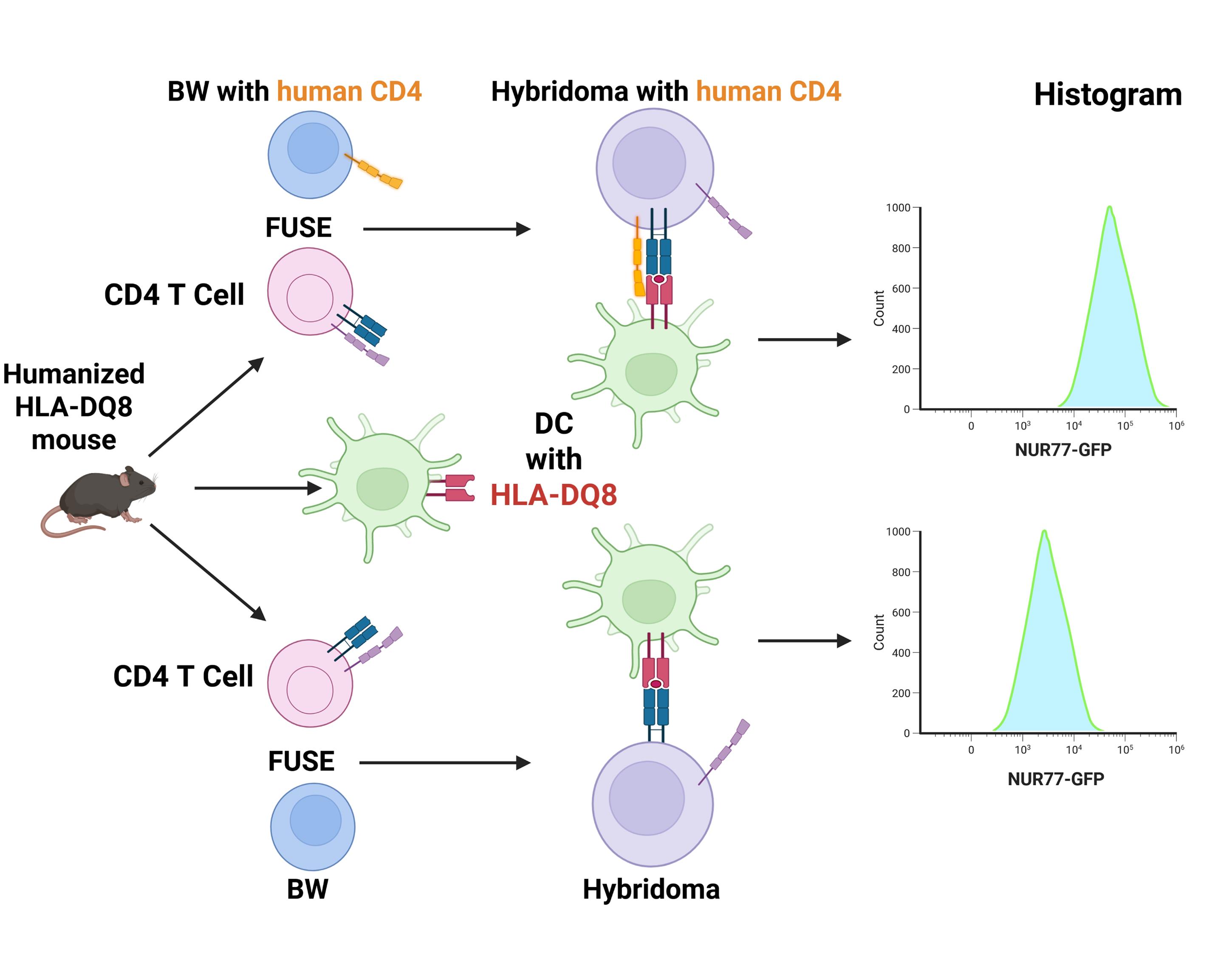
Hybridomas expressing optimized human CD4 are more responsive to human class II MHC/peptide complex. In this hybridoma-based system, antigen-specific responses are evaluated by co-culturing with autologous dendritic cells expressing human HLA-DQ8. Because conventional hybridomas retain murine CD4, the species mismatch with human MHC class II may limit TCR signaling efficiency. Engineered BW-GFP thymoma fusion partner expressing human CD4 addresses this limitation.
Background
In the normal immune repertoire, autoreactive CD4 T cells normally remain quiescent, as their activation is tightly restrained by central and peripheral tolerance. MHC class II molecules play a key role in shaping the repertoire [1]. Extensive genetic and immunological studies have shown that HLA-DQ8 is one of the strongest MHC class II susceptibility alleles for type 1 diabetes (T1D) and celiac disease (CD). HLA-DQ8 contributes to disease risk by preferentially presenting specific self or dietary peptides to CD4+ T cells, thereby promoting pathogenic immune activation [2–5]. Genetically engineered mice expressing human HLA-DQ8 in the absence of endogenous murine MHC class II (Ab-) molecules represent a robust model for investigating autoimmune disease pathogenesis and for characterizing HLA-restricted autoreactive CD4+ T-cell responses [6]. To define antigen specificity and T-cell receptor (TCR)-mediated activation in this context, T-cell hybridomas provide a stable, immortalized, and reproducible platform for antigen-specific functional assays, thereby overcoming the variability and maintenance limitations associated with primary T-cell cultures [7]. T-cell hybridomas are generated by fusing primary murine T cells with an immortal thymoma cell line, enabling stable propagation while preserving antigen-specific TCR expression. Our approach builds upon the hybridoma-based methodology used by several researchers, including Ignatowicz’s laboratory, which utilized BW5147-derived thymoma fusion partners to generate stable, MHC class II–restricted T-cell hybridomas for the analysis of TCR specificity and selection [8,9]. In our system, this platform is further enhanced through integration of a Nur77-GFP reporter into the BW5147 line, allowing highly sensitive recognition and quantification of antigen-induced TCR signaling [10,11]. Using this platform, we analyzed autoreactive responses in sorted CD4+Foxp3- conventional (non-Treg) and CD4+Foxp3+ (Treg) T cells isolated from various lymphatic organs of HLA-DQ8 humanized mice. Approximately 10% of hybridomas derived from CD4+Foxp3- cells exhibited autoreactivity measured by Nur77-GFP upregulation (unpublished data). In contrast, a prior study from our laboratory [12] demonstrated no detectable autoreactive CD4+ T-cell responses in C57BL/6 mice, which express endogenous murine MHC class II molecules and lack human HLA-DQ8. These findings suggest that expression of HLA-DQ8, rather than background genetic differences, drives the expanded autoreactive CD4+ T-cell repertoire.
In this hybridoma-based system, antigen-specific activation is assessed by co-culturing hybridomas with autologous dendritic cells expressing human MHC class II (HLA-DQ8), providing a physiologically relevant model of HLA-restricted antigen presentation and TCR signaling. Efficient activation depends on CD4-mediated stabilization of the TCR–peptide–MHC complex and initiation of proximal signaling events. However, murine CD4 may interact sub-optimally with human HLA-DQ8, which could potentially affect early signaling efficiency [13].
To address this limitation, we engineered a BW-GFP thymoma fusion partner expressing human CD4. The resulting hybridomas co-express murine and human CD4, which is expected to improve CD4–MHC class II compatibility and may lead to more efficient proximal TCR signaling.
We fused both Treg and conventional (non-Treg) CD4+ T cells isolated from HLA-DQ8 Ab- Foxp3-GFP mice with this novel hCD4-expressing BW-GFP line for comparison with the previous BW-GFP line.
Beyond correcting CD4-MHC interspecies incompatibility, this system provides a sensitive and standardized platform for analyzing HLA-DQ8-restricted antigen recognition. It enables precise epitope mapping, quantification of TCR signaling thresholds, and detailed assessment of antigen presentation efficiency in a humanized MHC context.
Moreover, this approach offers a versatile tool for preclinical screening of antigen-specific immunotherapies and can be readily adapted to other HLA-transgenic models to enhance the physiological relevance of humanized T-cell assays.
Materials and reagents
Biological materials
1. HLA-DQ8 Ab- Foxp3-GFP mice (our laboratory, 8–10 weeks of age)
2. Dendritic cells produced from the bone marrow of HLA-DQ8 Ab- Foxp3-GFP mice (our laboratory) [11]
3. BW-GFP thymoma (our laboratory) [11]
4. Phoenix-Eco packaging cells (ATCC, catalog number: CRL-3214)
Reagents
1. CaCl2 (Sigma, catalog number: 223506-25G)
2. HEPES (Sigma, catalog number: H-7006)
3. Na2HPO4 (Sigma, catalog number: S9763-100G)
4. Anti-hCD4 antibody (APC/eFire750; clone RPA-T4) (BioLegend, catalog number: 300559; RRID AB_2629692)
5. Isotype antibody (APC/eFire750; mouse IgG1, κ clone MOPC-21) (BioLegend, catalog number: 400195; RRID AB_2942001)
6. Anti-mCD4 antibody (APC, clone GK1.5) (BioLegend, catalog number: 100412, RRID: AB_312697)
7. Anti-TCR β antibody (Brilliant Violet 650, clone H57-597) (BioLegend, catalog number: 109251, RRID: AB_2810348)
8. Anti-mCD3 antibody (clone: 145-2C11) (BioLegend, catalog number: 100340, RRID: AB_11149115)
9. Anti-mCD28 antibody (clone: 37.51) (BioLegend, catalog number: 102116, RRID: AB_11147170)
10. PBS without calcium and magnesium (Fisher, catalog number: MT21040CV)
11. DAPI (Thermo Fisher Scientific, catalog number: 62247)
12. PMA (Sigma, catalog number: P1585-1MG)
13. Ionomycin (Sigma, catalog number: I0634-1MG)
14. EDTA (Invitrogen, catalog number: AM9260G)
15. FBS (R&D Systems, catalog number: S11150H)
16. MEM (Minimum Essential Medium Eagle with Earle’s salts & L-glutamine) (Corning, catalog number: 10-010-CV)
17. HAT supplement 50× (Gibco, catalog number: 21060-017)
18. Hank’s balanced salt solution (HBSS) without calcium and magnesium and phenol red (Corning, catalog number: 21-022-CM)
19. Polybrene (Sigma, catalog number: H9268-5G)
20. Boric acid (Fisher Chemical, catalog number: A73-500)
21. Recombinant murine IL2 (PEPROTECH, catalog number: 212-12-20UG)
22. Polyethylene glycol (PEG) 1450, Waxy Soft Solid (J.T.Baker, catalog number: U220-07)
23. Dextrose (Sigma, catalog number: G7021)
24. Glutamine (Sigma, catalog number: G-8540)
25. Essential amino acids (50×) (Gibco, catalog number: 111-30051)
26. Non-essential amino acids (100×) (Gibco, catalog number: 11140-050)
27. Sodium pyruvate (100×) (Gibco, catalog number: 11360-070)
28. Sodium bicarbonate (Sigma, catalog number: S-5761)
29. Gentamycin (Sigma, catalog number: G-3632)
30. Penicillin G (Sigma, catalog number: P3032)
31. Streptomycin sulfate (Sigma, catalog number: S-9137)
32. 2-Mercaptoethanol (14.3 M) (Sigma, catalog number: M-7522)
33. NaCl (Fisher Chemical, catalog number: S271-5000)
34. NaOH (Fisher Chemical, catalog number: SS267)
35. HCl (Fisher Chemical, catalog number: SA48-1)
36. Freund’s adjuvant, complete (Sigma, catalog number: F5881-6X 10 mL)
37. Freund’s adjuvant, incomplete (Sigma, catalog number: F5506-6X 10 mL)
Solutions
1. 2.5 M CaCl2 (see Recipes)
2. 2× HBS (see Recipes)
3. Borate buffer (see Recipes)
4. Tumor cocktail (see Recipes)
5. FACS buffer
Recipes
1. 2.5 M CaCl2
| Reagent | Final concentration | Quantity or volume |
|---|---|---|
| CaCl2 | 2.5 M | 27.2 g |
| H2O | n/a | 100 mL |
2. 2× HBS
| Reagent | Final concentration | Quantity or volume |
|---|---|---|
| NaCl | 1.6% | 0.8 g |
| HEPES | 1.3% | 0.65 g |
| Na2HPO4 | 2% | 1 mL |
| H2O | n/a | 49 mL |
| NaOH/HCl | n/a | n/a |
To make Na2HPO4 stock solution, add 0.525 g in 50 mL water. Adjust pH to 7.0 using NaOH or HCl. Bring the volume up to 50 mL. Check pH again.
Critical: The pH is very important; it must be exactly 7.0.
3. Borate buffer
| Reagent | Final concentration | Quantity or volume |
|---|---|---|
| Boric acid | 0.1 M | 0.6183 g |
| H2O | n/a | 100 mL |
| NaOH | n/a | n/a |
Adjust pH to 8.5 using NaOH. Bring the volume up to 100 mL. Check pH again.
4. Tumor cocktail
| Reagent | Final concentration | Quantity or volume |
|---|---|---|
| MEM | n/a | 351 mL |
| Dextrose | 11.21 mg/mL | 7.5 g |
| Glutamine | 4.87 mg/mL | 3.259 g |
| Essential amino acids (50×) | 11.21% v/v | 75 mL |
| Non-essential amino acids (100×) | 20.93% v/v | 140 mL |
| Sodium pyruvate (100×) | 14.95% v/v | 100 mL |
| Adjust pH to 7.0 with 10 N NaOH | ||
| Sodium bicarbonate | 12.71 mg/mL | 8.5 g |
| Gentamycin | 0.747 mg/mL | 500 mg |
| Penicillin G | 0.897 mg/mL | 600 mg |
| Streptomycin sulfate | 1.495 mg/mL | 1 g |
| 2-Mercaptoethanol (14.3 M) | 0.0508 μL/mL | 34 μL |
Filter through a 0.22 µm filter. Aliquot 30 mL per tube. Store at -20 °C.
5. FACS buffer
| Reagent | Final concentration | Quantity or volume |
|---|---|---|
| PBS | - | 1 L |
| EDTA | 1 mM | 372 mg |
| FBS | 1% (v/v) | 10 mL |
Laboratory supplies
1. 96-well round-bottom plate (Corning, catalog number: 3797)
2. 96-well flat-bottom plate (Corning, catalog number: 3596)
3. Falcon polypropylene tubes (Fisher, catalog number: 05-538-53D)
4. 40 μm strainer (Corning, catalog number: CLS431750)
5. 25 cm2 cell culture flask (Corning, catalog number: 430639)
Equipment
1. Flow cytometry analyzers and cell sorter (Beckman):
a. Analyzers: CytoFLEX LX (N3-V5-B3-Y5-R3-I2), 6 lasers (375, 405, 488, 561, 638, 808 nm), 21 parameters
b. Sorter: CytoFLEX SRT (V5-B2-Y5-R3); 4 lasers (405, 488, 561, 638 nm); 15 parameters; 4 -way sorter, capable of sorting single cells onto a 96 or 384 plate
2. Cell counter (Beckman, model: Z1 Coulter particle counter)
3. Centrifuge (Thermo Scientific, Sorvall Legend, model: XTR)
4. Pipette (PIPETMAN, model: GILSON)
5. Incubator (Thermo Scientific, model: HERACELL VIOS 160i CO2 Incubator)
6. Microscope (Leica, model: DMIL LED)
7. Biosafety cabinet class II (Thermo Scientific, model: 1300 Series A2)
Software and datasets
1. FlowJo (v10.10.0; BD)
2. Prism (v11.0.1; GraphPad)
Procedure
A. Production of retroviral particles
Note: This protocol is based on a modified version of the method reported by Nolan Lab [13].
The Phoenix system enables convenient, single-plasmid production of retroviral particles capable of infecting mouse cells. In this protocol, we used the calcium phosphate method for transfecting mammalian cells because it is cost-effective and relies on reagents commonly available in most laboratories. As alternatives to calcium phosphate, commercially available transfection reagents such as Fugene HD (Promega, catalog number: E2311) or Lipofectamine 3000 (Invitrogen, catalog number: L3000001) can also be used.
1. Plate 0.2–0.3 × 10⁶ Phoenix-Eco cells per well in 2.5 mL of culture medium without HEPES [e.g., Complete tumor medium (CTM) [14]: 500 mL of MEM + 50 mL of FBS + 30 mL of tumor cocktail] on a 6-well plate.
2. Allow cells to reach ~70% confluence within 24 h.
3. Immediately before transfection, remove the CTM and add 1 mL of fresh, warm CTM.
4. Combine the following in a single tube: Virus DNA (Figure 1), 5 μg; H2O (to bring the volume to 180 μL), 175.5 μL; 2.5 M CaCl2 (final CaCl2 concentration: 0.25 M), 20 μL.
(Optional) For use with 293T cells instead of Phoenix cells, add 2 μg of pCl Eco (Addgene, catalog number: 12371) and 1 μg of VSV G (Addgene, catalog number: 138479).
5. To the tube prepared above, add 200 μL of 2× HBS slowly, mixing gently to allow the calcium phosphate–DNA precipitate to form.
6. Transfer the DNA/CaCl2 mixture onto the cells and incubate at 37 °C.
Note: Calcium phosphate–DNA precipitates should be visible under the microscope.
7. After 8–16 h, replace the medium with fresh CTM.
8. Harvest viral supernatant at 48 and 72 h post-transfection.
9. Centrifuge at 1,000× g for 5 min to remove cells and debris.
10. Store clarified supernatant at -80 °C.
Optional (1): Concentrate viral particles using a 100 kDa Amicon column (Sigma, catalog number: UFC910008).
Optional (2): Assess transfection efficiency by staining Phoenix cells with anti-hCD4 antibody (clone RPA T4) and analyzing via flow cytometry (FACS) or fluorescence microscopy. Use supernatant from wells with the highest frequency of hCD4+ cells.

Figure 1. Diagram of retroviral vector encoding hCD4. The mutant human CD4 construct carrying the Q40Y and T45W mutations.
B. Transduction of BW5147 Nur77-GFP (BW-GFP) thymoma
Note: Production of BW-GFP cells has been previously described [11]. For introduction of hCD4, follow these steps.
1. Transfer 1 × 10⁵ BW-GFP cells to a 15 mL Falcon tube, centrifuge (500× g, 5 min), and wash twice with PBS.
2. Resuspend the cell pellet in 0.5–1 mL of viral supernatant.
3. Add polybrene to 2–4 μg/mL.
4. Centrifuge at 2,000× g for 90 min at 32 °C (to prevent overheating).
5. Remove viral supernatant, resuspend the pellet in 2 mL of warm CTM, and return the cells to the incubator.
6. Test transduction efficiency 2–3 days later by staining with anti-hCD4 antibody (clone RPA-T4).
7. Freeze cells and store at -80 °C if needed.
C. Transduction efficiency
Note: Transduction efficiency is assessed by flow cytometry (FACS). For a detailed FACS staining method, see [15].
1. Transfer 5 × 10⁴ cells/well into a 96-well round-bottom plate and centrifuge (500× g, 1 min).
2. Add titrated anti-hCD4 antibody (5–10 μL/well). Include one unstained control.
3. Incubate for 10–15 min at room temperature (or 30 min on ice) in the dark.
4. Wash cells once with PBS.
5. Resuspend in PBS, add a viability dye (e.g., DAPI), and analyze by flow cytometry.
6. Expand the well(s) containing the highest proportion of hCD4+ cells. Cells are selected based on the level of expression (MFI) of hCD4 (see Figures 2 and 3).
Optional: Confirm retention of inducible Nur77-GFP expression by stimulating cells overnight with PMA (5 ng/mL) and ionomycin (100 ng/mL), then assessing hCD4+GFP+ cells. Use unstimulated cells as the GFP baseline control.
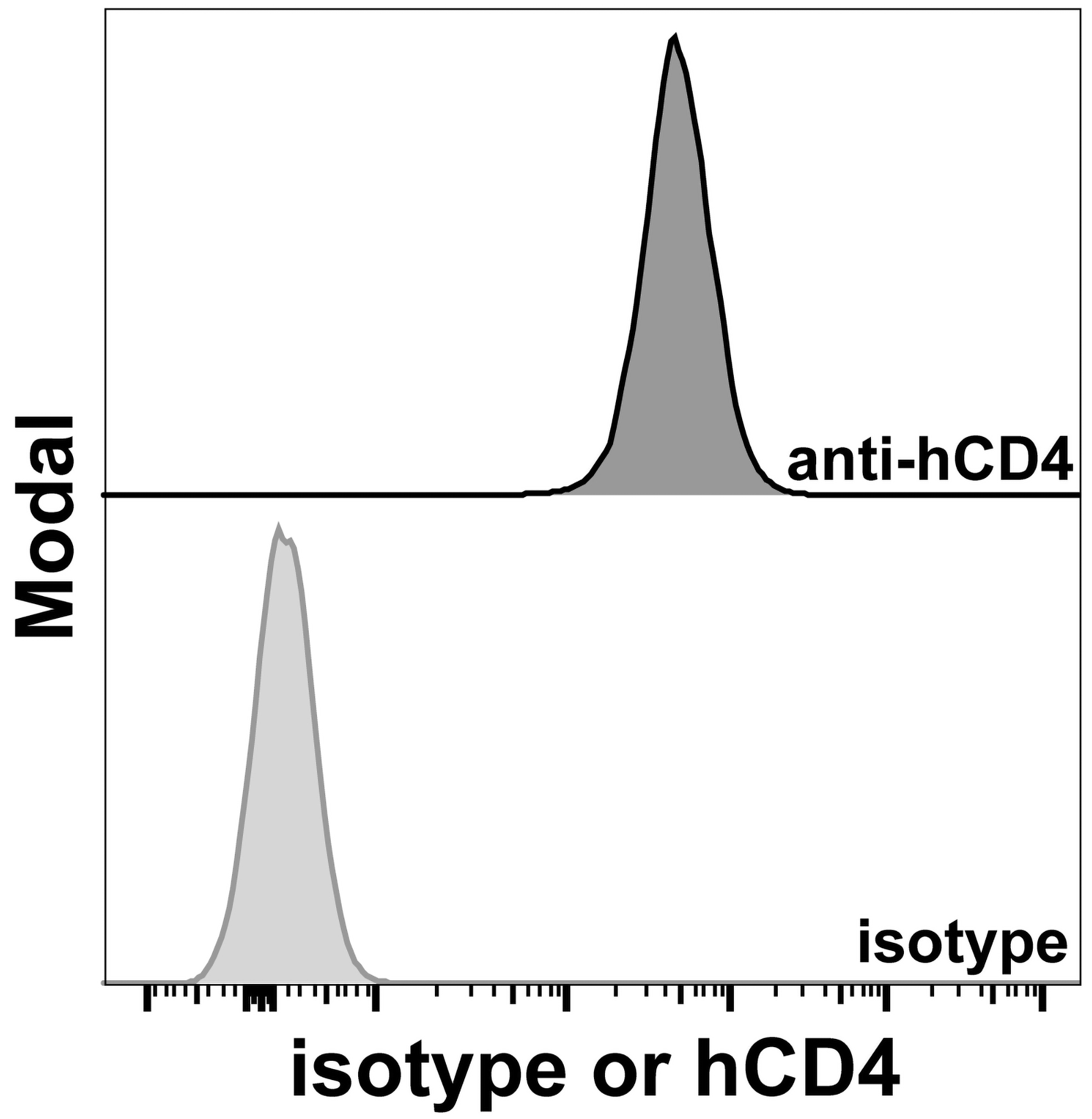
Figure 2. Transduction efficacy. Transduced BW5147 Nur77-GFP hCD4 cells were stained with an isotype control or an anti-hCD4 antibody. Expression of hCD4 is shown.
D. Preparation of BW5147 Nur77-GFP hCD4 (BW-GFP hCD4) cell line (single-cell FACS sorting)
1. Stimulate cells overnight with PMA + ionomycin (as above).
2. Collect cells (15 mL Falcon tube; 500× g, 5 min).
3. Wash with PBS, then resuspend in 0.5 mL of FACS buffer and filter through a 40 μm strainer.
4. Stain with anti-hCD4 antibody.
5. Wash once with 10 mL of PBS.
6. Resuspend cells in 2–3 mL of FACS buffer, add DAPI, and filter again through a 40 μm strainer.
7. Collect unstained and unstimulated cells on the sorter to establish gating.
8. Apply the stained sample and sort CD4+GFP+ cells, selecting cells with the highest expression.
9. Sort cells into FACS buffer or culture medium.
Note: Cells may be sorted directly as single cells into a 96-well round-bottom plate prefilled with 100 μL of CTM/well, or sorted in bulk and then re-sorted at 1 cell/well. Bulk-sorted material can be frozen as backup in case cloning needs to be repeated.
10. Return 96-well plates to the incubator. Add another 100 μL of CTM/well after 3–4 days. Culture until a visible pellet forms at the bottom of the well (inspecting multiple wells under a microscope may be time-consuming). Verify the clonal origin of each well.
11. Confirm hCD4 and GFP expressions as described above, using appropriate gating controls. Choose the clone with the highest expression of both markers.
Note: The authors assume that readers are familiar with flow cytometry, FACS sorting, and the use of FlowJo; for details, see [15] and other sources.
12. In our work, Clone 1.1 was selected as a final fusion partner.
Notes:
1. Transduced cells are sorted to high purity (>99%) by FACS as single-cell clones.
2. Stable integration results in sustained expression of hCD4 and Nur77-GFP.
3. Expression can be verified by flow cytometry before fusion experiments.
4. Re-sorting can be performed if needed upon restimulation with PMA/ionomycin, as described.
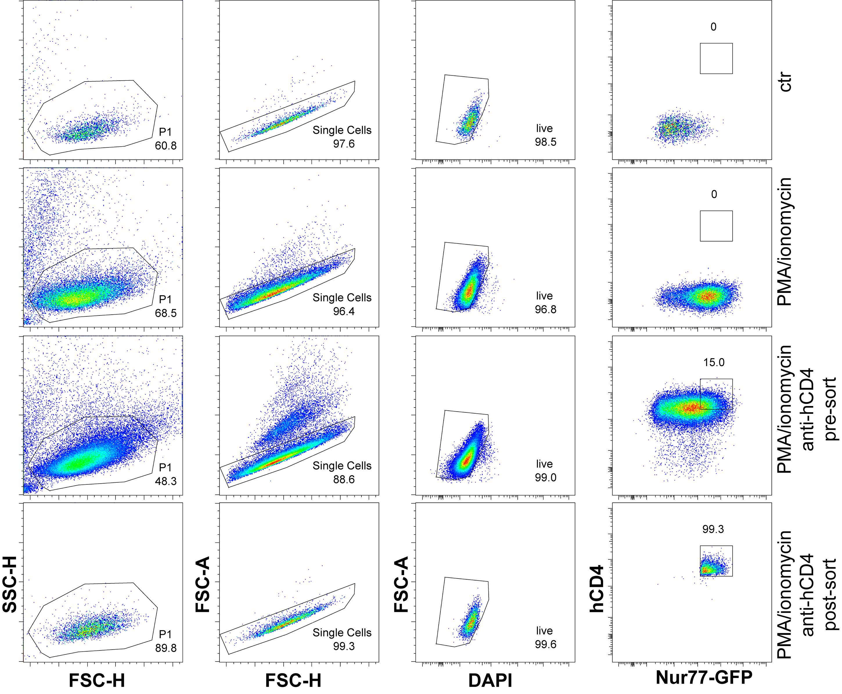
Figure 3. Generation of the cell line. Unstained and unstimulated or PMA/ionomycin-stimulated cells were used as controls to assign sorting gates. Pre- and post-sorting of CD4+GFP+ cell frequencies are shown. Bulk hCD4+Nur77-GFP+ cells were subsequently sorted as single cells.
E. Cell fusion/preparation of hybridomas
1. Immunize HLA-DQ8 Ab- Foxp3-GFP mice with complete Freund’s adjuvant (CFA) (50 μL + 50 μL PBS), followed by a booster immunization with incomplete Freund’s adjuvant (IFA; 50 μL + 50 μL PBS) one week later. Both injections were intraperitoneal. In our laboratory, the HLA-DQ8 Ab- Foxp3-GFP strain was generated by crossing HLA-DQ8 Ab- transgenic mice, which were kindly donated to our laboratory by Joseph Murray [16], with C57BL/6 Foxp3-GFP reporter mice [17] to generate this experimental strain. Following immunization, euthanize the mice under approved conditions and harvest mesenteric lymph nodes under sterile conditions.
Note: We evaluated this newly developed system only in CFA-immunized mice, with the intention of testing its performance under inflammatory conditions. While we reasoned that functionality in this context would support broader applicability, its use in other settings (e.g., naïve mice or cancer models) has not been directly assessed and will require independent validation.
2. Mechanically dissociate the lymph nodes in HBSS + 2% FBS to generate a single-cell suspension. Pass the suspension through a cell strainer to remove debris and obtain a uniform cell preparation.
3. Incubate cells with anti-mCD4 and anti-TCR antibodies for 30 min on ice. After incubation, add HBSS and centrifuge the cells to wash away excess antibody. Resuspend the cells in HBSS (10 × 106/mL).
4. Sort the cells using the cell sorter. Isolate CD4+ Foxp3- (conventional T cells) and CD4+Foxp3+ (regulatory T cells) populations.
5. Expand and fuse the cells according to the protocol [18]. Briefly culture sorted cells in antibody-coated flasks to induce activation.
6. For CD4+Foxp3- conventional T cells, coat flasks with anti-mCD3 (10 μg/mL) and anti-mCD28 (1 μg/mL) in 3 mL of PBS.
7. For CD4+Foxp3+ regulatory T cells, coat flasks with anti-mCD3 (5 μg/mL) and anti-mCD28 (5 μg/mL) prepared in 3 mL of borate buffer.
8. Add recombinant IL-2 (10 ng/mL) immediately to regulatory T-cell cultures. For conventional T cells, add IL-2 (10 ng/mL) after 24–48 h to support cell proliferation and expansion.
9. After activation and expansion, fuse 1 × 10⁶ T cells (Treg or non-Treg) with at least 10 × 106 BW-GFP hCD4 or BW-GFP thymoma cells by adding 1 mL of PEG (50%).
10. After fusion, distribute the cells into flat-bottom 96-well plates.
11. Determine cell concentration, dilute to ~1 cell/100 μL, and plate 100 μL per well (Poisson-based limiting dilution gives ~37% wells with exactly one cell [19,20]). Mix thoroughly at each dilution step and use fresh tips to ensure uniform distribution before dispensing into the 96-well plate. On the following day, add HAT selection medium (1×) and maintain the cultures under selection for approximately one week before screening the resulting hybridomas. Screen the hybridomas by checking expression of TCR, hCD4, and mCD4 (Figure 4). Transfer TCR+hCD4+mCD4+ hybridomas from a 96-well to a 24-well plate and add 500 μL of MEM + HAT to expand them. Check the hybridoma daily under the microscope.
Note: The time that cells become ready and reach ~50% confluence depends on the hybridoma. Do not let cells overgrow.
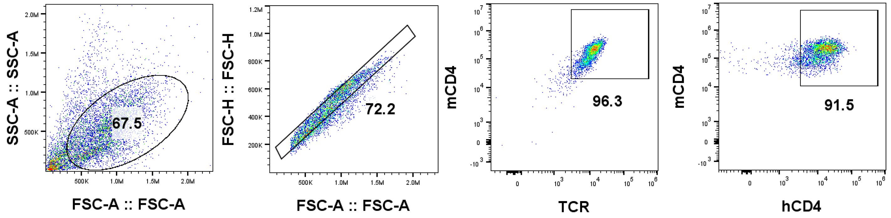
Figure 4. Flow cytometric screening of hybridomas for T cell receptor (TCR), human CD4 (hCD4), and murine CD4 (mCD4) expression. Clones co-expressing these markers were considered optimal for HLA-DQ8-restricted functional analyses.
F. Hybridoma testing
1. Plate 2 × 10⁵ hybridoma suspension onto the well of a round-bottom 96-well plate in triplicate. In the first well, seed hybridoma cells alone. In the second well, co-culture hybridomas with 2 × 10⁴ dendritic cells per well. In the third well, include 2 × 10⁴ dendritic cells per well with anti-mCD3 (1.5 μg/mL) antibody as a positive control for T-cell activation.
2. Incubate for 16 h and check the Nur77-GFP expression by flow cytometry (Figure 5).
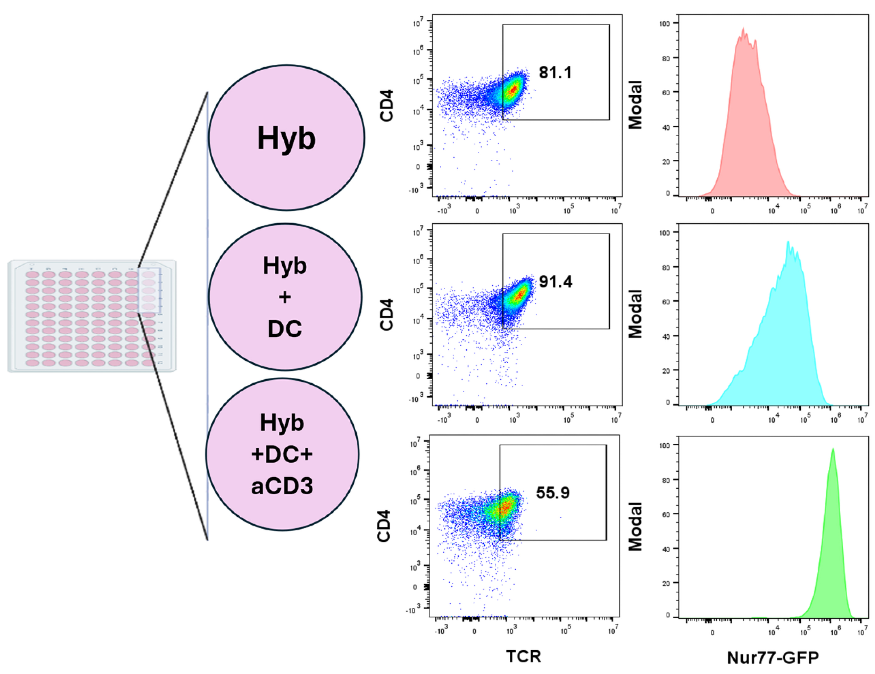
Figure 5. Experimental setup for hybridoma testing. Hyb, hybridoma; DC, dendritic cell; aCD3, anti-mCD3. Strong (aCD3) stimulation downregulates surface TCR [21].
3. Measure the mean fluorescence intensity (MFI) of the Nur77-GFP signal for each condition using FlowJo software. Calculate the response as the ratio of MFI in hybridomas stimulated with dendritic cells to MFI in unstimulated hybridomas (baseline Nur77 expression). This ratio represents fold change over background, with higher values indicating stronger responses (Figure 6).
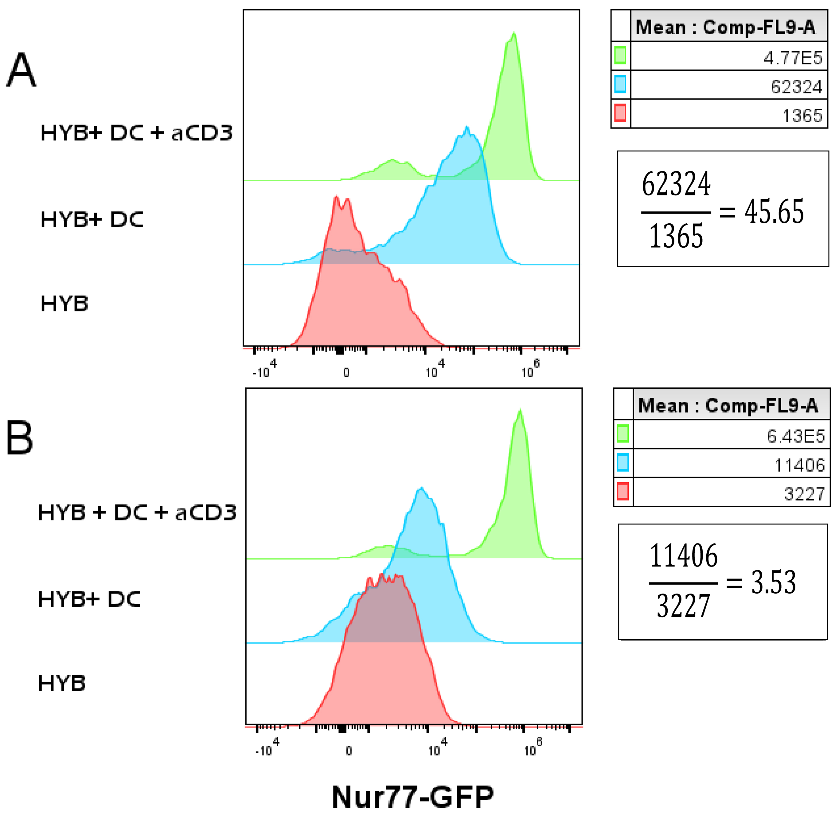
Figure 6. Typical Nur77 expression in responding and non-responding hybridomas. (A) Autoreactive hybridomas exhibit robust activation in response to autologous dendritic cells. (B) Non-autoreactive hybridomas display minimal or no response to autologous dendritic cells. HYB, hybridoma; DC, dendritic cell; aCD3, anti-mCD3.
G. Comparing Treg and non-Treg hybridomas
Perform the procedures separately for hybridomas prepared from CD4+ T-cell subsets (CD4+Foxp3- conventional T cells and CD4+Foxp3+ regulatory T cells), maintaining identical experimental conditions to ensure accurate comparison between populations. Test each hybridoma in three independent experimental runs under identical conditions (Figure 7).
This approach enables direct evaluation of differences in the responses of regulatory and effector CD4+ T cells to self-peptides in the presence or absence of human CD4, representing a key conceptual and technical advance of the study.
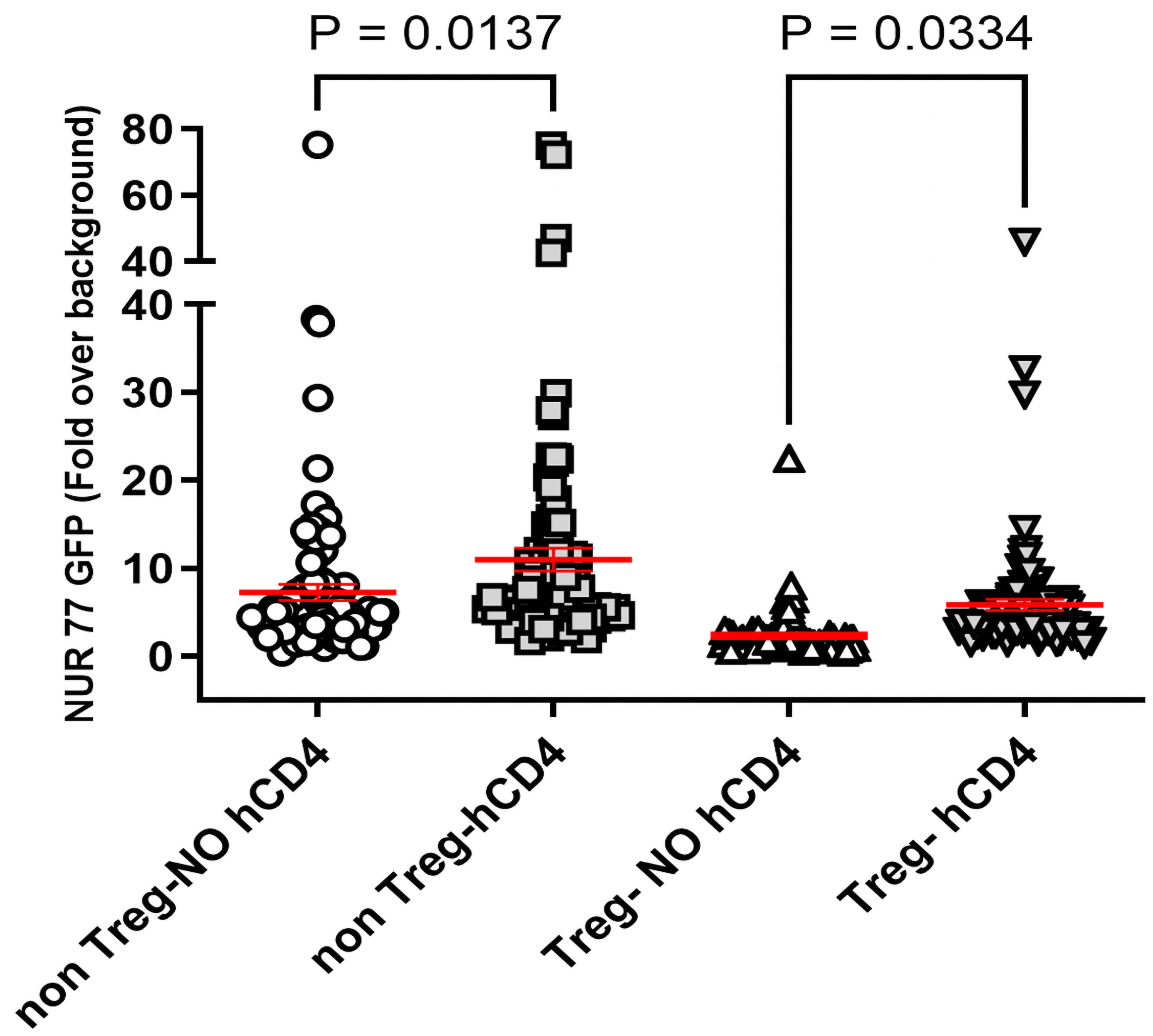
Figure 7. Fusion with hCD4-expressing BW cells increased antigen-specific responses in hybridomas derived from both Treg and conventional (non-Treg) CD4+ T cells, consistent with improved CD4-HLA-DQ8 compatibility and resolution of interspecies mismatch. Each data point represents the mean of three independent measurements of the mean fluorescence intensity (MFI) ratio per hybridoma. Sample sizes are as follows: non-Treg without hCD4 (n = 107), non-Treg with hCD4 (n = 92), Treg without hCD4 (n = 81), and Treg with hCD4 (n = 94) from four separate fusions, 3–5 mice in each fusion. Statistical comparisons among multiple groups were conducted using one-way analysis of variance (ANOVA), followed by Tukey’s post hoc test for pairwise comparisons in GraphPad Prism software. Error bars show the standard error of the mean (SEM).
Data analysis
Analyze all data using Microsoft Excel and GraphPad Prism software. Perform statistical comparisons among four groups using one-way analysis of variance (ANOVA), followed by Tukey’s post hoc test for multiple comparisons. Statistical significance was defined as p < 0.05. Test each hybridoma in three independent experimental runs under identical conditions. Use anti-mCD3 stimulation as a positive control because of its potent ability to induce T-cell activation. Hybridomas that failed to respond to anti-mCD3 stimulation should be excluded from subsequent analyses.
Validation of protocol
Information about the number of replicates, statistical tests applied, and controls is mentioned in the Data analysis section.
This protocol or parts of it have been used and validated in the following research article(s):
Kuczma et al. [11]. Commensal epitopes drive differentiation of colonic Tregs. Science advances. (Figures 1 and 2)
General notes and troubleshooting
Troubleshooting
Problem: Low transduction efficiency.
Possible cause: Viral-related or target cell–related issues.
Solutions:
1. Select high-expressing producer wells.
2. Perform serial dilutions of viral supernatant (e.g., 1:50–1:500) to test the titer and select optimal MOI for transduction efficiency.
3. Concentrate viral particles when transduction efficiency is low.
4. Sort cells with FACS cell sorter (best) or enrich cells (hCD4+) with magnetic beads (anti-hCD4-bead) using Miltenyi or StemCell Technology systems.
Please also see the guide in [13].
Acknowledgments
F.M., M.P.K., and L.I. contributed to conceptualization and original draft preparation. F.M. and M.P.K performed most of the experiments and analyzed the data. E.A.S. engineered the original BW5147 introducing Nur77-GFP [10,11]. E.A.S. and A.C. contributed to manuscript review and editing. L.I. and M.P.K. secured funding and supervised the study. We acknowledge Maki Nakayama for providing the retroviral vector encoding modified hCD4 [22] (supported by grant P30 DK116073).
This project was supported by Georgia State University intramural funding, NIH grant R01AG068875 to L.I., and CDA 833676 (Crohn’s and Colitis Foundation) to M.P.K. The protocol described in this manuscript was developed and validated in [11]. In the current report, the BW-GFP variant also includes hCD4 and was tested in different conditions: APCs with HLADQ8.
The authors further acknowledge the use of ChatGPT (OpenAI) for grammatical refinement and language editing of the manuscript.
Competing interests
The authors declare that there are no conflicts of interest related to this work.
Ethical considerations
Animals were maintained in a specific pathogen-free (SPF) environment within the animal facility at Georgia State University. All experimental procedures were conducted in accordance with the guidelines and approvals of the University’s Institutional Animal Care and Use Committee (IACUC), protocol number A25041, ensuring compliance with established ethical and welfare standards.
References
- Klein, L. and Petrozziello, E. (2024). Antigen presentation for central tolerance induction. Nat Rev Immunol. 25(1): 57–72. https://doi.org/10.1038/s41577-024-01076-8
- Lundin, K. E. and Sollid, L. M. (2014). Advances in coeliac disease. Curr Opin Gastroenterol 30(2): 154–162. https://doi.org/10.1097/MOG.0000000000000041.
- Lebwohl, B., Sanders, D. S. and Green, P. H. R. (2018). Coeliac disease. Lancet. 391(10115): 70-81. https://doi.org/10.1016/S0140-6736(17)31796-8.
- Pugliese, A. (2017). Autoreactive T cells in type 1 diabetes. J Clin Invest. 127(8): 2881–2891. https://doi.org/10.1172/jci94549
- Erlich, H., Valdes, A. M., Noble, J., Carlson, J. A., Varney, M., Concannon, P., Mychaleckyj, J. C., Todd, J. A., Bonella, P., Fear, A. L., et al. (2008). HLA DR-DQ Haplotypes and Genotypes and Type 1 Diabetes Risk. Diabetes. 57(4): 1084–1092. https://doi.org/10.2337/db07-1331
- Nabozny, G. H., Baisch, J. M., Cheng, S., Cosgrove, D., Griffiths, M. M., Luthra, H. S. and David, C. S. (1996). HLA-DQ8 transgenic mice are highly susceptible to collagen-induced arthritis: a novel model for human polyarthritis. J Exp Med. 183(1): 27–37. https://doi.org/10.1084/jem.183.1.27
- Kappler, J., Skidmore, B., White, J. and Marrack, P. (1981). Antigen-inducible, H-2-restricted, interleukin-2-producing T cell hybridomas. Lack of independent antigen and H-2 recognition. J Exp Med. 153(5): 1198–1214. https://doi.org/10.1084/jem.153.5.1198
- White, J., Blackman, M., Bill, J., Kappler, J., Marrack, P., Gold, D. P. and Born, W. (1989). Two better cell lines for making hybridomas expressing specific T cell receptors.. J Immunol. 143(6): 1822–1825. https://doi.org/10.4049/jimmunol.143.6.1822
- Ignatowicz, L., Kappler, J. and Marrack, P. (1996). The Repertoire of T Cells Shaped by a Single MHC/Peptide Ligand. Cell. 84(4): 521–529. https://doi.org/10.1016/s0092-8674(00)81028-4
- Kuczma, M. P., Szurek, E. A., Cebula, A., Ngo, V. L., Pietrzak, M., Kraj, P., Denning, T. L. and Ignatowicz, L. (2021). Self and microbiota-derived epitopes induce CD4+ T cell anergy and conversion into CD4+Foxp3+ regulatory cells. Mucosal Immunol. 14(2): 443–454. https://doi.org/10.1038/s41385-020-00349-4
- Kuczma, M. P., Szurek, E. A., Cebula, A., Chassaing, B., Jung, Y. J., Kang, S. M., Fox, J. G., Stecher, B. and Ignatowicz, L. (2020). Commensal epitopes drive differentiation of colonic T regs. Sci Adv. 6(16): eaaz3186. https://doi.org/10.1126/sciadv.aaz3186
- Cebula, A., Kuczma, M., Szurek, E., Pietrzak, M., Savage, N., Elhefnawy, W. R., Rempala, G., Kraj, P. and Ignatowicz, L. (2019). Dormant pathogenic CD4+ T cells are prevalent in the peripheral repertoire of healthy mice. Nat Commun. 10(1): 4882. https://doi.org/10.1038/s41467-019-12820-3
- Nolan lab. (Accessed on https://web.stanford.edu/group/nolan/_OldWebsite/protocols/pro_helper_dep.html)
- Kappler-Marrack Complete Tumor Medium. (Accessed on https://www.nationaljewish.org/getmedia/a8ad806e-21ca-46a5-8f6f-4cc6bbc10945/pdf-KMLab-CTM-Recipe.pdf)
- Mow, R., Kuczma, M., Shi, X., Merlin, D. and Yang, C. (2025). Tracking Oral Nanoparticle Uptake in Mouse Gastrointestinal Tract by Fluorescent Labeling and t-SNE Flow Cytometry. Bio Protoc. 15(1372): e5309. https://doi.org/10.21769/bioprotoc.5309
- Ju, J. M., Marietta, E. V. and Murray, J. A. (2015). Generating Transgenic Mouse Models for Studying Celiac Disease. Methods Mol Biol. 1326: 23–33. https://doi.org/10.1007/978-1-4939-2839-2_3
- Kuczma, M., Pawlikowska, I., Kopij, M., Podolsky, R., Rempala, G. A. and Kraj, P. (2009). TCR Repertoire and Foxp3 Expression Define Functionally Distinct Subsets of CD4+ Regulatory T Cells. J Immunol. 183(5): 3118–3129. https://doi.org/10.4049/jimmunol.0900514
- Singh, N., Pacholczyk, R., Iwashima, M. and Ignatowicz, L. (2011). Generation of T Cell Hybridomas from Naturally Occurring FoxP3+ Regulatory T Cells. Methods Mol Biol. 707: 39–44. https://doi.org/10.1007/978-1-61737-979-6_3
- Ye, M., Wilhelm, M., Gentschev, I. and Szalay, A. (2021). A Modified Limiting Dilution Method for Monoclonal Stable Cell Line Selection Using a Real-Time Fluorescence Imaging System: A Practical Workflow and Advanced Applications. Methods Protoc. 4(1): 16. https://doi.org/10.3390/mps4010016
- Waldmann, H. and Lefkovits, I. (1984). Limiting dilution analysis of cells of the immune system II: What can be learnt?. Immunol Today. 5(10): 295–298. https://doi.org/10.1016/0167-5699(84)90154-3
- San Jose, E., Borroto, A., Niedergang, F., Alcover, A. and Alarcon, B. (2000). Triggering the TCR complex causes the downregulation of nonengaged receptors by a signal transduction-dependent mechanism. Immunity. 12(2): 161–170. https://doi.org/10.1016/s1074-7613(00)80169-7
- Williams, T., Krovi, H. S., Landry, L. G., Crawford, F., Jin, N., Hohenstein, A., DeNicola, M. E., Michels, A. W., Davidson, H. W., Kent, S. C., et al. (2018). Development of T cell lines sensitive to antigen stimulation. J Immunol Methods. 462: 65–73. https://doi.org/10.1016/j.jim.2018.08.011
Article Information
Publication history
Received: Apr 7, 2026
Accepted: May 22, 2026
Available online: Jun 10, 2026
Published: Jul 5, 2026
Copyright
© 2026 The Author(s); This is an open access article under the CC BY-NC license (https://creativecommons.org/licenses/by-nc/4.0/).
How to cite
Readers should cite both the Bio-protocol article and the original research article where this protocol was used:
- Mousavinasab, F., Szurek, E. A., Cebula, A., Ignatowicz, L. and Kuczma, M. P. (2026). Improved Protocol for Establishing CD4+ Hybridomas Specific for Human Class II MHC/Peptide Complex. Bio-protocol 16(13): e5737. DOI: 10.21769/BioProtoc.5737.
- Kuczma, M. P., Szurek, E. A., Cebula, A., Chassaing, B., Jung, Y. J., Kang, S. M., Fox, J. G., Stecher, B. and Ignatowicz, L. (2020). Commensal epitopes drive differentiation of colonic T regs. Sci Adv. 6(16): eaaz3186. https://doi.org/10.1126/sciadv.aaz3186

 Download Citation in RIS Format
Category
Immunology > Immune cell function > Antigen-specific response
Immunology > Animal model > Mouse
Cell Biology > Cell engineering > Cell fusion
Do you have any questions about this protocol?
Post your question to gather feedback from the community. We will also invite the authors of this article to respond.
 Post a Question


