


- Protocols

- Articles and Issues
- For Authors
- About
- Become a Reviewer


A Versatile In Vitro Quantitative Assay for Macrophage Efferocytosis in Diverse Research Applications

(*contributed equally to this work) Published: Vol 16, Iss 10, May 20, 2026 DOI: 10.21769/BioProtoc.5690 Views: 1526
Reviewed by: Philipp WörsdörferScott McCombAnonymous reviewer(s)
 PDF
PDF  Ask a question
Original research article
The authors used this protocol in:
Jan 2026
Advertisement



Protocol Collections
Comprehensive collections of detailed, peer-reviewed protocols focusing on specific topics




Related protocols
Abstract
Macrophage efferocytosis is a previously unrecognized key pathogenic event, engulfing apoptotic targets, preventing inflammation and necrosis, and maintaining immune homeostasis. The phagocytic function can be disrupted by harmful factors and toxic substances. This protocol describes a versatile visualized in vitro method that can be used for the detection of general efferocytosis. This method is applicable to a wide range of research scenarios. As a representative application, it can be used to evaluate macrophage efferocytosis dysfunction in diseases linked to harmful exposures, including atherosclerosis, chronic inflammation, and malignant tumors. Among them, the detection of the effects of oxidized low-density lipoprotein (ox-LDL) and arsenite on macrophage efferocytosis capacity is an exemplary application of this protocol. Primary macrophages collected from mice were labeled with a cell-tracking dye and exposed to ox-LDL or arsenite, then co-cultured with apoptotic thymocytes or hepatocytes (labeled with another cell-tracking dye) for 2 h at a ratio of 5:1. Macrophage efferocytosis was visualized using a laser confocal microscope. The results indicate that arsenite impaired macrophage efferocytosis, leading to insufficient clearance of apoptotic thymocytes or hepatocytes. This method can be extended to subsequent studies, including those involving different types of phagocytes, apoptotic cell models, and research related to exposure to various factors.
Key features
• This protocol harvests primary mouse macrophages, treats them with different factors (ox-LDL, sodium arsenite), and assesses efferocytosis of apoptotic/necrotic thymocytes or hepatocytes.
• Dye-swap red–green staining eliminates fluorescence interference and staining bias and validates dye effects on efferocytosis.
Keywords: Efferocytosis Macrophages
Thymocytes
Hepatocytes
Apoptotic targets
Macrophages
Thymocytes
Hepatocytes
Apoptotic targets
Graphical overview
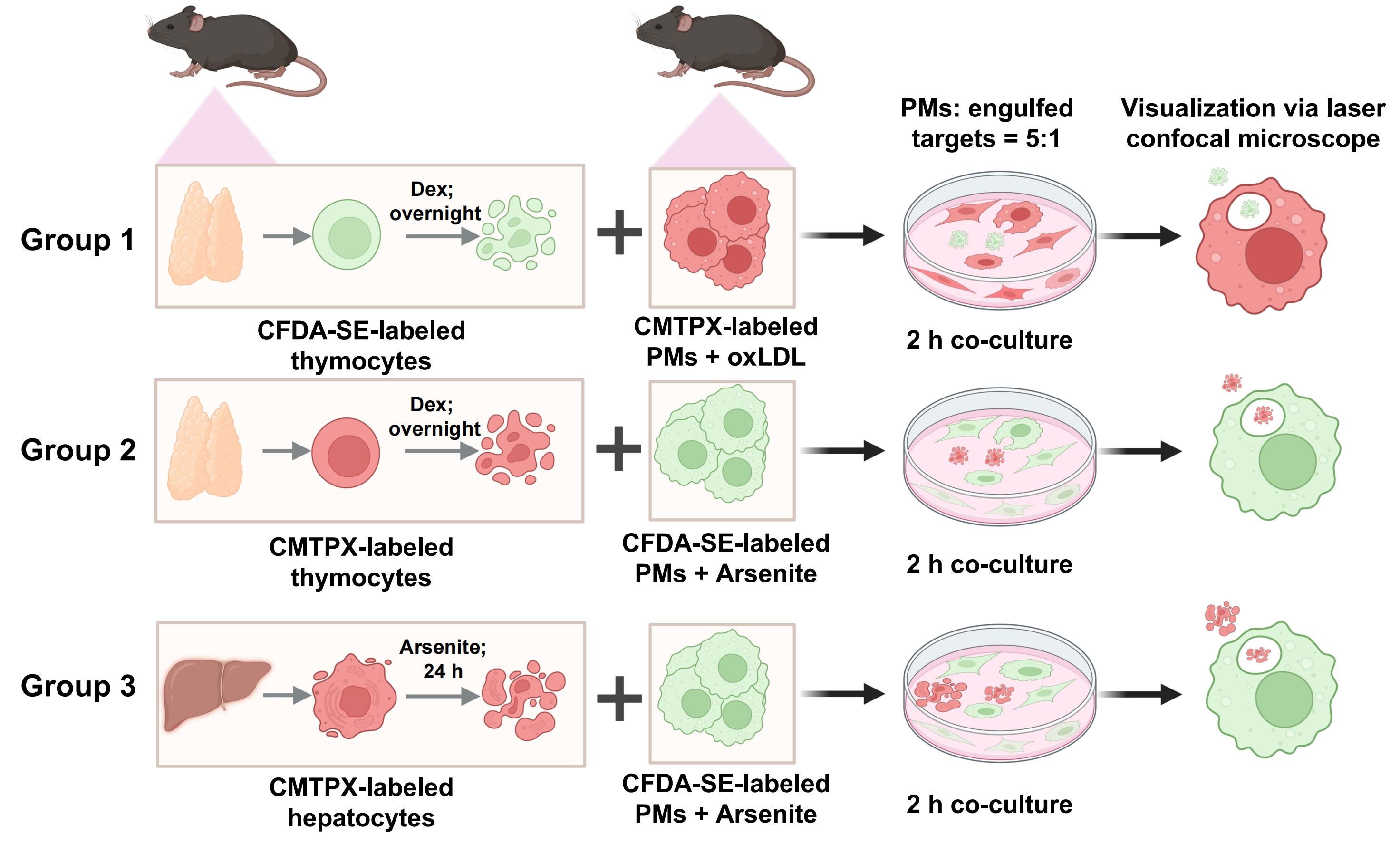
Confocal laser scanning microscopy is employed to evaluate the efferocytosis of various apoptotic cells by primary peritoneal macrophages (PMs) in response to different factors [oxidized low-density lipoprotein (ox-LDL) and sodium arsenite]. This schematic illustrates the step-by-step experimental procedure, including efferocytosis of dexamethasone-induced apoptotic/necrotic thymocytes by macrophages under ox-LDL exposure, efferocytosis of dexamethasone-induced apoptotic/necrotic thymocytes by macrophages under sodium arsenite exposure, and efferocytosis of sodium arsenite-induced apoptotic/necrotic hepatocytes by macrophages under sodium arsenite exposure.
Background
Efferocytosis, a specialized type of phagocytosis, is defined as the phagocytosis and clearance of apoptotic, aged, or injured cells [1,2]. Impaired efferocytosis causes an inflammatory response and leads to disease states [2,3]. Macrophages are the major component involved in efferocytosis [4]. Impaired macrophage efferocytosis exacerbates inflammation and tissue damage, thereby advancing the progression of various inflammatory disorders, such as atherosclerosis, autoimmune conditions, and cancer [2,5–8]. Under physiological conditions, macrophages residing in almost every organ and tissue are the major phagocytes to perform efferocytosis [9–11]. Some tissue-resident macrophages have specific names, such as alveolar macrophages in the lungs, microglia in the brain, and Kupffer cells in the liver [1]. All of these cells are involved in removing apoptotic cells through efferocytosis in different tissues [1].
Various factors (e.g., alkaloids, advanced glycation end products, modified low-density lipoproteins, heavy metals, pesticides, and pollutant residues) have a pathogenetic role in the development and progression of impaired efferocytosis-related diseases, including diabetes [12], cardiovascular diseases [13], and neurological disorders [14]. Previous studies have demonstrated that 7-ketocholesterol, 12(S)-hydroxyeicosatetraenoic acid, 20-OH-RvE4, SiO2, ethanol, and acetaldehyde impair the efferocytosis capacities of mouse or human macrophages [15–19]. Arsenic, classified as a metalloid, is a naturally occurring element that is widely distributed in the Earth's crust [20]. Environmental arsenic exposure correlates with multi-system disorders, including skin [21], nervous [22], respiratory [23], cardiovascular [24,25], and immune systems [26]. Arsenic is one of WHO’s 10 chemicals of major public health concern [27]. In this study, sodium arsenite was used as a representative factor to elucidate its effect on macrophage efferocytosis. Low-density lipoproteins (LDLs), especially oxidized LDL (ox-LDL), play a key role in promoting atherosclerosis [28,29]. High levels of ox-LDL are considered a risk factor for cardiovascular events due to their central role in atherosclerotic plaque formation [30]. In this study, ox-LDL was selected as another distinct representative factor to investigate its regulatory effects on macrophage efferocytosis.
In the present study, a visualized in vitro protocol is applied to evaluate the effects of different factors on efferocytosis. This approach involves isolating primary cells, co-culturing cell-tracking dye-labeled macrophages with apoptotic cells, and recording the results using laser confocal microscopy. This approach offers several advantages, including convenience and visualization, particularly of the intricate details of cellular processes [31,32]. The superior resolution and depth of field provided by confocal microscopy allow for the observation of intracellular structures, facilitating the analysis of morphological changes during efferocytosis, such as the formation of pseudopodia and the engulfment of apoptotic targets. These details enable researchers to directly observe the process of macrophages engulfing apoptotic targets and the nuances of their interactions.
This protocol presents an updated efferocytosis assay protocol, based on existing approaches [33,34]. The protocol details two distinct approaches—the first using two distinct representative factors (ox-LDL and arsenite) and the second using two kinds of apoptotic cells (thymocytes and hepatocytes). These approaches clarify the efferocytotic capacity of macrophages toward apoptotic cells generated under different conditions. Furthermore, they provide a reliable method for evaluating the effects of different factors on macrophage efferocytosis.
Materials and reagents
Reagents
1. Isoflurane (RWD, catalog number: R510-22-10)
2. Annexin V-FITC/PI Apoptosis Detection kit (Elabscience, catalog number: E-CK-A211)
3. CellTraceTM CMTPX Red cell tracer (Yeasen Biotechnology, catalog number: 40717ES)
4. CFDA SE Cell Proliferation and Cell Tracking kit (Yeasen Biotechnology, catalog number: 40714ES), comprising CFDA-SE fluorescent probe (40714-A), CFDA-SE solvent (40714-B), and 5× cell staining buffer (40714-C)
5. Collagenase Type IV (Sigma, catalog number: C5138, specific activity ≥125 CDU/mg)
6. Dexamethasone (GlpBio, catalog number: GC40775)
7. EDTA (Invitrogen, catalog number: 91222920)
8. M199 medium (GlpBio, catalog number: C11150500BT)
9. Oxidized LDL (ox-LDL) (Yiyuan Biotech, catalog number: YB-002)
10. Penicillin-streptomycin solution (pen/strep) (Procell Life Science & Technology, catalog number: PB180120)
11. RPMI 1640 culture medium (Gibco, catalog number: C11875500BT)
12. Sodium arsenite (Sigma-Aldrich, catalog number: S9663)
13. Thioglycollate medium (Sigma-Aldrich, catalog number: T9032)
14. Phosphate buffer saline (PBS), pH 7.4 (Biopico Life Sciences, catalog number: 090501)
15. 1 M HEPES solution (Procell Life Science & Technology, catalog number: PB180325)
16. 10× Hanks’ balanced salt solution (HBSS), Ca2+/Mg2+-free (Sigma-Aldrich, catalog number: H4641)
17. Calcium chloride, anhydrous (CaCl2) (Sangon Biotech, catalog number: A501330)
18. Red blood cell lysis buffer (Elabscience, catalog number: E-CK-A105)
19. Fetal bovine serum (FBS) (Procell Life Science & Technology, catalog number: 164210)
20. Sodium pyruvate solution (Procell Life Science & Technology, catalog number: PB180422)
21. GlutaMAX (Gibco, catalog number: 35050061)
22. Puralube vet ointment (Dechra, catalog number: 1703321138)
Solutions
1. 3% (w/v) thioglycollate medium (see Recipes)
2. 1,000× CFDA-SE stock solution (see Recipes)
3. CFDA-SE working solution (see Recipes)
4. Wash buffer (see Recipes)
5. Collagen buffer (see Recipes)
Recipes
1. 3% (w/v) thioglycollate medium
Prepare fresh 3% (w/v) thioglycollate medium aseptically in a biosafety cabinet by dissolving 0.03 g of thioglycollate medium powder (4 °C storage) in 1 mL of sterile PBS. Immediately apply the prepared solution to maintain optimal induction efficiency.
2. 1,000× CFDA-SE stock solution
To prepare the 1,000× CFDA-SE stock solution, add 500 μL of CFDA-SE solvent to one tube of CFDA-SE fluorescent probe (1.4 mg) and mix thoroughly to yield a clear stock solution with a concentration of 2.8 mg/mL. Aliquot the stock solution immediately after preparation and store it at ≤ -20 °C (or -70 °C for an extended shelf life) under dry and light-protected conditions; repeated freeze-thaw cycles must be strictly avoided. The stock solution is optimal for use within 1 month and has a maximum shelf life of 2 months when stored as specified above.
3. CFDA-SE working solution
Prepare 1× cell staining buffer (the diluent for CFDA-SE) by taking the supplied 5× cell staining buffer and diluting it 1:5 with sterile deionized water (e.g., 2 mL of 5× buffer mixed with 8 mL of sterile deionized water) with thorough agitation. Store the diluted 1× buffer at 4 °C, protected from light, and use it within 1 week for optimal performance. For the 2× CFDA-SE working solution, take the 1,000× CFDA-SE stock solution thawed to 20–25 °C, dilute it 1:500 with freshly prepared 1× cell staining buffer (e.g., 2 μL of 1,000× stock added to 1 mL of 1× buffer), and mix gently to form a homogeneous 2× working solution.
Critical: The working solution must be freshly prepared immediately before use, as CFDA-SE is susceptible to hydrolysis in aqueous solutions; all preparation steps should be performed in the dark to prevent fluorophore quenching. All kit components are pre-sterilized, and no additional sterilization is required.
4. Wash buffer
| Reagent | Final concentration | Volume |
|---|---|---|
| 1 M HEPES | 50 mM | 5 mL |
| 0.5 M EDTA (pH 7.98) | 5 mM | 1 mL |
| 10× HBSS | 1× | 10 mL |
| ddH2O | 84 mL | |
| Total | 100 mL |
Prepare the solution according to the table above and autoclave before use.
5. Collagen buffer
| Reagent | Final concentration | Volume |
|---|---|---|
| 1 M HEPES | 50 mM | 1.5 mL |
| CaCl2 (50 mM) | 5 mM | 3 mL |
| 10× HBSS | 1× | 3 mL |
| ddH2O | 22.5 mL | |
| Total | 30 mL |
Prepare the solution according to the table above and autoclave prior to use.
Note: The 50 mM CaCl2 working solution is prepared by diluting 2 mL of 1 M CaCl2 stock solution (11.1 g of CaCl2 dissolved in 100 mL of ddH2O) into 38 mL of ddH2O. Due to the poor aqueous stability of collagenase, add 30 mg of collagenase powder to 30 mL of the pre-autoclaved solution immediately before primary hepatocyte isolation, yielding a working concentration of 1 mg/mL (≥125 CDU/mL) to ensure on-site preparation and immediate application.
Laboratory supplies
1. Scissors (Beyotime, catalog number: FS500)
2. Tweezers (Beyotime, catalog number: FS500)
3. 1 and 5 mL syringes (Heshou, Changzhou Yuekang Medical Devices Co., Ltd.)
4. 70 μm cell strainer (BIOFIL, catalog number: 01.TC.010.0053)
5. 15 mL centrifuge tube (BIOFIL, catalog number: CFT011150)
6. P1250 pipette tips (A-gen Biotechnology, catalog number: T-1250-B)
7. P200 pipette tips (Biosharp, catalog number: BS-200-T)
8. P10 pipette tips (Axygen, catalog number: T-300)
9. 24 G catheter (Yikang group)
10. 35 mm non-treated glass-bottom cell culture dish for laser confocal microscopy (BIOFIL, catalog number: BDD012035)
11. 60 mm cell culture dish (BIOFIL, catalog number: TCD000060)
Equipment
1. Automatic cell counter (Thermo Fisher Scientific, model: Countess 3)
2. Confocal laser scanning microscope (Nikon, model: A1R)
3. Flow cytometer (BD, model: FACSCelesta)
Software and datasets
1. FlowJo software (Version X; TreeStar, Ashland, OR, USA)
2. GraphPad Prism 5 software (GraphPad Software Inc., San Diego, CA, USA)
Procedure
A. Isolation of primary peritoneal macrophages
1. Inject 8–12-week-old mice with 3% (w/v) thioglycollate medium intraperitoneally for two consecutive days [35]. Thioglycollate medium induces controlled sterile peritoneal inflammation and efficiently elicits and enriches murine peritoneal macrophages [36]. On the first day, inject 2 mL of 3% thioglycollate medium. On the second day, inject 1 mL of 3% thioglycollate medium.
2. On the third day, euthanize mice by carbon dioxide (CO2) asphyxiation (following an approved IACUC euthanasia protocol). Alternatively, use a method that has been approved by the local animal care and use committee. Use 75% alcohol to disinfect the surface of the mice. Primary peritoneal macrophages can be extracted on an ultra-clean bench.
3. Inject 10 mL of RPMI 1640 culture medium containing 0.5% (w/v) EDTA through the peritoneal wall into the peritoneal cavity of mice using a sterile 5 mL syringe. Do not puncture the intestine or any other organ.
4. Massage the abdominal cavity gently for 10 min.
5. Aspirate the abdominal fluid with a sterile 1 mL syringe and collect it into a 15 mL centrifuge tube. Ensure that the extracted liquid is clear. If the liquid is cloudy, there may be contamination from the intestines.
6. Inject 5 mL of culture medium again, massage for 5 min, then aspirate and collect the medium into the 15 mL centrifuge tube.
7. Centrifuge the abdominal fluid suspension at 400× g for 10 min at room temperature (RT) [37]. Resuspend the collected fluid in 1 mL of red blood cell lysis buffer to lyse red blood cells (RBCs). Repeat this step until the RBCs disappear. Ensure that the final lysed cell precipitate is white without RBCs.
8. Add 2 mL of culture medium to resuspend the final lysed precipitate in the culture medium. Pass the suspended cells through a 70 μm cell strainer into a new sterile 15 mL tube.
9. Count cells with an automatic cell counter or manual trypan blue exclusion assay. Use only samples with viability ≥85% for all downstream functional assays. Control the cell density of macrophages from the experimental group to be the same as that from the control group, both at 1 × 10⁶/mL. Then, place the cells into a 35 mm dish specifically designed for laser confocal microscopy.
10. Culture peritoneal macrophages (PMs) in RPMI 1640 medium supplemented with 10% fetal bovine serum (FBS), 1 mM sodium pyruvate, 100 U/mL penicillin, and 100 μg/mL streptomycin at 37 °C in a 5% CO2 incubator (Video 1). After approximately 2 h, when peritoneal macrophages have fully adhered to the dish, wash away non-adherent cells with 1 mL of sterile PBS and add new culture medium.

11. Before co-culturing with apoptotic cells, incubate PMs with 50 μg/mL ox-LDL for 6 h or with 1 μM arsenite for 24 h. The control group is not subjected to these treatments.
Note: Surgical instruments used in ultra-clean benches require high-pressure sterilization. Primary peritoneal macrophages are isolated with thioglycollate elicitation. PMs are cultured with RPMI 1640 medium supplemented with 10% (v/v) heat-inactivated FBS, 1 mM sodium pyruvate, 100 U/mL penicillin, and 100 μg/mL streptomycin.
B. Isolation of thymocytes and induction of apoptotic thymocytes
B1. Isolation of primary thymocytes
1. Euthanize 8–12-week-old mice by carbon dioxide asphyxiation (following an approved IACUC euthanasia protocol). Alternatively, use a method that has been approved by the local animal care and use committee to euthanize the mice. Use 75% alcohol to disinfect the surface of the mice. Thymocytes can be extracted on an ultra-clean bench.
2. To isolate thymocytes, initiate the procedure by using tissue forceps to elevate the xiphoid process, followed by the upward shearing of the bilateral ribs to the sternoclavicular joint. Then, expose the milky-white, bilobed thymus located superior to the heart. Use fine pointed forceps to pull out the thymus.
3. Place the thymus into a centrifuge tube containing 10 mL of sterile PBS. Wash away the blood cells and tissue fragments sticking to the thymus surface.
4. Place the entire thymus between the frosted ends of two microscope slides and then move the slides in a back-and-forth motion to grind the thymus in a 35 mm dish containing 2 mL of RPMI 1640 medium (Figure 1).
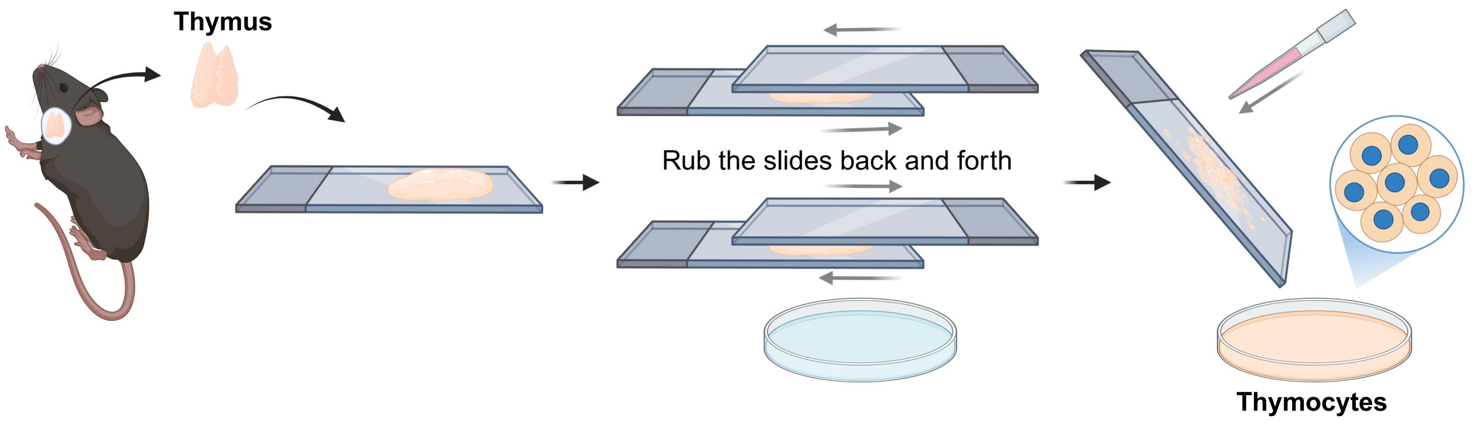
Figure 1. Isolation of primary thymocytes
Note: For thymus grinding, the two microscope slides require disinfection. The slides are thoroughly sterilized by autoclaving prior to tissue processing.
5. Collect the tissue grinding fluid and filter the suspension into a 15 mL tube through a 70 μm cell strainer.
6. Centrifuge the filtered suspension at 360× g for 15 min at RT. Discard the supernatant and resuspend the cell precipitate with 1 mL of red blood cell lysis buffer to lyse the RBCs. Repeat this step to ensure RBCs disappear.
7. After the final centrifugation to lyse red blood cells, discard the supernatant. Following this step, the harvested thymocytes are divided into two portions: one is used for subsequent differential fluorescent labeling (CFDA-SE or CMTPX), and the other is subjected to apoptosis induction for the measurement of apoptosis rate.
B2. Labeling of thymocytes with CFDA-SE Green cell tracer
1. Add 1 mL of 1× cell staining buffer to a 15 mL tube to resuspend the thymocytes. Count cells with an automatic cell counter. Dilute the thymocytes to a concentration of 5 × 10⁶/mL with 1× cell staining buffer.
2. Configure thymocyte dye: Prepare 1 mL of CFDA-SE working solution (5.6 μg/mL) by diluting 2 μL of CFDA-SE Green cell tracer in 1 mL of 1× cell staining buffer.
Note: CFDA-SE Green cell tracer is engineered to be non-toxic or exhibit low toxicity to cells. This concentration of CFDA-SE does not affect cell viability [38,39].
3. Add the prepared CFDA-SE working solution to the 15 mL tube from step B2.1 and gently resuspend to ensure homogenous mixing.
4. Incubate thymocytes in the dark at RT for 10 min (CFDA-SE Green cell tracer is used to track thymocytes). At the end of incubation, add 10 mL of RPMI 1640 medium containing 10% heat-inactivated FBS to stop the reaction. Collect cells by centrifugation at 360× g for 15 min at RT.
5. Wash the labeled cells again in 5 mL of RPMI 1640 medium containing 10% heat-inactivated FBS and centrifuge. Resuspend the cells with 5 mL of RPMI 1640 medium containing 10% heat-inactivated FBS. Incubate the cells at 37 °C for 10 min to facilitate the retention of CFDA-SE within the cells and to allow any unreacted CFDA-SE to enter the culture medium.
6. Collect cells by centrifugation and wash the labeled cells again in 5 mL of sterile PBS. Centrifuge tubes at 360× g for 15 min at RT, discard the supernatant, and add 5 mL of RPMI 1640 medium containing 10% heat-inactivated FBS. Perform the apoptotic induction steps described in section B4.
B3. Labeling of thymocytes with CMTPX Red Tracer
1. Count thymocytes with an automatic cell counter. Dilute the thymocytes to a concentration of 5 × 10⁶/mL with FBS-free RPMI 1640 medium. Supplement an aliquot of 1 mL of cell suspension with 0.5 μL of CMTPX Red cell tracer. Incubate the thymocytes at RT in the dark for 30 min. After incubation, add 1 mL of RPMI 1640 medium containing 10% heat-inactivated FBS to terminate the staining reaction.
2. Aspirate the supernatant, wash the thymocytes twice with 1 mL of sterile PBS, and then add 1 mL of RPMI 1640 medium containing 10% heat-inactivated FBS for standby. Perform the apoptotic induction steps described in section B4.
B4. Induction of apoptotic thymocytes
1. Count thymocytes with an automatic cell counter or manual trypan blue exclusion assay. Use only samples with viability ≥85% for all downstream functional assays.
2. Dilute the thymocytes to a concentration of 5 × 10⁶/mL with RPMI 1640 medium containing 10% heat-inactivated FBS.
3. Add dexamethasone to the cells at a final concentration of 1 μM. Culture the thymocytes with dexamethasone overnight in the incubator.
C. Isolation of hepatocytes and induction of apoptotic hepatocytes
1. Prewarm wash buffer (200 mL per mouse) and collagenase buffer (30 mL: 1× HBSS, 50 mM HEPES, 5 mM CaCl2, and 30 mg of collagenase type IV). Prepare sterilized forceps and scissors for dissection.
2. Anesthetize 8–12-week-old mice using isoflurane (800 mL/min O2, 3%–5% isoflurane) [40,41]. Apply Puralube vet ointment onto the eyes to prevent eye dryness [42,43]. Confirm proper anesthetization by performing loss-of-reflex tests, such as the toe pinch. Maintain anesthesia by lowering isoflurane to 2%–2.5%. Continuous isoflurane anesthesia is preferred.
3. Place the anesthetized mice on a dissection mat and fix the mouse in a supine position using waterproof tape. Disinfect the surface of the abdomen liberally with 75% ethanol.
4. Open the abdominal cavity by making a U-shape incision, approximately 2–3 cm in length, on the abdominal layers using sharp scissors and toothed forceps. Expose the inferior vena cava (IVC), liver vein, and portal vein clearly by moving the intestines and colon toward the left side of the mouse [44].
5. Insert a 24 G catheter into the IVC and avoid puncture. Start pumping the wash buffer and cut the portal vein open to allow the wash buffer to flow out from the liver to prevent expansion of the liver.
6. Continue pumping the wash buffer to remove as much blood as possible from the liver by making the buffer reach every corner of the liver. Stop pumping wash buffer when discoloration of the liver is observed. Perfuse with an entire 15 mL solution of collagenase buffer for digestion [45]. Be careful to avoid air bubbles entering the IVC.
Note: The perfusion parameters applied in this protocol are as follows: approximately 30 mL per mouse at a flow rate of 5 mL/min; digestion typically completed within 4 min, terminated when the liver turns pale, becomes flaccid, and the hepatic parenchyma dissociates. Guidance for lot-to-lot variation management: Retain the certificate of analysis and product lot number; adjust the collagenase dosage accordingly to maintain the targeted final enzymatic activity (CDU/mL) in working solution.
7. After removing the gallbladder gently using straight-tipped forceps, isolate the whole liver and transfer it into a cell culture dish containing pre-cooled M199 medium. Then, tear the liver up into pieces with forceps to release primary hepatocytes into the medium.
Note: The following steps are performed in the cell culture hood.
8. Gently blow the hepatocyte suspension through a 100 μm cell strainer into a 50 mL centrifuge tube using a sterile syringe plunger or a 1 mL pipette tip.
Note: No grinding is performed to avoid mechanical damage to the hepatocytes.
9. Centrifuge tubes at 30× g for 3 min at 4 °C. Discard the supernatant and resuspend the cells in 30 mL of M199 medium.
10. Centrifuge tubes at 30× g for 3 min at 4 °C. Remove the supernatant and resuspend the pellet with 30 mL of M199 medium.
11. Count the cell number with an automated cell counter or manual trypan blue exclusion assay. Use only samples with viability ≥85% for all downstream functional assays.
12. Use M199 medium with pen/strep, GlutaMAX, and 10% FBS to resuspend cells. Seed 2 × 10⁶ live cells in each 10 cm plate. Keep the plate in an incubator at 37 °C for 2–3 h. Then, remove floating cells and continue to culture the cells using an M199 medium containing pen/strep and GlutaMAX.
13. The next day, primary mouse hepatocytes are ready for experimental use (Video 2).
14. Add a final concentration of 10 μM arsenite to the hepatocytes and incubate for 24 h to induce apoptosis. Label cells with 1 μM CMTPX working solution by diluting 2.5 μL of CMTPX Red cell tracer in 5 mL of M199 medium. Similarly to CFDA-SE, 1 μM CMTPX does not affect cell viability. Terminate the reaction by adding culture medium containing 10% heat-inactivated FBS.
D. Measurement of apoptosis
1. Use an Annexin V-FITC/PI Apoptosis Detection kit to stain apoptotic thymocytes and hepatocytes with Annexin V and propidium iodide (PI) and analyze them on a flow cytometer.
2. Determine the apoptosis index by calculating the ratio of Annexin V-positive/PI-negative events (early apoptotic cells) or Annexin V-positive/PI-positive events (late apoptotic cells) to the total number of events analyzed.
Note: Prior to CFDA-SE/CMTPX labeling in the formal efferocytosis assay, Annexin V–FITC/PI staining should be performed on separate aliquots of unlabeled cells to assess the efficiency of apoptosis induction. Cells that are singly positive for Annexin V are indicative of early apoptotic cells. Cells that are doubly positive for both Annexin V and PI are characteristic of necrotic or late apoptotic cells, while cells that are singly positive for PI are indicative of bare nuclei.
E. Co-culture of macrophage with apoptotic thymocytes or hepatocytes
E1. Reagent preparation
1. CMTPX working solution preparation: Prepare with RPMI 1640 medium. For each 35 mm culture dish, dissolve 0.5 μL of CMTPX Red cell tracer in 1 mL of RPMI 1640 medium.
2. CFDA-SE working solution preparation: see Recipe 3 (CFDA-SE working solution).
3. Auxiliary reagents: RPMI 1640 medium containing 10% heat-inactivated FBS, 1× cell staining buffer, and sterile PBS.
E2. Grouped staining
Note: This is aligned with the three efferocytosis models shown in the graphical abstract.
1. Group 1. Co-culture of ox-LDL-treated macrophages with apoptotic thymocytes
a. Staining of ox-LDL-treated primary peritoneal macrophages (CMTPX Red Tracer): Add 1 mL of the prepared CMTPX working solution to each culture dish containing ox-LDL-treated primary peritoneal macrophages. Incubate the macrophages at RT in the dark for 30 min. After incubation, add 1 mL of RPMI 1640 medium containing 10% heat-inactivated FBS to terminate the staining reaction.
b. Aspirate the supernatant, wash the primary peritoneal macrophages twice with 1 mL of sterile PBS, and then add 1 mL of RPMI 1640 medium containing 10% heat-inactivated FBS for standby (ready for co-culture).
c. Co-culture the ox-LDL-treated peritoneal macrophages (CMTPX Red cell tracer) with prepared apoptotic thymocytes (CFDA-SE Green cell tracer) at a ratio of 5:1 for 2 h.
2. Group 2. Co-culture of arsenite-treated macrophages with apoptotic thymocytes
a. Staining of sodium arsenite–treated primary peritoneal macrophages (CFDA-SE Green Tracer): Add 500 μL of 1× cell staining buffer and 500 μL of the prepared CFDA-SE working solution to each culture dish containing sodium arsenite–treated primary peritoneal macrophages. Incubate the macrophages at RT in the dark for 10 min. After incubation, add 1 mL of RPMI 1640 medium containing 10% heat-inactivated FBS to terminate the reaction.
b. Aspirate the supernatant with 1 mL of sterile PBS. Then, add 1 mL of RPMI 1640 medium containing 10% heat-inactivated FBS and incubate the cells at 37 °C for 10 min to promote the retention of CFDA-SE in the cells and facilitate unreacted CFDA-SE to enter the culture medium.
c. Aspirate the supernatant, wash the primary peritoneal macrophages twice with 1 mL of sterile PBS, and add 1 mL of RPMI 1640 medium containing 10% heat-inactivated FBS for standby (ready for co-culture).
d. Co-culture the arsenite-treated peritoneal macrophages (CFDA-SE Green cell tracer) with prepared apoptotic thymocytes (CMTPX Red cell tracer) at a ratio of 5:1 for 2 h.
Note: Before flexibly assigning fluorescent dyes to label effector macrophages and apoptotic target cells interchangeably, dye-swap controls are required to validate the reliability of the detection system.
3. Group 3. Co-culture of arsenite-treated macrophages with apoptotic hepatocytes
a. Staining of sodium arsenite–treated primary peritoneal macrophages (CFDA-SE Green Tracer): Perform the staining according to the same steps as the staining of sodium arsenite–treated macrophages in Group 2 (steps E2.2a–c) and set aside after staining.
b. Co-culture the arsenite-treated peritoneal macrophages (CFDA-SE Green cell tracer) with prepared apoptotic hepatocytes (CMTPX Red cell tracer) at a ratio of 5:1 for 2 h.
E3. Post-co-culture treatment and result recording
1. After co-culture, place the culture dishes on a shaker and wash them three times with sterile PBS, for 5 min each time, to remove floating apoptotic cells.
2. Evaluate efferocytosis by laser confocal scanning microscopy and record the experimental results.
Note: In this study, laser confocal scanning microscopy was applied to evaluate the efferocytosis capacity of macrophages toward various apoptotic cells following exposure to different factors (ox-LDL and sodium arsenite), ensuring experimental reproducibility and methodological standardization.
Data analysis
1. Record macrophage efferocytosis using a laser confocal microscope. Each glass dish should capture at least five microscopic fields without repetition.
2. Quantitative method 1: Calculate the number of macrophages engulfing apoptotic targets and the total number of macrophages in each field. The ratio of the two is the efferocytic index. Multiply the ratio by 100% to convert it to a percentage.
3. Quantitative method 2: Count the number of engulfed apoptotic cells within macrophages in each field by confocal microscopy after 2 h of efferocytosis. Classify macrophages based on the number of engulfed apoptotic targets and calculate their proportions separately.
Validation of protocol
Primary peritoneal macrophages from C57BL/6 mice were co-cultured for 2 h with apoptotic thymocytes that were labeled with a cell-tracking dye. The apoptotic thymocytes were induced with dexamethasone. Other apoptosis inducers could also be used. Figure 2 shows a representative scattergram of early (Annexin V+ PI-) and late (Annexin V+ PI+) apoptotic thymocytes induced by dexamethasone. Compared with the control group, the majority of thymocytes subjected to dexamethasone treatment were identified to be in the late stages of apoptosis. The rate of apoptosis in thymocytes was determined to be 97.1%. Laser confocal microscopy was then performed to assess the efficacy of macrophage efferocytosis of apoptotic/necrotic thymocytes. Macrophages were grouped by engulfed cell count for proportional distribution analysis. The macrophage efferocytosis assay revealed that macrophages efficiently engulfed apoptotic/necrotic thymocytes labeled with CFDA-SE Green cell tracer. However, ox-LDL treatment induced no significant differences in the number of apoptotic thymocytes internalized by macrophages (Figure 3).
In this study, the results of the macrophage–target cell dye-swap assay confirmed that dye interchange exerted no significant influence on efferocytosis efficiency between groups (Figure 4). The ability of macrophage efferocytosis of apoptotic/necrotic thymocytes treated with or without arsenite was compared. Suppressed efferocytic capacity was observed in arsenite-treated primary peritoneal macrophages (Figure 5). Based on the macrophage efferocytosis of apoptotic target data, it could be speculated that arsenite may inhibit the phagocytosis of apoptotic/necrotic hepatocytes to promote liver injury. To verify that result, primary hepatocytes were isolated, and apoptosis was induced by a high concentration of arsenite (10 μM). Figure 6 shows a representative scattergram of early (Annexin V+ PI-) and late (Annexin V+ PI+) apoptotic hepatocytes induced by high-concentration arsenite. The majority of hepatocytes induced by arsenite were found to be in the early apoptosis stage. The rate of apoptosis in hepatocytes was determined to be 36.12%. Then, these apoptotic hepatocytes were co-cultured with peritoneal macrophages treated with an environmentally relevant concentration of arsenite (1 μM). Within a span of 2 h, primary peritoneal macrophages successfully engulfed apoptotic/necrotic hepatocytes. Arsenite exposure reduced the capacity of peritoneal macrophages to phagocytose (Figure 7).
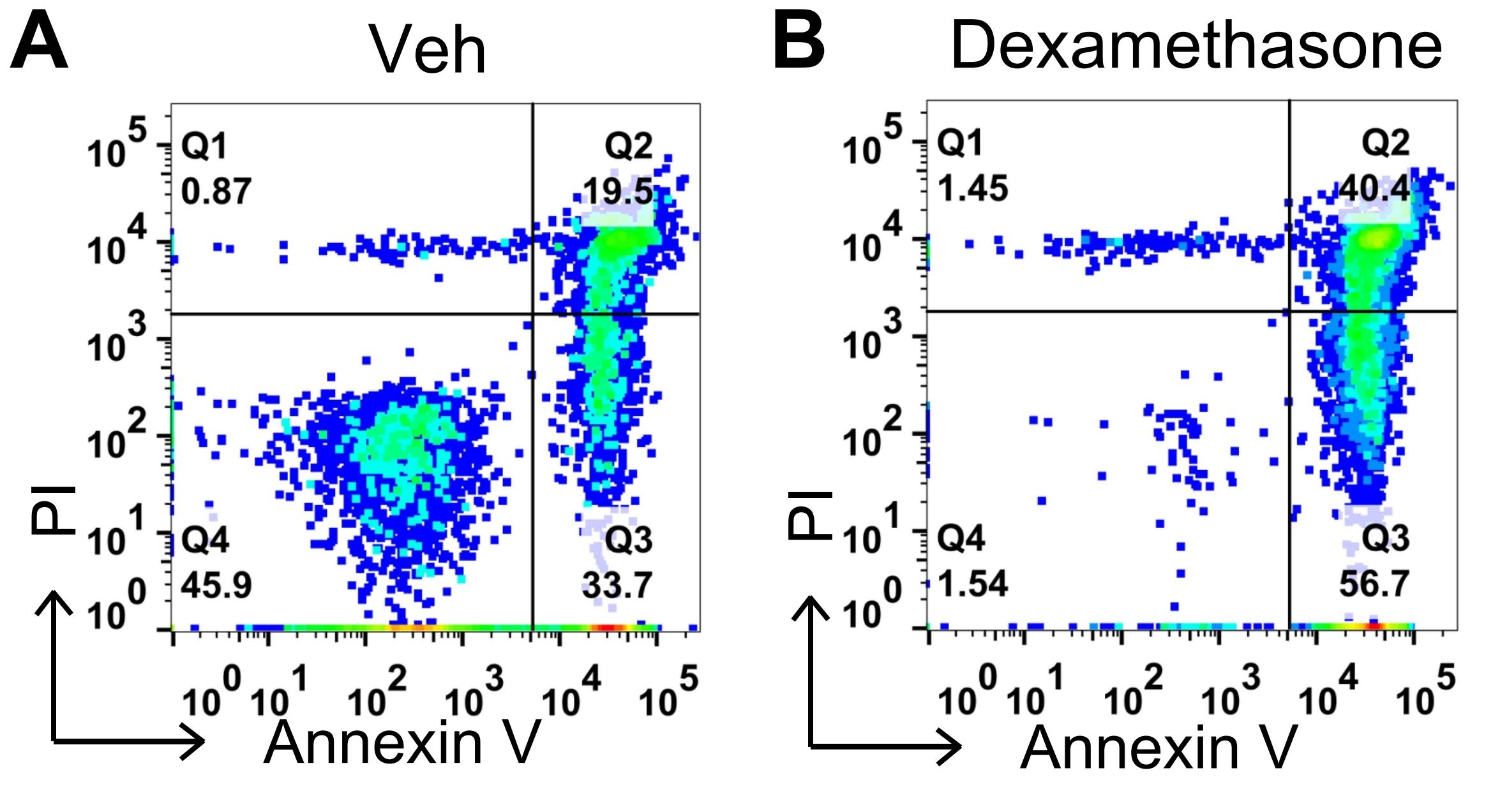
Figure 2. Flow cytometric analysis of thymocyte apoptosis induced by dexamethasone. Thymocytes were treated (A) without (Vehicle, Veh) or (B) with 1 μM dexamethasone overnight. Cells were then stained with propidium iodide (PI) and Annexin V. Representative scattergrams of cells in early (Q3, Annexin V+ PI-) and late (Q2, Annexin V+ PI+) apoptosis were analyzed by flow cytometry.
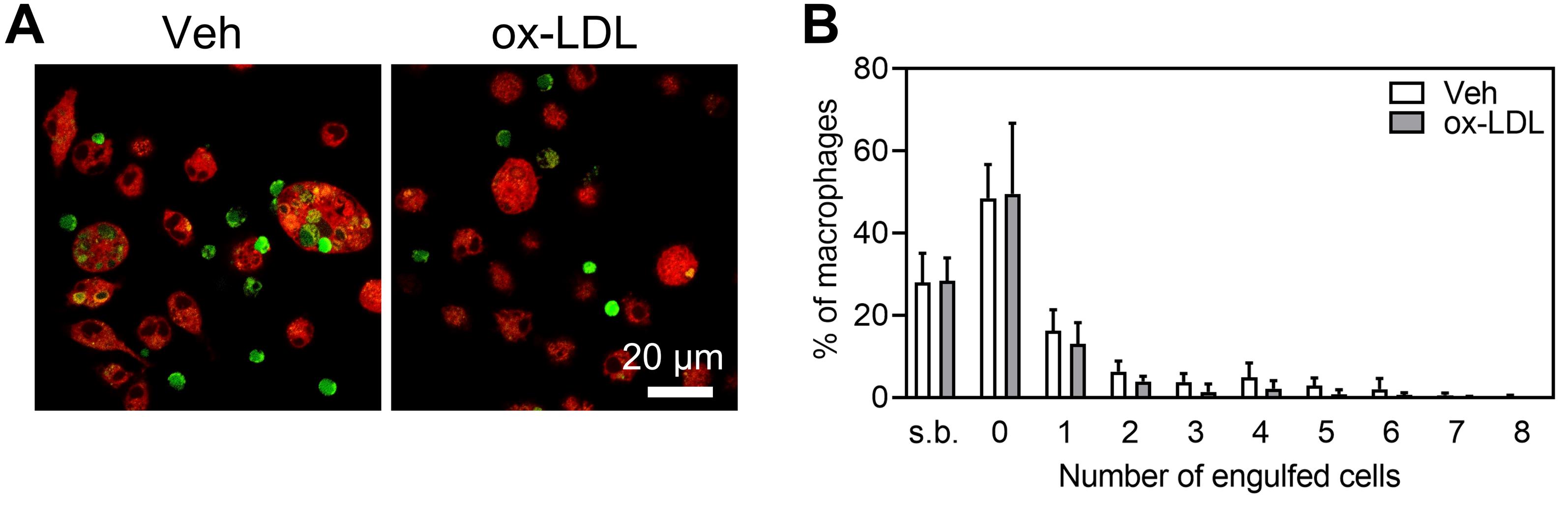
Figure 3. Effect of oxidized low-density lipoprotein (ox-LDL) treatment on the efferocytosis of apoptotic/necrotic thymocytes by mouse macrophages. (A) Elicited peritoneal macrophages were treated without (Vehicle, Veh) or with ox-LDL (50 μg/mL) for 6 h. Then, CMTPX (red)-labeled peritoneal macrophages were co-cultured with CFDA-SE (green)-labeled apoptotic mouse thymocytes for 2 h. The efferocytosis of apoptotic/necrotic thymocytes by mouse macrophages was determined by confocal microscopy. (B) Number of engulfed CFDA-SE-labeled apoptotic targets within CMTPX-labeled macrophages counted by confocal microscopy after 2 h of efferocytosis (n = 3). Scale bar = 20 μm. Results are expressed as mean ± SD of three independent experiments. s.b., surface-bound cells.
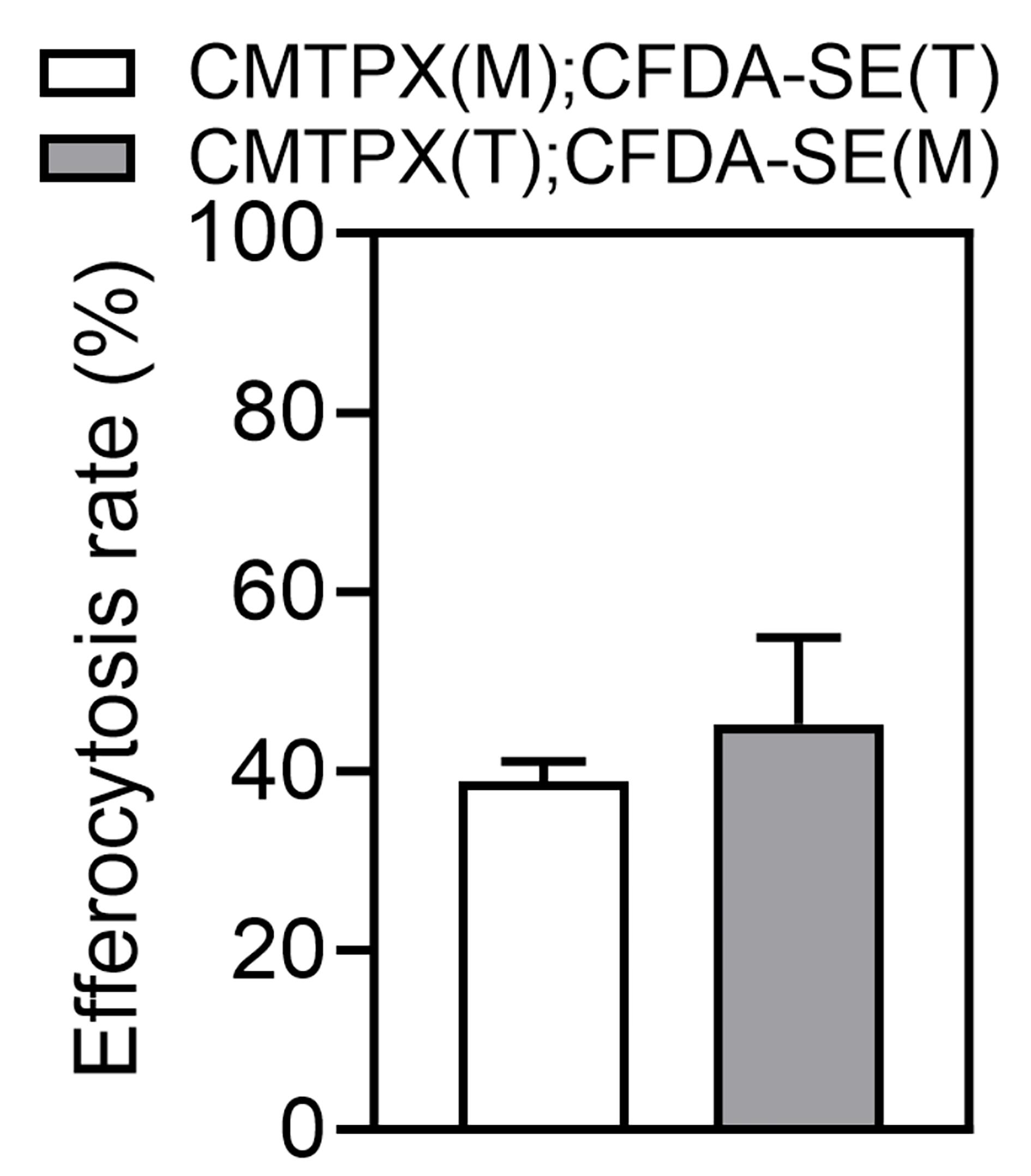
Figure 4. CMTPX-labeled peritoneal macrophages (M) were co-cultured with CFDA-SE-labeled apoptotic mouse thymocytes (T) for 2 h. In parallel, peritoneal macrophages (labeled with CFDA-SE) were co-cultured with apoptotic thymocytes (labeled with CMTPX) for 2 h. Macrophage efferocytosis of apoptotic/necrotic thymocytes was determined by confocal microscopy. Results are expressed as mean ± SD of three independent experiments, n = 3.
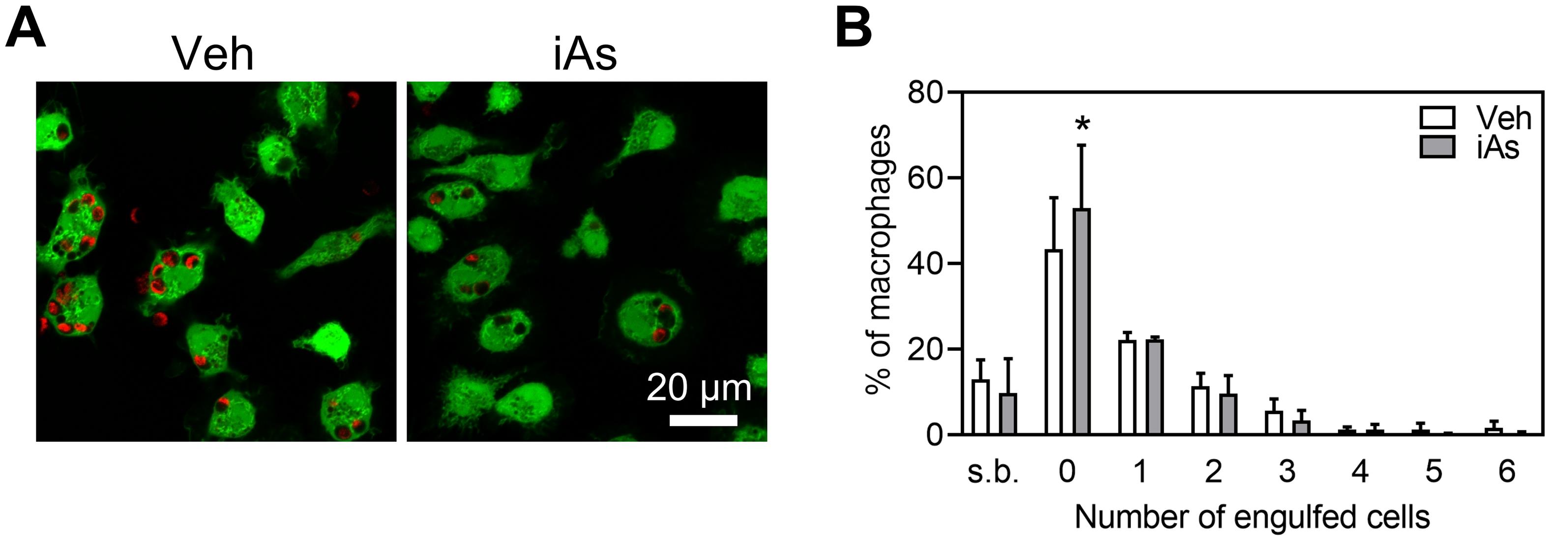
Figure 5. Arsenite (iAs) exposure suppressed efferocytosis of apoptotic/necrotic thymocytes in mouse macrophages. (A) Primary peritoneal macrophages were treated with arsenite (1 μM) for 24 h or without (Vehicle, Veh). Then, peritoneal macrophages (labeled with CFDA-SE) were co-cultured with apoptotic thymocytes (labeled with CMTPX) for 2 h. The efferocytosis of apoptotic/necrotic thymocytes by mouse macrophages was determined by confocal microscopy. (B) Number of engulfed CMTPX-labeled apoptotic targets within CFDA-SE-labeled macrophages counted by confocal microscopy after 2 h of efferocytosis (n = 3). Scale bar = 20 μm. *, compared with Veh, P < 0.05, two-tailed Student’s t-test. Results are expressed as mean ± SD of three independent experiments. s.b., surface-bound cells.
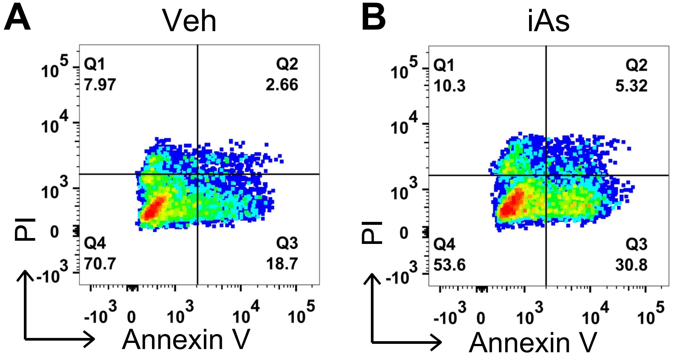
Figure 6. Flow cytometric analysis of hepatocytes apoptosis induced by arsenite. Hepatocytes were prepared as described in section C of the Procedure, and treated (A) without (Vehicle, Veh) or (B) with 10 μM arsenite for 24 h. Cells were then stained with PI and Annexin V. Representative scattergram of cells in early (Q3, Annexin V+ PI-) and late (Q2, Annexin V+ PI+) apoptosis was analyzed by flow cytometry.
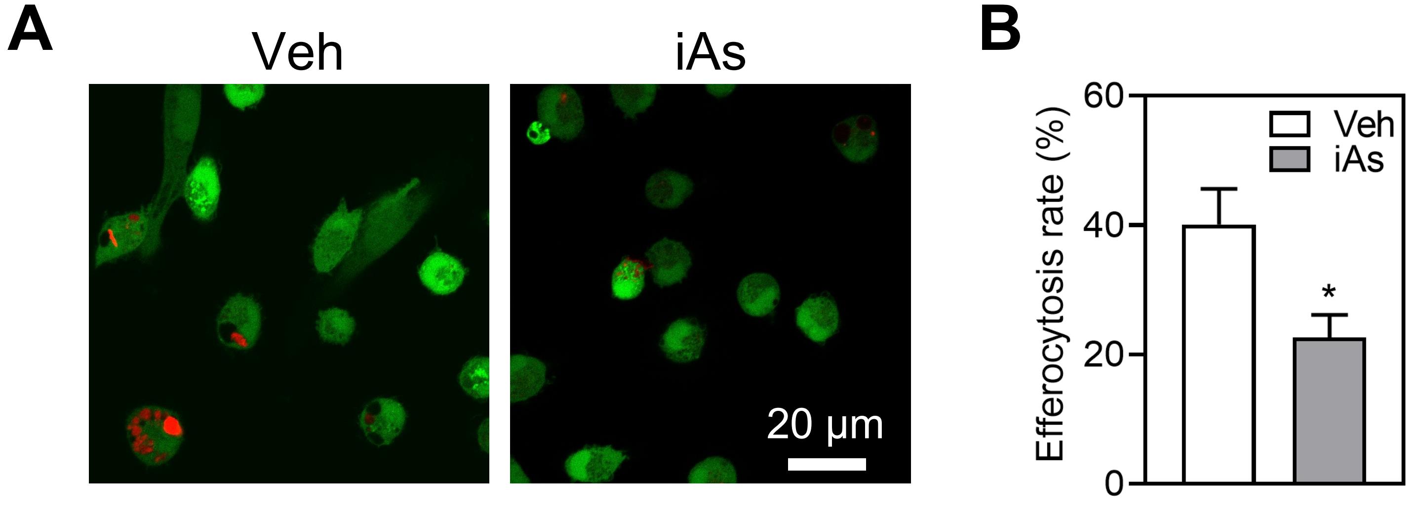
Figure 7. Arsenite exposure inhibited efferocytosis of apoptotic/necrotic hepatocytes in mouse macrophages. (A) Primary peritoneal macrophages were treated with arsenite (1 μM) for 24 h or without (Vehicle, Veh). Then, peritoneal macrophages (labeled with CFDA-SE) were co-cultured with apoptotic hepatocytes (labeled with CMTPX) for 2 h. Apoptotic hepatocytes were induced by treating cells with arsenite (10 μM) for 24 h. The efferocytosis of apoptotic/necrotic hepatocytes by mouse macrophages was determined by confocal microscopy. (B) Percent efferocytosis was quantified as the number of macrophages with engulfed apoptotic/necrotic hepatocytes as a percentage of total macrophages (n = 3). Scale bar = 20 μm. *, compared with Veh, P < 0.05, two-tailed Student’s t-test.
This protocol or parts of it has been used and validated in the following research article:
• Xu et al. [48]. Nrf2 deficiency in myeloid cells accelerates atherosclerosis by promoting the inflammatory response and impairing efferocytosis. J Adv Res (Figure 7H, I). https://doi.org/10.1016/j.jare.2026.01.005
General notes and troubleshooting
General notes
1. Prior to co-culture, perform cell counting using an automated cell counter or the manual trypan blue exclusion assay. For all downstream functional assays, only utilize samples with a cell viability of ≥85%.
2. Primary hepatocytes, thymocytes, and macrophages are recommended to be freshly isolated for each experiment to ensure optimal cell viability and physiological function. The use of cryopreserved primary cells is not recommended, as repeated freeze–thaw cycles may impair cell viability, recovery rate, adherence rate, phagocytic capacity, and normal biological responses [46,47]. If cryopreserved cells must be used, first it is essential to validate whether frozen and fresh cells yield comparable results for the main experimental endpoints.
3. Appropriate negative controls should be included in the apoptosis induction assay.
4. In the efferocytosis assay, 40×, 60×, and 100× objectives on a confocal microscope are all applicable. The 40× objective represents the minimum imaging configuration required to clearly visualize the number of apoptotic cells internalized within macrophages. The use of oil immersion objectives helps to achieve higher resolution.
5. Conventional fluorescence microscopy is not recommended, as it cannot provide sufficiently clear images.
6. A minimum of 200 cells in total should be analyzed per experimental group.
7. Prior to CFDA-SE/CMTPX labeling in the formal efferocytosis assay, Annexin V–FITC/PI staining should be performed on separate aliquots of unlabeled cells to assess the efficiency of apoptosis induction.
8. Sodium arsenite must be handled strictly in accordance with the protocols for highly toxic reagents throughout the experiment. All preparation and dosing are performed in a biosafety cabinet with full personal protection, dual-key access, and complete record keeping. Arsenic-containing liquid and solid waste is collected separately, clearly labeled, temporarily stored, and disposed of by licensed professional institutions following standard hazardous chemical and waste management guidelines.
Troubleshooting
Problem: Quantified efferocytosis efficiency is inaccurate and unstable across replicates.
Possible causes: Inaccurate timing and incomplete removal of excess floating apoptotic cells.
Solutions:
1. Since efferocytosis is a dynamic biological process, perform confocal laser scanning microscopy immediately upon completion of co-culture.
2. Gently rinse samples before imaging to remove unbound free apoptotic cells, minimizing false-positive deviations caused by nonspecific adhesion.
3. Strictly standardize the co-culture duration, incubation conditions, cell ratio, and culture temperature for all experimental groups.
Acknowledgments
Authors’ contribution
Conceptualization, X.X., T.S., H.W.; Investigation, X.X., T.S., Q.S.; Writing—Original Draft, X.X., T.S., H.W.; Writing—Review & Editing, X.W., Y.L., H.W.; Funding acquisition, H.W.; Supervision, Y.L., H.W.
This research was funded by the National Natural Science Foundation of China 82574068 and 82073513; Liaoning Revitalization Talents Program XLYC2203028; Natural Science Foundation of Liaoning Province 2022-MS-215; the Educational Department of Liaoning Province JYTMS20230125; Shenyang Science and Technology Bureau of Support Program for Young Innovation Scholar RC210296; Innovation Team Support from China Medical University CXTD2022004; and High-level Talents Support Foundation of China Medical University 2400022052.
This protocol was used in [48].
The following figures were created using BioRender: Graphical overview, https://BioRender.com/bewcl0q; Figure 1, https://BioRender.com/v9ydgit.
Competing interests
The authors declare no conflicts of interest.
Ethical considerations
All procedures were performed in accordance with protocols approved by the Institutional Animal Care and Use Committee of China Medical University.
References
- Boada-Romero, E., Martinez, J., Heckmann, B. L. and Green, D. R. (2020). The clearance of dead cells by efferocytosis. Nat Rev Mol Cell Biol. 21(7): 398–414. https://doi.org/10.1038/s41580-020-0232-1
- Doran, A. C., Yurdagul, A. and Tabas, I. (2020). Efferocytosis in health and disease. Nat Rev Immunol. 20(4): 254–267. https://doi.org/10.1038/s41577-019-0240-6
- Abdolmaleki, F., Farahani, N., Gheibi Hayat, S. M., Pirro, M., Bianconi, V., Barreto, G. E. and Sahebkar, A. (2018). The Role of Efferocytosis in Autoimmune Diseases. Front Immunol. 9: 1645. https://doi.org/10.3389/fimmu.2018.01645
- Arandjelovic, S. and Ravichandran, K. S. (2015). Phagocytosis of apoptotic cells in homeostasis. Nat Immunol. 16(9): 907-917. https://doi.org/10.1038/ni.3253
- Qiu, H., Shao, Z., Wen, X., Liu, Z., Chen, Z., Qu, D., Ding, X. and Zhang, L. (2023). Efferocytosis: An accomplice of cancer immune escape. Biomed Pharmacother. 167: 115540. https://doi.org/10.1016/j.biopha.2023.115540
- Seneviratne, A. N., Edsfeldt, A., Cole, J. E., Kassiteridi, C., Swart, M., Park, I., Green, P., Khoyratty, T., Saliba, D., Goddard, M. E., et al. (2017). Interferon Regulatory Factor 5 Controls Necrotic Core Formation in Atherosclerotic Lesions by Impairing Efferocytosis. Circulation. 136(12): 1140–1154. https://doi.org/10.1161/CIRCULATIONAHA.117.027844
- Wang, X., He, Q., Zhou, C., Xu, Y., Liu, D., Fujiwara, N., Kubota, N., Click, A., Henderson, P., Vancil, J., et al. (2023). Prolonged hypernutrition impairs TREM2-dependent efferocytosis to license chronic liver inflammation and NASH development. Immunity. 56(1): 58–77.e11. https://doi.org/10.1016/j.immuni.2022.11.013
- Mehrotra, P. and Ravichandran, K. S. (2022). Drugging the efferocytosis process: concepts and opportunities. Nat Rev Drug Discov. 21(8): 601–620. https://doi.org/10.1038/s41573-022-00470-y
- Ma, Y., Mouton, A. J. and Lindsey, M. L. (2018). Cardiac macrophage biology in the steady-state heart, the aging heart, and following myocardial infarction. Transl Res. 191: 15–28. https://doi.org/10.1016/j.trsl.2017.10.001
- A-Gonzalez, N., Quintana, J. A., García-Silva, S., Mazariegos, M., González de la Aleja, A., Nicolás-Ávila, J. A., Walter, W., Adrover, J. M., Crainiciuc, G., Kuchroo, V. K., et al. (2017). Phagocytosis imprints heterogeneity in tissue-resident macrophages. J Exp Med. 214(5): 1281–1296. https://doi.org/10.1084/jem.20161375
- Roberts, A. W., Lee, B. L., Deguine, J., John, S., Shlomchik, M. J. and Barton, G. M. (2017). Tissue-Resident Macrophages Are Locally Programmed for Silent Clearance of Apoptotic Cells. Immunity. 47(5). https://doi.org/10.1016/j.immuni.2017.10.006
- Prasad, A., Bekker, P. and Tsimikas, S. (2012). Advanced glycation end products and diabetic cardiovascular disease. Cardiol Rev. 20(4): 177–183. https://doi.org/10.1097/CRD.0b013e318244e57c
- Del Turco, S. and Basta, G. (2012). An update on advanced glycation endproducts and atherosclerosis. Biofactors. 38(4): 266–274. https://doi.org/10.1002/biof.1018
- Li, J., Liu, D., Sun, L., Lu, Y. and Zhang, Z. (2012). Advanced glycation end products and neurodegenerative diseases: mechanisms and perspective. J Neurol Sci. 317(1–2): 1–5. https://doi.org/10.1016/j.jns.2012.02.018
- Lu, S. M. and Fairn, G. D. (2018). 7-Ketocholesterol impairs phagocytosis and efferocytosis via dysregulation of phosphatidylinositol 4,5-bisphosphate. Traffic. 19(8): 591–604. https://doi.org/10.1111/tra.12576
- Manega, C. M., Fiorelli, S., Porro, B., Turnu, L., Cavalca, V., Bonomi, A., Cosentino, N., Di Minno, A., Marenzi, G., Tremoli, E., et al. (2019). 12(S)-Hydroxyeicosatetraenoic acid downregulates monocyte-derived macrophage efferocytosis: New insights in atherosclerosis. Pharmacol Res. 144: 336–342. https://doi.org/10.1016/j.phrs.2019.03.012
- Reinertsen, A. F., Libreros, S., Nshimiyimana, R., Serhan, C. N. and Hansen, T. V. (2023). Metabolization of Resolvin E4 by ω-Oxidation in Human Neutrophils: Synthesis and Biological Evaluation of 20-Hydroxy-Resolvin E4 (20-OH-RvE4). ACS Pharmacol Transl Sci. 6(12): 1898–1908. https://doi.org/10.1021/acsptsci.3c00201
- Lescoat, A., Ballerie, A., Lelong, M., Augagneur, Y., Morzadec, C., Jouneau, S., Jégo, P., Fardel, O., Vernhet, L. and Lecureur, V. (2020). Crystalline Silica Impairs Efferocytosis Abilities of Human and Mouse Macrophages: Implication for Silica-Associated Systemic Sclerosis. Front Immunol. 11: 219. https://doi.org/10.3389/fimmu.2020.00219
- Schopf, R. E., Trompeter, M., Bork, K. and Morsches, B. (1985). Effects of ethanol and acetaldehyde on phagocytic functions. Arch Dermatol Res. 277(2): 131–137. https://doi.org/10.1007/bf00414111
- De Francisco, P., Martín-González, A., Rodriguez-Martín, D. and Díaz, S. (2021). Interactions with Arsenic: Mechanisms of Toxicity and Cellular Resistance in Eukaryotic Microorganisms. Int J Environ Res Public Health. 18(22): 12226. https://doi.org/10.3390/ijerph182212226
- Chen, C. J. (2014). Health hazards and mitigation of chronic poisoning from arsenic in drinking water: Taiwan experiences. Rev Environ Health. 29: 13–19. https://doi.org/10.1515/reveh-2014-0007
- O'Bryant, S. E., Edwards, M., Menon, C. V., Gong, G. and Barber, R. (2011). Long-term low-level arsenic exposure is associated with poorer neuropsychological functioning: a Project FRONTIER study. Int J Environ Res Public Health. 8(3): 861–874. https://doi.org/10.3390/ijerph8030861
- Parvez, F., Chen, Y., Yunus, M., Olopade, C., Segers, S., Slavkovich, V., Argos, M., Hasan, R., Ahmed, A., Islam, T., et al. (2013). Arsenic exposure and impaired lung function. Findings from a large population-based prospective cohort study. Am J Respir Crit Care Med. 188(7): 813–819. https://doi.org/10.1164/rccm.201212-2282OC
- Stea, F., Bianchi, F., Cori, L. and Sicari, R. (2014). Cardiovascular effects of arsenic: clinical and epidemiological findings. Environ Sci Pollut Res Int. 21(1): 244–251. https://doi.org/10.1007/s11356-013-2113-z
- Wu, M. M., Kuo, T. L., Hwang, Y. H. and Chen, C. J. (1989). Dose-response relation between arsenic concentration in well water and mortality from cancers and vascular diseases. Am J Epidemiol. 130(6): 1123–1132. https://doi.org/10.1093/oxfordjournals.aje.a115439
- Mohammed Abdul, K. S., Jayasinghe, S. S., Chandana, E. P., Jayasumana, C. and De Silva, P. M. C. (2015). Arsenic and human health effects: A review. Environ Toxicol Pharmacol. 40(3): 828–846. https://doi.org/10.1016/j.etap.2015.09.016
- Cantoni, O., Zito, E., Fiorani, M. and Guidarelli, A. (2021). Arsenite impinges on endoplasmic reticulum-mitochondria crosstalk to elicit mitochondrial ROS formation and downstream toxicity. Semin Cancer Biol. 76: 132–138. https://doi.org/10.1016/j.semcancer.2021.06.002
- Wang, Y.-C., Lee, A.-S., Lu, L.-S., Ke, L.-Y., Chen, W.-Y., Dong, J.-W., Lu, J., Chen, Z., Chu, C.-S., Chan, H.-C., et al. (2018). Human electronegative LDL induces mitochondrial dysfunction and premature senescence of vascular cells in vivo. Aging Cell. 17(4): e12792. https://doi.org/10.1111/acel.12792
- Wang, J., Bai, Y., Zhao, X., Ru, J., Kang, N., Tian, T., Tang, L., An, Y. and Li, P. (2018). oxLDL-mediated cellular senescence is associated with increased NADPH oxidase p47phox recruitment to caveolae. Biosci Rep. 38(3): e1042/bsr20180283. https://doi.org/10.1042/bsr20180283
- Gao, S., Zhao, D., Wang, M., Zhao, F., Han, X., Qi, Y. and Liu, J. (2017). Association Between Circulating Oxidized LDL and Atherosclerotic Cardiovascular Disease: A Meta-analysis of Observational Studies. Can J Cardiol. 33(12): 1624–1632. https://doi.org/10.1016/j.cjca.2017.07.015
- Miksa, M., Komura, H., Wu, R., Shah, K. G. and Wang, P. (2009). A novel method to determine the engulfment of apoptotic cells by macrophages using pHrodo succinimidyl ester. J Immunol Methods. 342(1–2): 71–77. https://doi.org/10.1016/j.jim.2008.11.019
- Wootton, D. G., Diggle, P. J., Court, J., Eneje, O., Keogan, L., Macfarlane, L., Wilks, S., Woodhead, M. and Gordon, S. B. (2016). Recovery from pneumonia requires efferocytosis which is impaired in smokers and those with low body mass index and enhanced by statins. Thorax. 71(11): 1052–1054. https://doi.org/10.1136/thoraxjnl-2016-208505
- Taruc, K., Yin, C., Wootton, D. G. and Heit, B. (2018). Quantification of Efferocytosis by Single-cell Fluorescence Microscopy. J Visualized Exp.: e3791/58149. https://doi.org/10.3791/58149
- Yurdagul, A. (2022). Dual-Fluorescence In Vitro Efferocytosis Assay. Methods Mol Biol. 2419: 293–299. https://doi.org/10.1007/978-1-0716-1924-7_18
- Layoun, A., Samba, M. and Santos, M. M. (2015). Isolation of murine peritoneal macrophages to carry out gene expression analysis upon Toll-like receptors stimulation. J Vis Exp (98): e52749. https://doi.org/10.3791/52749
- Li, Y. M., Baviello, G., Vlassara, H. and Mitsuhashi, T. (1997). Glycation products in aged thioglycollate medium enhance the elicitation of peritoneal macrophages. J Immunol Methods. 201(2): 183–188. https://doi.org/10.1016/s0022-1759(96)00224-4
- Zhang, Y., Wang, G., Lu, J., Xu, L.-M. and Xiong, J.-Y. (2019). Using Enhanced Green Fluorescence Protein-expressing Escherichia Coli to Assess Mouse Peritoneal Macrophage Phagocytosis. J Vis Exp. (143). https://doi.org/10.3791/58751
- Rödel, F., Franz, S., Sheriff, A., Gaipl, U., Heyder, P., Hildebrandt, G., Schultze-Mosgau, S., Voll, R. E. and Herrmann, M. (2005). The CFSE distribution assay is a powerful technique for the analysis of radiation-induced cell death and survival on a single-cell level. Strahlenther Onkol. 181(7): 456–462. https://doi.org/10.1007/s00066-005-1361-3
- Dumitriu, I. E., Mohr, W., Kolowos, W., Kern, P., Kalden, J. R. and Herrmann, M. (2001). 5,6-carboxyfluorescein diacetate succinimidyl ester-labeled apoptotic and necrotic as well as detergent-treated cells can be traced in composite cell samples. Anal Biochem. 299(2): 247–252. https://doi.org/10.1006/abio.2001.5415
- Korelova, K., Jirouskova, M., Sarnova, L. and Gregor, M. (2019). Isolation and 3D Collagen Sandwich Culture of Primary Mouse Hepatocytes to Study the Role of Cytoskeleton in Bile Canalicular Formation In Vitro. J Visualized Exp.: e3791/60507. https://doi.org/10.3791/60507
- Breuer, E. and Humar, B. (2022). Isolation of Regenerating Hepatocytes after Partial Hepatectomy in Mice. J Visualized Exp.: e3791/64493–v. https://doi.org/10.3791/64493-v
- Pineda-Torra, I., Gage, M., de Juan, A. and Pello, O. M. (2015). Isolation, Culture, and Polarization of Murine Bone Marrow-Derived and Peritoneal Macrophages. Methods Mol Biol. 1339: 101–109. https://doi.org/10.1007/978-1-4939-2929-0_6
- Cisney, E. D., Fernandez, S., Hall, S. I., Krietz, G. A. and Ulrich, R. G. (2012). Examining the role of nasopharyngeal-associated lymphoreticular tissue (NALT) in mouse responses to vaccines. J Vis Exp. (66): 3960. https://doi.org/10.3791/3960
- Salem, E. S., Murakami, K., Takahashi, T., Bernhard, E., Borra, V., Bethi, M. and Nakamura, T. (2018). Isolation of Primary Mouse Hepatocytes for Nascent Protein Synthesis Analysis by Non-radioactive L-azidohomoalanine Labeling Method. J Visualized Exp.: e3791/58323. https://doi.org/10.3791/58323
- Berry, M. N. and Friend, D. S. (1969). High-yield preparation of isolated rat liver parenchymal cells: a biochemical and fine structural study. J Cell Biol. 43(3): 506–520. https://doi.org/10.1083/jcb.43.3.506
- Luu, A. M., Shepardson, K. M. and Rynda-Apple, A. (2024). A Comprehensive Protocol for the Collection, Differentiation, Cryopreservation, and Resuscitation of Primary Murine Bone Marrow Derived Macrophages (BMDM). Immunol Invest. 53(7): 1001–1012. https://doi.org/10.1080/08820139.2024.2382805
- Tomás, R. M. F., Dallman, R., Congdon, T. R. and Gibson, M. I. (2023). Cryopreservation of assay-ready hepatocyte monolayers by chemically-induced ice nucleation: preservation of hepatic function and hepatotoxicity screening capabilities. Biomater Sci. 11(23): 7639–7654. https://doi.org/10.1039/d3bm01046e
- Xu, X., Liu, C., Bo, J., Wang, H., Zhang, Y., Xu, Y., Fu, J., Pi, J. and Wang, H. (2026). Nrf2 deficiency in myeloid cells accelerates atherosclerosis by promoting the inflammatory response and impairing efferocytosis. J Adv Res.: e005. https://doi.org/10.1016/j.jare.2026.01.005
Article Information
Publication history
Received: Feb 14, 2026
Accepted: Apr 9, 2026
Available online: Apr 24, 2026
Published: May 20, 2026
Copyright
© 2026 The Author(s); This is an open access article under the CC BY-NC license (https://creativecommons.org/licenses/by-nc/4.0/).
How to cite
Xu, X., Su, T., Sun, Q., Wang, X., Liu, Y. and Wang, H. (2026). A Versatile In Vitro Quantitative Assay for Macrophage Efferocytosis in Diverse Research Applications. Bio-protocol 16(10): e5690. DOI: 10.21769/BioProtoc.5690.

 Download Citation in RIS Format
Category
Immunology > Immune cell function > Macrophage
Cell Biology > Cell-based analysis > Cytosis
Environmental science
Do you have any questions about this protocol?
Post your question to gather feedback from the community. We will also invite the authors of this article to respond.
 Post a Question


