


- Protocols

- Articles and Issues
- For Authors
- About
- Become a Reviewer


Plasmid Curing of Pseudoalteromonas haloplanktis TAC125 Using Homologous Recombination and PTasRNA Gene Silencing

(§Technical contact: angelica.severino@unina.it) Published: Vol 16, Iss 10, May 20, 2026 DOI: 10.21769/BioProtoc.5687 Views: 1059
Reviewed by: Alba BlesaAmar Chandra MahathaAnonymous reviewer(s)
 PDF
PDF  Ask a question
Original research article
The authors used this protocol in:

Feb 2025
Advertisement



Protocol Collections
Comprehensive collections of detailed, peer-reviewed protocols focusing on specific topics




Related protocols

Abstract
Pseudoalteromonas haloplanktis TAC125 is a psychrophilic marine bacterium widely used to study cold adaptation and increasingly exploited as a non-conventional platform for biotechnological applications. The strain harbors the endogenous megaplasmid pMEGA (64.7 kb), whose presence may limit its exploitation as a cell factory, making its elimination advantageous to strain engineering. Traditional plasmid-curing approaches based on chemical and physical agents are often inefficient and unsuitable for stable endogenous replicons, such as pMEGA. Here, we describe a targeted protocol for pMEGA curing in P. haloplanktis TAC125 that combines homologous recombination with paired-termini antisense RNA (PTasRNA) gene silencing. First, a selectable marker cassette is inserted into pMEGA by homologous recombination using a suicide vector, enabling selective discrimination between plasmid-positive and plasmid-cured bacteria. Next, PTasRNA gene silencing technology is applied to target a gene essential for the replication of pMEGA, thereby transiently interfering with its replication and promoting its loss. This approach provides a specific method to cure a highly stable endogenous megaplasmid in a psychrophilic non-conventional bacterium, enabling improved functional studies and strain optimization, establishing a broadly applicable framework for targeted curing across diverse bacterial systems.
Key features
• Enables targeted curing of stable endogenous plasmids lacking selectable markers.
• Combines replication silencing and homologous recombination for targeted plasmid elimination without permanent chromosomal modification.
• Adaptable framework for non-model bacteria with limited genetic toolkits, such as marine psychrophiles.
Keywords: Pseudoalteromonas haloplanktis TAC125 Plasmid curing
Homologous recombination
PTasRNA technology
Marine psychrophilic bacteria
Megaplasmid
Strain engineering
Plasmid curing
Homologous recombination
PTasRNA technology
Marine psychrophilic bacteria
Megaplasmid
Strain engineering
Graphical overview

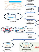
Graphical overview of pMEGA plasmid curing in Pseudoalteromonas haloplanktis TAC125
Background
Plasmid curing is a fundamental strategy in microbial genetics used to dissect plasmid contributions to cellular functions and improve bacterial hosts for basic and applied research [1,2]. Plasmids are widespread across bacteria and often encode traits that provide selective advantages in natural ecosystems, but their stable maintenance can complicate functional genomics analyses and limit the rational engineering of microbial strains [3]. Over the past decades, plasmid curing has relied on nonspecific approaches such as chemical agents (e.g., intercalating dyes, detergents, etc.) or physical treatments (exposure to UV or elevated temperatures) [2,4,5]. Nevertheless, these methods are frequently inefficient, poorly reproducible, and highly species-dependent, particularly for low copy number or highly stable plasmids. These limitations are particularly evident in non-conventional organisms, such as marine psychrophiles, where restricted genetic toolkits hinder classical genetic manipulation.
Pseudoalteromonas haloplanktis TAC125 (PhTAC125) is considered a model for studying cold adaptation strategies and is a promising host for biotechnological applications [6–11]. It has a multipartite genome composed of two chromosomes and two endogenous plasmids: the cryptic plasmid pMtBL and the pMEGA megaplasmid [12,13]. pMEGA, which is 64,758 bp in size and contains 52 open reading frames (ORFs), is a non-conjugative and low-copy-number plasmid. It encodes genes required for autoreplication, stability, and plasmid partitioning functions [14]. Recent studies have shown that the elimination of pMEGA does not impair bacterial growth under standard laboratory conditions but leads to measurable phenotypic changes, including increased resistance to oxidative stress and reduced biofilm formation [15]. These observations suggest that, while not essential for viability, pMEGA contributes to stress response and surface-associated behaviors, likely conferring adaptive advantages in natural environments. However, the absence of a native selectable marker and its high stability under laboratory conditions make pMEGA refractory to conventional curing strategies; thus, a highly specific and controlled approach is necessary to eliminate it [14]. The protocol described here addresses this challenge by combining i) homologous recombination to introduce a selectable marker necessary to distinguish wild-type from cured bacteria [16] and ii) targeting the essential replication initiator gene (repB) on pMEGA with paired-termini antisense RNA (PTasRNA) to interfere with pMEGA plasmid replication [10,15]. This strategy enables efficient and reproducible curing of pMEGA without permanently altering the host chromosome. This modular and targeted framework overcomes key limitations of traditional curing methods and can be adapted to other stable plasmids and genetically recalcitrant bacterial species.
Materials and reagents
Biological materials
1. Escherichia coli TOP10 [mcrA, Δ(mrr-hsdRMS-mcrBC), ϕ80lacZ (del) M15, ΔlacX74, deoR, recA1, araD139, Δ(ara-leu)—7697, galU, galK, rpsL (SmR), endA1, nupG] [17] (available in the laboratory)
2. Escherichia coli S17-1(λpir) [thi, pro, hsd (r− m+) recA:: RP4- 2TcR::Mu KmR::Tn7 TpR SmR λpir] [18] (available in the laboratory)
3. Pseudoalteromonas haloplanktis TAC125 (PhTAC125) KrPL strain [8] (available in the laboratory)
4. PhTAC125 KrPL insPolV (generated in this work)
5. PhTAC125 KrPL2 (generated in this work)
Reagents
1. Bacto-agar (BD DifcoTM Bacteriological Agar, catalog number: 214530)
2. Tryptone (Biolife, catalog number: 4122902)
3. Yeast extract (Biolife, catalog number: 4122202)
4. Sodium chloride (Biochem Chemopharma, catalog number: 319121000)
5. L-glutamic acid monosodium salt monohydrate (Molekula, catalog number: 40359230)
6. D-gluconic acid sodium salt (Molekula, catalog number: 50444955)
7. Ammonium nitrate (NH4NO3) (Sigma-Aldrich, catalog number: A3795)
8. Potassium phosphate anhydrous (KH2PO4) (Thermo Scientific, catalog number: A12142)
9. Magnesium sulphate heptahydrate (MgSO4·7H2O) (Honeywell, Riedel-de Haën, catalog number: 13142)
10. Iron (II) sulphate heptahydrate (FeSO4·7H2O) (VWR AnalaR NORMAPUR, catalog number: 24244.298)
11. Calcium chloride powder (CaCl2) (PanReac AppliChem, catalog number: A3652)
12. Trizma base (PanReac AppliChem, catalog number: A1379)
13. EDTA (VWR Chemicals, catalog number: 20301.290)
14. Glacial acetic acid (Carlo Erba, catalog number: 401392)
15. NaOH pellets (BioFroXX, catalog number: 1180)
16. Phusion High-Fidelity DNA polymerase (New England Biolabs, catalog number: M0530)
17. Taq DNA polymerase (New England Biolabs, catalog number: M0273)
18. Primers (Table 1) (Eurofins Genomics, Ebersberg, Germany)
Table 1. Primers employed for vector construction and pMEGA curing analysis.
Underlined sequences indicate restriction sites as specified in the primer name.
| Primers | Sequence 5'–3' |
|---|---|
| umuC_NotI fw | TGCGGCCGCTAATCAGCCCAGTCATTAATG |
| umuC_AscI rv | AGGCGCGCCATGGATGTATTTGTGCCATAT |
| NdeI_eGFP fw | ctgCATATGGGTGGAGGGAATTCAAGC |
| KpnI_eGFP rv | ctGGTACCTTATTTGTAGAGCTCATCCATGCC |
| 5'umuC fw | GCTTGCAGCGAATATCACGAGTCGTTTTGC |
| OriC rv | CAGCGTGAGCTATGAGAAAGCGCC |
| XhoI-asRNArepB fw | ATTTCTCGAGAATTAGAGAGTATATTTTAATGTC |
| PstI-asRNArepB rv | ATTCTGCAGTTTCTCATCGATACTCATTTC |
| prom7 fw | CCTTTATTCAGCGTGTTGGCGAGC |
| prom7 rv | GTTATCAGGGTCGGGCGTATCGG |
| CDS49 fw | AACTGACTGTGGTGCTCTTC |
| CDS49 rv | ACTGGTCCCTATTTGTTTATGCT |
19. Deoxynucleotide (dNTP) solution mix (New England BioLabs, catalog number: N0447)
20. T4 DNA ligase (New England BioLabs, catalog number: M0202)
21. Genomic DNA of PhTAC125 (available upon request at this laboratory)
22. NotI-HF (New England BioLabs, catalog number: R3189)
23. AscI (New England BioLabs, catalog number: R0558)
24. PstI (New England BioLabs, catalog number: R0140)
25. XhoI (New England BioLabs, catalog number: R0146)
26. GelRed® nucleic acid gel stain (Millipore, catalog number: SCT123)
27. Gel loading dye, purple (6×) (New England BioLabs, catalog number: B7024)
28. Agarose basic for molecular biology (BioFroXX, catalog number: 1100)
29. QIAquick® Gel Extraction kit (QIAGEN®, catalog number: 28706)
30. Monarch® Spin Plasmid Miniprep kit (New England BioLabs, Monarch®, catalog number: T1110)
31. E.Z.N.ATM Bacterial DNA kit (OMEGA bio-tek, catalog number: D3350-02)
32. Isopropyl β-d-1-thiogalactopyranoside (IPTG) (BioSynth, catalog number: EI05931)
33. Lambda DNA/EcoRI + HindIII ladder (Promega, catalog number: G173A)
34. 1 kb Plus DNA ladder (New England BioLabs, catalog number: N3200S)
35. 1 kb DNA ladder (New England BioLabs, catalog number: N3232)
36. Plasmids (Table 2)
Table 2. Plasmids used in this work
| Plasmids | Relevant characteristics | Size | Source |
|---|---|---|---|
| pAT-eGFP | Suicide plasmid, AmpR | 4,934 bp | [19] |
| pAT-VS-HRumuC | Suicide plasmid, CamR | 4,862 bp | This study |
| pB40-79-PTasRNAlon | pMtBL-derived, containing lon asRNA, AmpR | 5,036 bp | This study |
| pB40-79C-PTasRNAlon | pMtBL-derived, containing lon asRNA, CamR | 4,838 bp | [10] |
| pB40-79-PTasRNArepB | pMtBL-derived, containing repB asRNA, AmpR | 5,036 bp | This study |
37. High-purity MilliQ water
38. Glycerol >99% (Fisher Chemical, catalog number G065017)
39. Kanamycin (PanReac AppliChem, catalog number: A4789)
40. Chloramphenicol (Acros organics, catalog number: 227920250)
41. Ampicillin (PanReac AppliChem, catalog number: A6352)
42. Ethanol 100% (Carlo Erba Reagents, catalog number: 414605)
Solutions
1. Media
a. TYP liquid medium (see Recipes)
b. LB liquid medium (see Recipes)
c. GG liquid medium (see Recipes)
2. Schatz salts stock solutions
a. 100 g/L KH2PO4 solution (100×) (see Recipes)
b. 60 g/L MgSO4 solution (300×) (see Recipes)
c. 2.5 g/L FeSO4 solution (500×) (see Recipes)
d. 15 mg/L CaCl2 solution (3,000×) (see Recipes)
3. Antibiotics
a. Chloramphenicol 50 mg/mL (see Recipes)
b. Kanamycin 50 mg/mL (see Recipes)
c. Ampicillin 100 mg/mL (see Recipes)
4. IPTG 1 M sterile solution (see Recipes)
5. 80% glycerol solution (see Recipes)
6. 0.1 M CaCl2 (see Recipes)
7. NaOH 0.5 M (see Recipes)
8. NaOH 5 M (see Recipes)
9. TAE 50× (see Recipes)
10. TAE 1× (see Recipes)
11. EDTA 0.5 M (see Recipes)
12. Agarose gel electrophoresis (see Recipes)
13. E. coli chemically competent cells (see Recipes)
14. E. coli transformation via heat shock treatment (see Recipes)
15. Intergeneric conjugation of PhTAC125 strain (see Recipes)
Recipes
1. Media
a. TYP liquid medium
i. Weigh 16 g of tryptone.
ii. Weigh 16 g of yeast extract.
iii. Weigh 10 g of NaCl.
iv. Put the mixture in a plastic cylinder.
v. Add MilliQ water to a final volume of 1 L.
vi. Stir until dissolved.
vii. Autoclave at 121 °C for 20 min at 1 bar and cool down before use.
b. LB liquid medium
i. Weigh 8 g of tryptone.
ii. Weigh 5 g of yeast extract.
iii. Weigh 10 g of NaCl.
iv. Put the mixture in a plastic cylinder.
v. Add MilliQ water to a final volume of 1 L.
vi. Stir until dissolved.
viii. Autoclave at 121 °C for 20 min at 1 bar and cool down before use.
c. GG liquid medium
i. Weigh 10 g of L-glutamic acid monosodium salt monohydrate.
ii. Weigh 10 g of D-gluconic acid sodium salt.
iii. Weigh 10 g of NaCl.
iv. Weigh 1 g of NH4NO3.
v. Put the mix in a glass cylinder and add 500 mL of MilliQ water.
vi. Stir until dissolved.
vii. Using NaOH 5 M, adjust the pH to 7.8.
viii. Add MilliQ water to a final volume of 1 L.
ix. Autoclave at 121 °C for 20 min.
x. Cool down and, in sterile conditions, add the following Schatz salts: KH2PO4 100× solution to a concentration of 1 g/L, MgSO4 300× solution to a concentration of 200 mg/L, FeSO4 500× solution to a concentration of 5 mg/L, and CaCl2 3,000× solution to a concentration of 5 mg/L.
Note: When required, add 15 g/L of Bacto-agar to the TYP or LB liquid media to cast 1.5% w/v TYP agar or LB agar plates. Store at room temperature (RT).
2. Schatz salts stock solutions
a. 100 g/L KH2PO4 (100×)
i. Weigh 100 g of KH2PO4.
ii. Add 500 mL of MilliQ water.
iii. Stir until dissolved.
iv. Add NaOH 5 M until the pH is 7.
v. Add MilliQ water to a final volume of 1 L.
b. 60 g/L MgSO4 (300×)
i. Weigh 60 g of MgSO4 heptahydrate powder.
ii. Add MilliQ water to a final volume of 1 L.
iii. Stir until dissolved.
c. 2.5 g/L FeSO4 (500×)
i. Weigh 2.5 g of FeSO4 heptahydrate powder.
ii. Add 500 mL of MilliQ water.
iii. Stir until dissolved.
iv. Add NaOH 0.5 M until the pH is 4.5.
v. Add MilliQ water to a final volume of 1 L.
d. 15 mg/L CaCl2 (3,000×)
i. Weigh 15 g of CaCl2 dihydrate powder.
ii. Add 500 mL of MilliQ water.
iii. Stir until dissolved.
iv. Add NaOH 0.5 M until the pH is 7.
v. Add MilliQ water to a final volume of 1 L.
Note: All Schatz salts must be filter sterilized using a 0.22 μm sterile filter. Store at RT.
3. Antibiotics
a. Chloramphenicol 50 mg/mL
i. Weigh 500 mg of chloramphenicol powder under a safety hood.
ii. Add ethanol 100% to a final volume of 10 mL.
iii. Mix thoroughly until completely dissolved.
iv. Prepare 1 mL aliquots and store at -20 °C.
b. Kanamycin 50 mg/mL
i. Weigh 500 mg of kanamycin powder under a safety hood.
ii. Add MilliQ water to a final volume of 10 mL.
iii. Mix the solution evenly until completely dissolved.
iv. Sterilize the solution with a 0.22 μm syringe filter.
v. Prepare 1 mL aliquots and store at -20 °C.
c. Ampicillin 100 mg/mL
i. Weigh 1 g of ampicillin under a safety hood.
ii. Add MilliQ water to a final volume of 10 mL.
iii. Mix the solution evenly until completely dissolved.
iv. Sterilize the solution with a 0.22 μm syringe filter.
v. Prepare 1 mL aliquots and store at -20 °C.
4. IPTG 1 M solution (10 mL)
a. Weigh 2.38 g of IPTG powder in a glass cylinder.
b. Add 5 mL of MilliQ water and dissolve.
c. When it is completely dissolved, add MilliQ water to the final volume of 10 mL.
d. Sterilize the solution with a 0.22 μm syringe filter.
e. Prepare 1 mL aliquots and store at -20 °C.
5. 80% glycerol solution
a. Pour 80 mL of 100% glycerol into a glass cylinder.
b. Add 20 mL of MilliQ water to reach 100 mL final volume.
c. Mix thoroughly until completely homogeneous.
d. Autoclave at 121° C for 20 min at 1 bar to sterilize the solution.
6. 0.1 M CaCl2
a. Weigh 5.55 g of CaCl2 powder.
b. Add MilliQ water to a final volume of 500 mL.
c. Mix until completely dissolved.
d. Autoclave to sterilize the solution.
e. Store at 4 °C.
7. NaOH 0.5 M
a. Dissolve 2 g of NaOH pellets in 80 mL of MilliQ water in a glass cylinder.
b. Add water to the final volume of 100 mL.
8. NaOH 5 M
a. Dissolve 20 g of NaOH pellets in 80 mL of MilliQ water in a glass cylinder.
Note: Add the pellets slowly, one at a time, as the dissolution is strongly exothermic and the solution will heat rapidly.
Tip: The solution can be prepared under refrigerated conditions or in an ice bath to limit temperature increase.
b. When cooled, add water to the final volume of 100 mL.
9. TAE 50×
a. Weigh 242 g of Trizma base and dissolve in 500 mL of MilliQ water.
b. Add 57.1 mL of glacial acetic acid.
c. Add 100 mL of EDTA 0.5 M.
d. Mix the solution until completely dissolved.
e. Add MilliQ water to a final volume of 1 L.
Note: The pH of the solution is 8.5. Do not add acids or bases.
10. TAE 1×
a. Measure 20 mL of TAE 50× using a graduated cylinder.
b. Add 980 mL of MilliQ water and mix thoroughly.
11. EDTA 0.5 M
a. Add 73.06 g of EDTA to a beaker, fill with 300 mL of MilliQ water, and mix.
b. Using a pH meter, measure the pH of the solution with continuous mixing. The pH should be around 3 at this point but needs to be at 8.
c. Add NaOH pellets until the pH is near 7.8 (the solution will be clear around pH 7.5).
f. Carefully arrive at pH 8.
e. Once the solution has a pH of 8, add MilliQ water to a final volume of 500 mL.
f. Filter using a 0.22 μm filter.
12. Agarose gel electrophoresis
a. Pouring agarose gel
i. Weigh the appropriate amount of agarose (1.2% w/v: 1.2 g; 1% w/v: 1 g; 0.8% w/v: 0.8 g).
ii. Mix agarose powder with 100 mL of 1× TAE in a microwavable flask (see Recipe 10).
iii. Microwave for 1–3 min until the agarose is completely dissolved (but do not overboil the solution; microwave in pulses, swirling the flask occasionally as the solution heats up).
iv. Let the agarose solution cool down to approximately 50 °C for about 5 min.
v. Add 5 μL of GelRed® nucleic acid gel stain to a final concentration of 1:20,000.
vi. Pour the agarose into a gel tray with the well comb in place.
Note: If bubbles appear, remove them with the help of a pipette tip.
vii. Let it cool down and polymerize for 30 min at RT until it has completely solidified.
b. Prepare samples and run the electrophoresis
i. Place the gel in the electrophoresis chamber and fill the tank with TAE 1× until the gel is covered.
ii. Add loading buffer to each of your DNA samples and mix well.
iii. Carefully load a molecular weight ladder (1 kb or Lambda DNA/EcoRI + HindIII) into the first lane of the gel.
iv. Carefully load your samples into the additional wells of the gel.
v. Run the gel at 100 V until the dye line is approximately 80% of the way down the gel.
vi. Stop the run and visualize your DNA fragments using a UV-light Transilluminator.
13. E. coli chemically competent cells
a. Streak E. coli (TOP10 or S17-1 λpir strain) on LB agar using a sterile loop.
b. Incubate at 37 °C overnight.
c. Inoculate a single colony of E. coli (TOP10 or S17-1 λpir strain) in 3 mL of LB media.
d. Incubate at 37 °C overnight with shaking (220 rpm).
e. Inoculate the saturated culture to 0.1 OD/mL in 200 mL of LB liquid medium in a sterile 1 L Erlenmeyer flask.
f. Incubate at 37 °C with shaking (220 rpm) until the cells reach an OD600 = 0.4–0.6.
g. Transfer 200 mL culture into four 50 mL sterile Falcon tubes and incubate on ice for 20 min.
Note: From this step on, always keep cells cold.
h. Centrifuge at 2,500× g for 5 min at 4 °C (pre-cooled centrifuge) and discard the supernatant.
i. Carefully resuspend each pellet in 25 mL of ice-cold 0.1 M CaCl2 and incubate on ice for 20 min.
j. Centrifuge at 2,500× g for 10 min at 4 °C (pre-cooled centrifuge) and discard the supernatant.
k. Carefully suspend the pellets in a total volume of 10.4 mL of ice-cold 0.1 M CaCl2.
l. Add 2.4 mL of sterile 80% glycerol and gently mix the solutions (final concentration of glycerol: 15% v/v).
m. Pipette 200 μL aliquots in sterile 1.5 mL tubes (work on ice) and store at -80 °C.
Note: You can shock-freeze the cells in liquid nitrogen or in dry ice before storage.
14. E. coli transformation via heat shock treatment
a. Mix 80 μL of E. coli chemically competent cells with 20 μL of ligation mixture (or 100 ng of plasmid DNA) in a sterile tube.
b. Keep the reaction mix on ice for 30 min.
c. Incubate the cells at 42 °C for 1 min.
d. Keep the cells on ice for 1 min.
e. Add 900 μL of LB media to the reaction mix and incubate the cells at 37 °C with vigorous shaking (220 rpm) for 1 h.
f. Plate 100 μL of the transformation mixture on LB agar plates containing the appropriate antibiotic (100 μg/mL ampicillin or 34 μg/mL chloramphenicol). Centrifuge the remaining 900 μL at 3,000× g for 3 min at 4 °C. Discard the supernatant and resuspend the cell pellet in 100 μL of LB medium. Plate on selective agar plates.
g. Incubate at 37 °C overnight.
15. Intergeneric conjugation of PhTAC125 strain
Note: This protocol is an adaptation of the intergeneric conjugation of PhTAC125 described by Duilio et al. [20]. E. coli S17-1(λpir) strain is used as a donor strain, while PhTAC125 KrPL and its derivatives are the recipient cells. Therefore, before DNA mobilization to the psychrophilic host, the donor is transformed with the desired plasmid using standard techniques (see Recipe 14 for E. coli transformation via heat shock treatment).
a. Streak a loop of the glycerol stock of PhTAC125 KrPL strain (KrPLinsPolV or KrPL2 strains) on a TYP agar plate (35 mL).
b. Incubate at 15 °C for three days.
c. Inoculate a single colony of PhTAC125 in 3 mL of TYP media.
d. Incubate PhTAC125 culture at 15 °C overnight with vigorous shaking (200 rpm).
e. On the same day, transform 100 μL of E. coli S17-1(λpir) harboring the plasmid of interest as described in Recipe 14.
f. Incubate recombinant E. coli S17-1(λpir) plates at 37 °C overnight with shaking (220 rpm).
g. Perform a dilution 1:1,000 of PhTAC125 overnight culture in 3 mL of TYP media.
h. Incubate at 15 °C overnight with vigorous shaking (200 rpm).
i. In parallel, inoculate recombinant E. coli S17-1(λpir) in 3 mL of LB media supplemented with the appropriate antibiotics overnight at 37 °C with shaking (220 rpm).
j. Mix 100 μL of PhTAC125 culture and 100 μL of recombinant E. coli S17-1(λpir) in a sterile 1.5 mL tube.
k. Spot a 100 μL drop of the mixture on a TYP agar plate (35 mL).
l. Let it dry for 5 min.
m. Incubate for 15–24 h at 15 °C with the Petri dish cap facing upward.
n. Scrape co-culture spots with the help of a sterile loop and resuspend the co-culture in 100 μL of sterile TYP media. Be careful to gently resuspend the co-culture evenly and entirely.
o. Perform serial dilutions of the initial suspension (1:10, 1:100, and 1:1,000) in 100 μL of TYP.
p. Spread each diluted suspension on TYP agar plates (35 mL) supplemented with the appropriate selective agent (100 μg/mL ampicillin, 12.5 μg/mL chloramphenicol, or 3.75 μg/mL chloramphenicol) and 50 μg/mL kanamycin.
q. Incubate at 15 °C for 4 days with shaking (200 rpm).
r. Perform a replica plating of at least 10 colonies from the selection plates on a master TYP agar plate containing the selective agent (100 μg/mL ampicillin, 12.5 μg/mL chloramphenicol, or 6.25 μg/mL chloramphenicol) and 50 μg/mL kanamycin at 15 °C for 24 h.
s. Inoculate the clones in 3 mL of TYP media supplemented with the appropriate selective agent (100 μg/mL ampicillin or 25 μg/mL chloramphenicol).
t. Incubate at 4 °C for 3 days with shaking (200 rpm).
u. Collect 750 μL of saturated culture and mix it with 250 μL of 80% glycerol to make a glycerol stock (20% final concentration of glycerol).
v. Store at -80 °C.
w. Use 500 μL of the remaining saturated culture for plasmid DNA extraction using the Monarch® Plasmid Miniprep kit.
x. Confirm the successful mobilization of the plasmid of interest by loading the extracted plasmid DNA on a 0.8% agarose gel and running electrophoresis.
Note: The choice and concentration of the antibiotic during selection depend on the experimental design. Ampicillin (100 μg/mL) or chloramphenicol (12.5 μg/mL) is used for the selection of transconjugant, following canonical plasmid transfer. A lower concentration of chloramphenicol (3.75 μg/mL or 6.25 μg/mL) is used in homologous recombination experiments to facilitate the recovery of recombinants (see section A2). Kanamycin (50 μg/mL) represents an episomal resistance for PhTAC125 KrPL and its derivative strains, thus it is included in all cases of intergeneric conjugation.
Laboratory supplies
1. 10 μL pipette tips (Sarstedt, catalog number: 703010)
2. 200 μL pipette tips (Sarstedt, catalog number: 703030)
3. 1,000 μL pipette tips (Sarstedt, catalog number: 703050)
4. 1.5 mL microcentrifuge tubes (Eppendorf, catalog number: 72706)
5. 0.2 mL thin-wall PCR tubes round cap (Eppendorf, catalog number: 72737002)
6. Petri dish 90 mm × 20 mm (SPL Life Sciences, catalog number: 330007)
7. Sterile loop (Biosigma, Clearline, catalog number: BSV120)
8. L-spreader (Biosigma, Clearline, catalog number: BSM0235)
Equipment
1. Pipetman P1000 (Gilson, catalog number: FA10001M), Pipetman P200 metal ejector (Gilson, catalog number: FA10002M), Pipetman P20L metal ejector (Gilson, catalog number: FA10003M), Pipetman P10 metal ejector (Gilson, catalog number: FA10005M), Pipetman P2L metal ejector (Gilson, catalog number: FA10006M)
2. Thermocycler for PCR (e.g., SimpliAmp Thermal Cycler, Applied Biosystem by Thermo Fisher Scientific, catalog number: A24812)
3. Transilluminator (e.g., ChemiDoc MP Imaging system, Bio-Rad, serial number: 731BR02807, model: Universal Hood III, catalog number: 12003154)
4. Agarose chamber, gel caster, combs, and power supply (Bio-Rad, Mini-sub-Cell GT system & Power pack, catalog number: 1645050)
5. MilliQ water purification system (e.g., Merck Millipore Q-Paks System, catalog number: ZMQS6000Y)
6. DNA sequencing service (e.g., Eurofins Genomics, Ebersberg, Germany)
7. -80 °C freezer for storage of strains (any commercially available freezer is suitable)
8. -20 °C freezer (any commercially available freezer is suitable)
9. Autoclave (any commercially available freezer is suitable)
10. Centrifuge, low temperature, up to 13,000 rpm (e.g., Eppendorf centrifuge 5910 Ri)
11. Water bath, temperature scale at least 30–60 °C (e.g., VWR International, catalog number: 462-0556)
12. Vertical laminar flow hood (e.g., Nuve, model: LN120)
13. Incubator [e.g., Nuve, model: EN120(H)]
14. Orbital shaker (Biosan, model: PSU-20i)
Software and datasets
1. RNAFold web server (ViennaRNA Web Services)
2. Genomes search - National Center for Biotechnology Information (NCBI) (https://www.ncbi.nlm.nih.gov/)
3. Basic Local Alignment Search Tool (BLAST®) (https://blast.ncbi.nlm.nih.gov/Blast.cgi, Blastn)
Procedure
The sequential genetic scheme to cure pMEGA is developed in three steps:
(A) Insertion of an antibiotic selection marker via homologous recombination
(B) Gene silencing of plasmid replication element repB by PTasRNA technology
(C) Selection of cured clones by following the loss of the selection marker introduced in section A
A. Insertion of chloramphenicol selection marker via homologous recombination
The first step is designed to introduce a chloramphenicol selection marker (cmR) within pMEGA, which lacks a proper one. Thus, the novel mutant strain of PhTAC125 named KrPL insPolV, resistant to the chloramphenicol antibiotic, was obtained through homologous recombination harnessing the suicide vector pAT-VS-HRumuC.
A1. Design and construction of the suicide vector pAT-VS-HR-umuC
1. Select the homology region of interest located on the pMEGA plasmid (NZ_MN400773.1) of approximately 300 bp (e.g., HRumuC, 259 bp, PSHA_00030 gene) as described in Giuliani et al. 2012 (Dataset S1) [16].
2. Design a couple of forward and reverse primers to amplify the HRumuC region using Taq DNA polymerase (NEB) and 1 ng of genomic DNA of PhTAC125, which includes the pMEGA plasmid, as the DNA template (see Table 1; e.g., umuC_NotI fw; umuC_AscI rv). Include the NotI and AscI restriction sites at the 5′ and 3′ end of the primer sequence (Figure 1A).
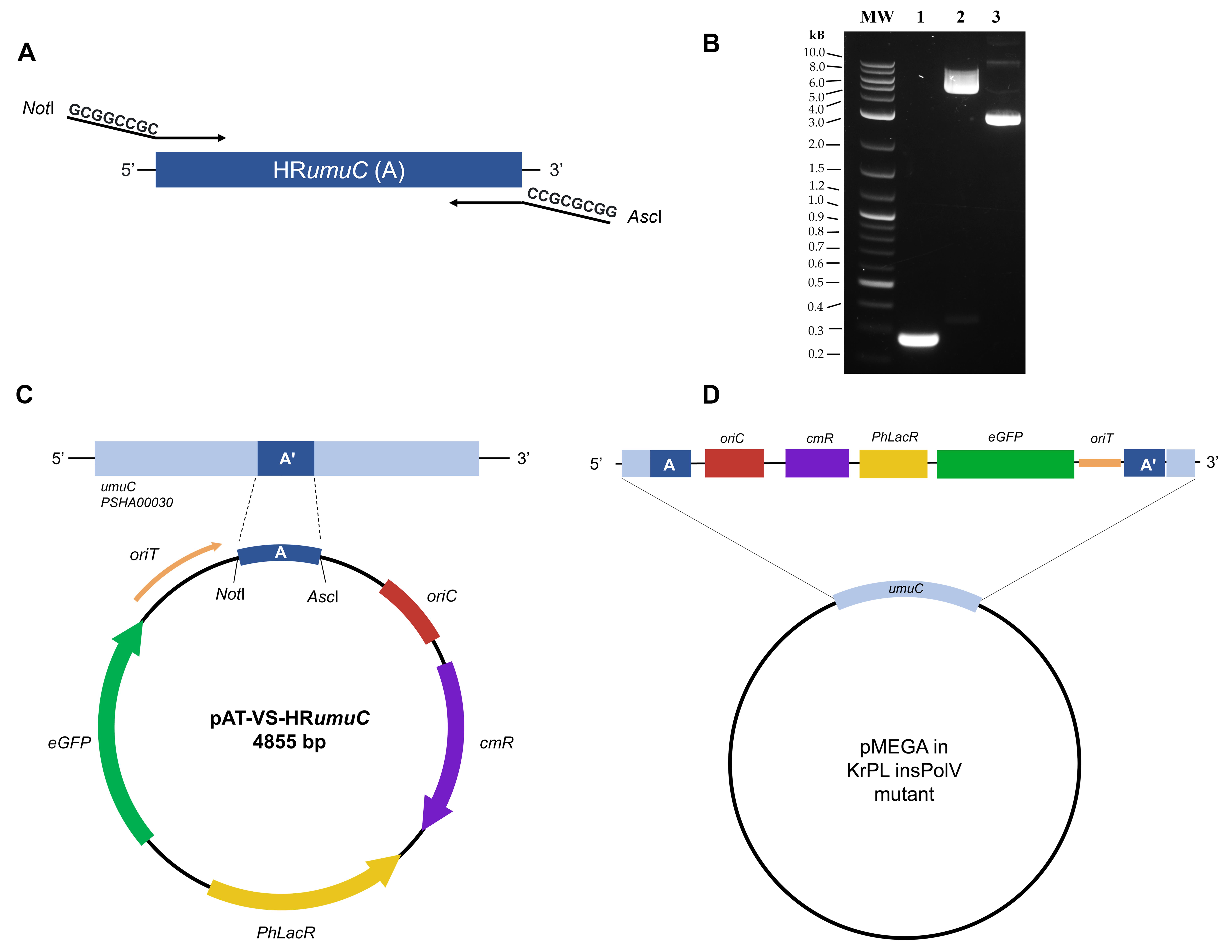
Figure 1. Insertion of the cmR selection marker in pMEGA. (A) Design of the primers for the amplification of the HRumuC region. The forward and reverse primers include, respectively, the NotI and AscI restriction sites at the 5′ and 3′ ends of the homology region. (B) 1% agarose gel electrophoresis of the PCR fragment and the digested pAT-egfp vector. MW = 1 kb Plus DNA ladder; 1 = HRumuC PCR fragment (275 bp); 2 = AscI-pAT-egfp-NotI linear fragment (4,587 bp); 3 = pAT-egfp plasmid DNA (negative control). (C) Schematic illustration of the suicide vector components. The umuC homology (259 bp) region A (dark blue) is inserted between NotI and AscI restriction sites; oriC is the origin of replication used to propagate the plasmid in E. coli strains (in red); oriT is responsible for the initiation of the conjugative transfer (in orange); PhLacR encodes the regulator of pLacZ promoter (not shown); eGFP represents the GFP reporter under the pLacZ promoter (in green); cmR represents the chloramphenicol resistance marker (in purple) used for selective pressure during the homologous recombination process. (D) Representation of the inserted pMEGA plasmid in KrPL insPolV mutants. The suicide vector is inserted at the umuC gene (in light blue) of the pMEGA plasmid. The umuC is disrupted, gaining the cmR resistance marker and the eGFP reporter as key features.
3. Order primers to a concentration of 100 μM from the Eurofins Genomics service.
4. Perform a PCR amplification following the manufacturer’s instructions and set the PCR program cycle as follows:
a. Initial denaturation, 95 °C, 30 s.
b. 25 cycles of denaturation, 95 °C, 15 s; annealing, 63 °C, 15 s; extension, 68 °C, 30 s.
c. Final extension, 68 °C, 5 min.
5. Load the PCR products on 1% w/v agarose gel and run electrophoresis (Figure 1B).
6. Extract the PCR fragment, NotI-HRumuC-AscI, from the gel using the QIAquick Gel Extraction kit and elute with 30 μL of double-distilled water (ddH2O).
7. In the following reaction, digest 1 μg of the extracted NotI-HRumuC-AscI fragment and 1 μg of the backbone pAT-egfp vector using NotI and AscI restriction enzymes following the manufacturer’s instructions.
Note: Perform DNA enzymatic digestion in two separate reaction mixes.
8. Incubate at 37 °C for at least 2 h to achieve proper digestion of both samples.
Note: Prior to DNA purification of the backbone pAT-egfp, check for successful DNA digestion by loading a small amount of digested DNA (100 ng) on a 0.8% agarose gel.
9. Load the linearized pAT-egfp vector on 1% w/v agarose gel and run electrophoresis (see Recipe 12) (Figure 1B).
10. Extract the linearized AscI-pAT-egfp-NotI fragment (4,587 bp) using the QIAquick Gel Extraction kit following the manufacturer’s instructions.
11. Elute the DNA with 30 μL of ddH2O.
12. Purify NotI-umuC-AscI (275 bp) using the QIAquick Gel Extraction kit and elute with 30 μL of ddH2O.
13. Perform a ligation reaction of both the NotI-HRumuC-AscI (25 ng) and the AscI-pAT-egfp-NotI (100 ng) fragments using T4 DNA ligase.
14. Set up the reaction mix as suggested by the manufacturer and incubate at 16 °C overnight. The resulting plasmid will be pAT-VS-HRumuC (Figure 1C).
15. Chill on ice and transform via heat-shock 20 μL of the reaction into 80 μL of E. coli TOP10 competent cells (see Recipes 13 and 14).
16. Plate the cells on LB agar plates containing 34 μg/mL of chloramphenicol antibiotic, as described in Recipe 14.
17. Incubate at 37 °C overnight.
18. Inoculate a single colony (10 independent clones) in 3 mL of LB medium supplemented with 34 μg/mL chloramphenicol and incubate at 37 °C with vigorous shaking (220 rpm, overnight).
19. Using 1 mL of overnight culture, perform plasmid DNA extraction using the Monarch® Plasmid Miniprep kit following the manufacturer’s instructions.
20. Elute the DNA using 100 μL of ddH2O.
21. Submit for Sanger sequencing to confirm the successful cloning of the HRumuC region inside the pAT-VS-HRumuC vector using the OriC rv primer listed in Table 1.
A2. Insertion of pAT-VS-HR-umuC suicide vector into pMEGA via homologous recombination
This procedure uses intergeneric conjugation to transfer the suicide vector into the recipient PhTAC125 KrPL strain, followed by a crossover event to insert the cmR selection marker into pMEGA (Figure 1D).
1. Transform 100 μL of E. coli S17-1 λpir donor strain using 50 ng of pAT-VS-HRumuC plasmid (see Recipe 14).
2. Plate the cells on LB agar plates containing 34 μg/mL of chloramphenicol.
3. Perform intergeneric conjugation using the E. coli S17-1 λpir as donor strain (harboring pAT-VS-HRumuC) and the PhTAC125 KrPL as recipient strain (see Recipe 15).
4. Spread the cultures on TYP solid medium supplemented with 50 μg/mL kanamycin and 3.75 μg/mL chloramphenicol to select the KrPL insPolV transconjugant strain.
5. Incubate at 15 °C for 7 days.
6. Perform a replica plating on TYP solid medium plates (35 mL) supplemented with 50 μg/mL kanamycin and increase the concentration of chloramphenicol to 6.25 μg/mL to enhance mutant selection.
7. Incubate at 15 °C overnight for 5 days.
A3. PCR screening
1. Inoculate each independent colony in 3 mL of TYP media supplemented with 25 μg/mL chloramphenicol and incubate at 4 °C for 3 days with shaking (200 rpm).
2. After incubation, make a glycerol stock by collecting 750 μL of saturated culture and mixing it with 250 μL of 80% glycerol.
3. Store at -80 °C.
4. With the remaining culture, collect 2 OD600 of the cell pellet.
5. Perform DNA extraction using the Bacterial DNA kit following the manufacturer’s instructions. The extracted DNA will serve as a DNA template for the following PCR screening. Store the DNA at -20 °C.
6. To confirm the insertion of the suicide vector into the target region (umuC) on pMEGA plasmid, perform PCR screening using Taq DNA Polymerase (NEB) following the manufacturer’s instructions and apply the PCR program described in step A1.4 with the following adjustments:
a. Annealing of 62 °C for 15 s for the couple of oligos NdeI_eGFP fw and KpnI_eGFP rv, and an extension time of 45 s to amplify the eGFP gene (Figure 2A).
b. Annealing of 61 °C for 15 s for the couple of oligos 5′umuC fw, oriC rv, and an extension time of 1 min and 10 s to amplify the pAT-VS-HRumuC insertion site (Figure 2B).
7. Verify the mutants (i.e., KrPL insPolV) by loading 10 μL of each PCR on a 1% w/v agarose gel and running electrophoresis (see Recipe 12) (Figure 2A, B).
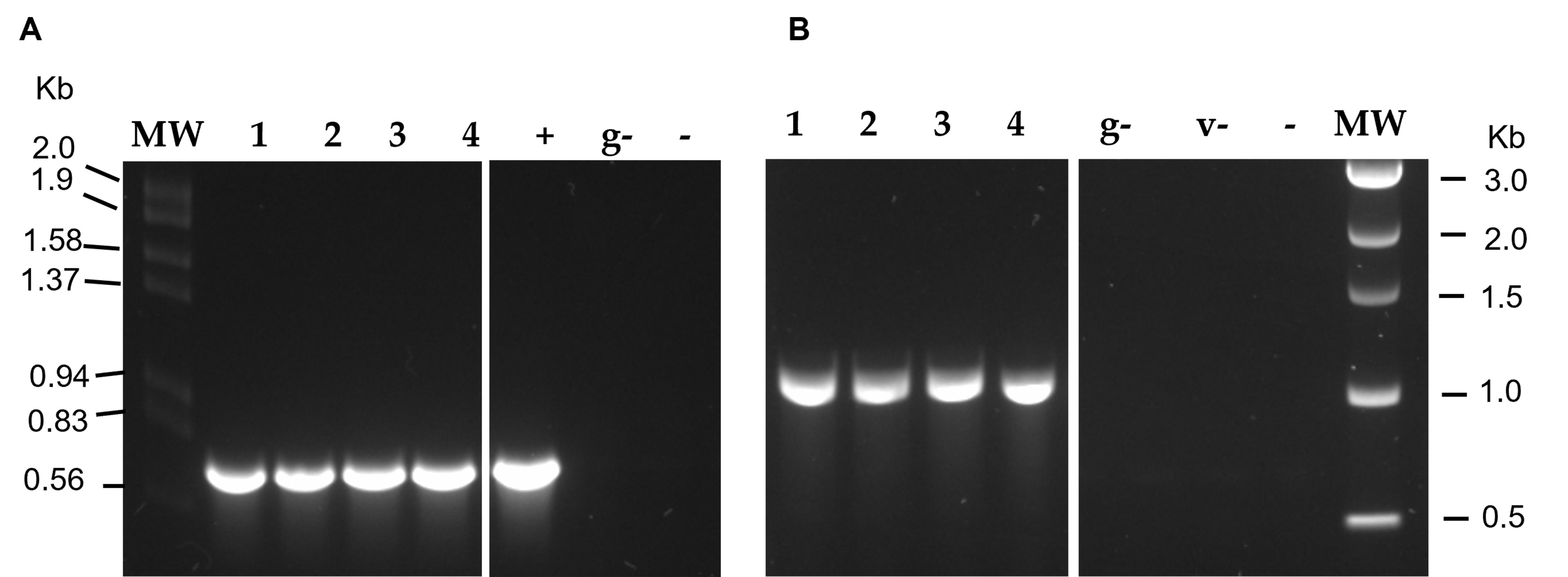
Figure 2. Validation of suicide vector integration. (A) PCR amplification of the eGFP gene (738 bp). MW = Lambda DNA EcoRI/HindIII ladder; + = pAT-VS-HRumuC vector as positive control. (B) PCR analysis of the suicide vector insertion site (1,044 bp). MW = 1 kb DNA ladder v- = pAT-VS-HRumuC vector negative control. g- = PhTAC125 KrPL bacterial DNA. Negative controls (-) performed without a DNA template were set up for both PCR analyses. Lanes 1–4 represent the isolated mutant clones of PhTAC125 KrPL pAT-VS-HR umuC; lanes separated by a thin white line, which indicates the splice, were removed for clarity.
B. Gene silencing of repB by PTasRNA technology
Note: This step is critical to the curing of pMEGA through PTasRNA technology. Thus, the choice of the target and the careful design of the paired termini antisense RNA are of key importance. The target must be a gene that is essential for pMEGA plasmid replication, maintenance, and stability, such as repB, which encodes for replication initiator protein RepB [13,14].
B1. Design of the antisense RNA
1. Download from NCBI the GenBank file (.gb) of pMEGA features (accession number NZ_MN400773.1) and identify the RepB protein as the PSHA_p00052 gene.
2. Retrieve the FASTA file, which includes the nucleotide sequence of the PSHA_p00052 gene to determine the ribosome-binding site (RBS) and the start codon (Dataset S2).
3. Design the sequence of the antisense (asRNArepB), including the RBS and the start codon of the gene inside its sequence (Figure 3A). The complementary reverse strand is the asRNArepB (Dataset S3).
4. Insert the antisense within the loop region of the PTasRNA scaffold. This placement ensures that the antisense sequence is accessible for targeting the transcript mRNA (Figure 3A, B) [10].
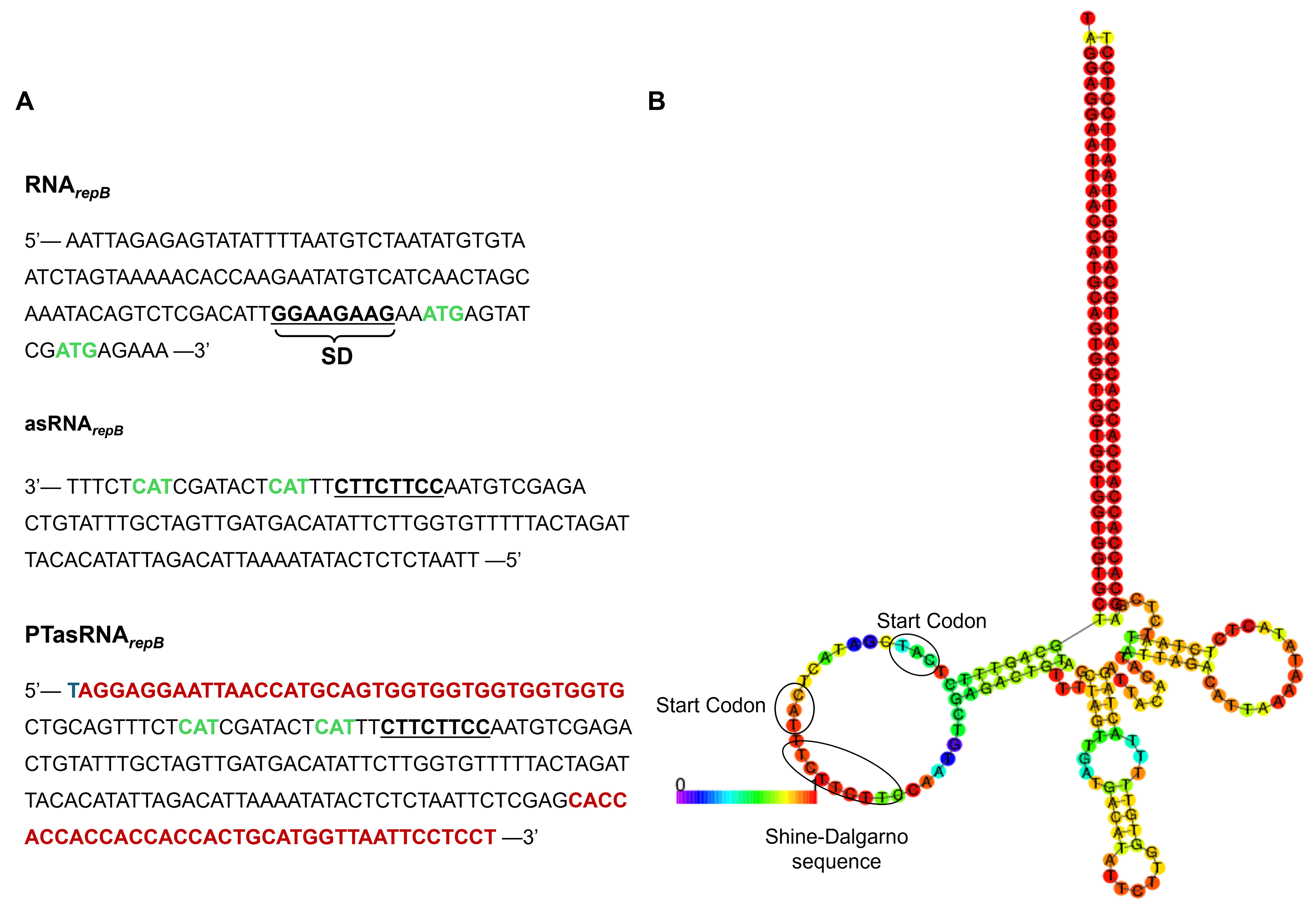
Figure 3. Design of the PTasRNArepB. (A) asRNArepB and PTasRNArepB nucleotide sequences. Underlined and in bold are reported the putative Shine–Dalgarno (SD), whereas in green, ATG start codons are highlighted. In red are reported the paired termini (PT) sequences flanking the asRNArepB RNA. (B) Predicted secondary structure of the PTasRNArepB through the mFold tool. Start codons of translation and putative SD sequence are pointed out by a circle. Two potential translation initiation sites are predicted. Uracil (U) is reported as thymine (T) to simplify visualization.
5. Perform a BLAST analysis of the asRNArepB against the whole genome of PhTAC125 using the NCBI nBlast tool (chromosome I: CR954246.1; chromosome II: CR954247.1; pMEGA: NZ_MN400773.1).
Note: This step is critical to ensure that the curing strategy does not induce any off-target effects, and repB is the unique target.
6. Use the RNAFold web server to perform a structure prediction of the full PTasRNArepB sequence.
7. Set parameters as default and calculate the minimum free energy (MFE) and partition function (Figure S1) [21].
8. A negative ∆G correlates to a higher thermodynamic stability of the designed asRNA (e.g., asRNArepB MFE is -100.50 kcal/mol).
Note: More negative values indicate a more stable mRNA molecule.
B2. Construction of the pB40-79C-PTasRNArepB vector
The antisense sequence is cloned into the psychrophilic expression vector pB40-79-PTasRNAlon in between the paired-termini sequence, exchanging the existing asRNAlon sequence with the newly designed asRNArepB. The resulting vector will have the PTasRNArepB. The backbone contains i) a psychrophilic origin of replication (OriR), ii) the ampicillin resistance cassette (ampR) for selection, and iii) the PTasRNA expression cassette, which includes the promoter and terminator flanking the exchangeable asRNA sequence.
1. Design the primers to amplify the asRNArepB sequence.
2. Add in the primer sequences the PstI and XhoI restriction sites at the 5′ and 3′ ends of the asRNArepB sequence.
3. Order primers to a concentration of 100 μM from the Eurofins Genomics service.
4. Prepare the vector backbone in which the asRNArepB will be cloned by digesting the pB40-79-PTasRNAlon plasmid with PstI and XhoI restriction enzymes following the manufacturer’s instructions.
5. Incubate at 37 °C for at least 2 h to achieve proper digestion.
6. Load the linearized pB40-79-PTasRNAlon vector on a 1.2% w/v agarose gel and run electrophoresis (Figure 4A).
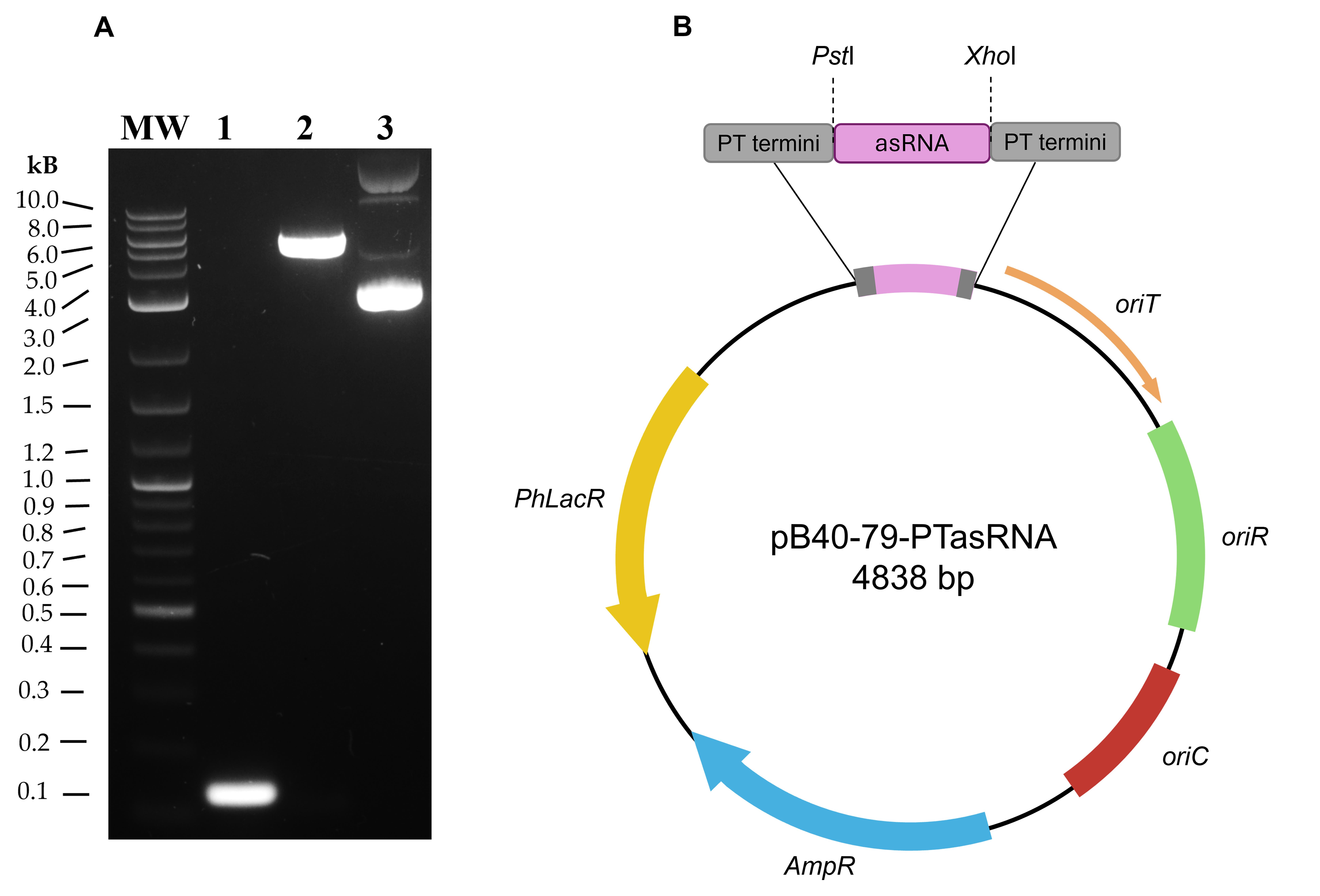
Figure 4. Cloning of the pB40-79C-PTasRNArepB vector. (A) 1.2% agarose gel electrophoresis of the PCR fragment and the digested pB40-79-PTasRNAlon vector. MW = 1 kb Plus DNA ladder; 1 = asRNArepB PCR fragment (110 bp); 2 = XhoI-pB40-79-PT-PstI linear fragment (4,979 bp); 3 = pB40-79-PTasRNAlon plasmid DNA (negative control). (B) Map of the PTasRNA vector. The diagram illustrates the modular organization of the silencing vector. The cloning site designed for the antisense sequence (pink) is flanked by PstI and XhoI restriction sites located between the two paired termini sequences (in gray). The vector includes two origins of replication: oriC for the propagation of the plasmid in E. coli strains (in red) and oriR for propagation in PhTAC125 (in green); oriT is pivotal for the conjugative transfer (in orange); PhLacR encodes the regulator of pLacZ promoter (not shown); ampR represents the ampicillin resistance marker (in blue).
7. Extract the linearized fragment XhoI-pB40-79-PT-PstI of 4979 bp using the QIAquick Gel Extraction kit following the manufacturer’s instructions.
8. Elute the DNA with 30 μL of ddH2O.
9. PCR-amplify the asRNArepB using Phusion High-Fidelity DNA polymerase and the pair of forward and reverse primers listed in Table 1 (XhoI-asRNArepB fw, PstI-asRNArepB rv).
10. Use 5 ng of the PhTAC125 genomic DNA, which includes pMEGA plasmid, as template.
11. Perform PCR following the manufacturer’s instructions as follows:
a. Initial denaturation, 98 °C, 30 s.
b. 25 cycles of denaturation, 98 °C, 10 s; annealing, 64 °C, 30 s; extension, 72 °C, 10 s.
c. Final extension, 72 °C, 10 min.
12. Load the PCR fragment on a 1.2% agarose gel and run electrophoresis (Figure 4A).
13. Purify the PCR fragment, PstI-asRNArepB-XhoI (110 nt), using the QIAquick Gel Extraction kit and elute with 30 μL of ddH2O.
14. Digest the extracted PstI-asRNArepB-XhoI fragment using PstI and XhoI restriction enzymes following the manufacturer’s instructions.
15. Purify the digested fragment using the QIAquick Gel Extraction kit.
16. Elute the DNA with 30 μL of ddH2O.
17. Perform a ligation reaction using both the PstI-asRNArepB-XhoI (15 ng) fragment and XhoI-pB40-79-PT-PstI (100 ng) fragments using T4 DNA ligase. The resulting vector will be the pB40-79-PTasRNArepB (Figure 4B, Dataset S4).
18. Set up the reaction mix as suggested by the manufacturer and incubate at 15 °C overnight.
19. Chill on ice and transform via heat-shock 20 μL of the reaction into 80 μL of E. coli TOP10 competent cells (see Recipe 14).
20. Plate the cells on LB agar plates containing 100 μg/mL of ampicillin.
21. Incubate at 37 °C overnight.
22. Inoculate a single colony in 3 mL of LB medium (see Recipes) and incubate at 37 °C with vigorous shaking (220 rpm, overnight).
23. Using 1 mL of overnight culture, perform plasmid DNA extraction using the Monarch® Plasmid Miniprep kit following the manufacturer’s instructions.
24. Elute the DNA using 100 μL of ddH2O.
25. With the remaining cultures, make a glycerol stock of E. coli TOP10 (pB40-79-PTasRNArepB), collecting 750 μL of saturated culture and mixing it with 250 μL of 80% glycerol.
26. Store at -80 °C.
27. Submit for Sanger sequencing to confirm the successful cloning of the PTasRNArepB cassette.
28. Transform 100 μL of E. coli S17-1 λpir donor strain using 50 ng of pB40-79-PTasRNArepB plasmid extracted in step B2.23 (see Recipe 14).
29. Plate the cells on LB agar plates containing 100 μg/mL of ampicillin.
30. Perform intergeneric conjugation using the E. coli S17-1 λpir as donor strain (harboring pB40-79-PTasRNArepB) and the PhTAC125 KrPL insPolV as recipient strain (see Recipe 15).
31. Select the transconjugants by plating on TYP solid medium plates (35 mL) supplemented with 50 μg/mL kanamycin and 100 μg/mL ampicillin to select the KrPL insPolV strain harboring the pB40-79-PTasRNArepB plasmid.
32. Incubate at 15 °C for 4 days.
33. Follow the intergeneric conjugation of the PhTAC125 protocol. As a final step, use 750 μL of saturated culture and mix it with 250 μL of 80% glycerol to make a glycerol stock.
34. Store at -80 °C.
C. Selection of cured clones by following the loss of the selection marker introduced in section A
C1. Gene silencing of repB with PTasRNA interference
1. Streak a loop of the glycerol stock of PhTAC125 KrPL insPolV strain harboring the pB40-79-PTasRNArepB on TYP agar plates supplemented with 100 μg/mL ampicillin. Use the non-recombinant KrPL insPolV strain as a negative control for the experiment, grown in non-selective conditions.
2. Incubate at 15 °C for three days.
3. Inoculate a single colony in 3 mL of TYP medium supplemented with 100 μg/mL ampicillin at 15 °C overnight using a 25 mL sterile glass tube with shaking (200 rpm).
4. Dilute the cultures to 0.2 OD600 in 10 mL of GG medium supplemented with Schatz salts and 100 μg/mL ampicillin in a 100 mL Erlenmeyer flask.
5. Incubate at 15 °C with shaking (200 rpm) overnight.
6. Perform a dilution to 0.35 OD600 in 20 mL of GG medium supplemented with Schatz salts and 100 μg/mL ampicillin in a 100 mL Erlenmeyer flask.
7. Incubate the diluted culture at 15 °C for 8 h with shaking (200 rpm).
8. In the evening, inoculate the cultures at 0.1 OD600 in 20 mL of GG medium supplemented with Schatz salts and 100 μg/mL ampicillin in a 100 mL Erlenmeyer flask.
9. Incubate at 15 °C for approximately 15 h with vigorous shaking (220 rpm).
10. When cells reach a value of 1.5 OD600, induce the production of the PTasRNArepB with 10 mM IPTG previously sterilized via filtration.
11. Collect culture samples equal to 0.1 OD600 (1–2 × 10⁷ CFU/mL) at three different time points: 8, 24, and 32 h post-induction.
12. Perform serial dilution of the 0.1 OD600 cultures up to 1:104 and spread 100 μL on non-selective TYP agar plates.
13. Incubate at 15 °C for 3 days.
14. Pick at least 100 colonies grown from each plate (8, 24, and 32 h post-induction) and replicate on a 70 mL TYP agar plate in the presence and absence of 12.5 μg/mL chloramphenicol as a selective agent.
Note: Plasmid loss was observed at low frequency under our experimental conditions (~4% of colonies screened). Therefore, screening at least 100 colonies is recommended to ensure recovery of cured clones.
15. Incubate at 15 °C for 24 h.
C2. Screening of mutants
The clones that produce PTasRNArepB can replicate easily on TYP agar without the selective agent but cannot grow if chloramphenicol is provided because they have lost the pMEGA plasmid. Then, clones are subjected to PCR colony screening.
1. Pick a single colony from the replica plates and resuspend the sample in 30 μL of sterile deionized MilliQ water.
2. Boil the sample for 10 min and spin down at maximum speed (13,000 rpm) for 10 min.
3. The supernatant will serve as a DNA template (2 μL) for PCR colony reactions.
4. Use the primers listed in Table 1 to amplify, respectively, the prom7 fragment (80 bp) (prom7 fw, prom 7 rv), the CDS49 fragment (100 bp) (CDS49 fw, CDS49 rv), and the umuC gene (259 bp) (5′umuC fw, umuC_AscI rv).
Note: The CDS49 and umuC genes are both located on the pMEGA plasmid.
5. Perform PCR screening using Taq DNA Polymerase according to the manufacturer’s instructions and apply the following PCR program:
a. Initial denaturation, 95 °C, 10 min.
b. 30 cycles of denaturation, 95 °C, 15 s; annealing, see step C2.6; extension, see step C2.6.
c. Final extension, 68 °C, 5 min.
6. Adjust annealing and extension steps according to the following parameters:
a. Annealing at 59 °C for 15 s for the couple of oligos prom7 fw and prom 7 rv, and an extension at 68 °C for 10 s.
b. Annealing at 52 °C for 15 s for the couple of oligos CDS49 fw and CDS49 rv, and an extension of 68 °C for 10 s.
c. Annealing at 63 °C for 15 s for the couple of oligos 5′umuC fw and umuC_AscI rv, and an extension of 68 °C for 30 s.
7. Verify the mutants (e.g., KrPL2) by loading 10 μL of each PCR on a 1.2% w/v agarose gel and running electrophoresis (Figure 5).
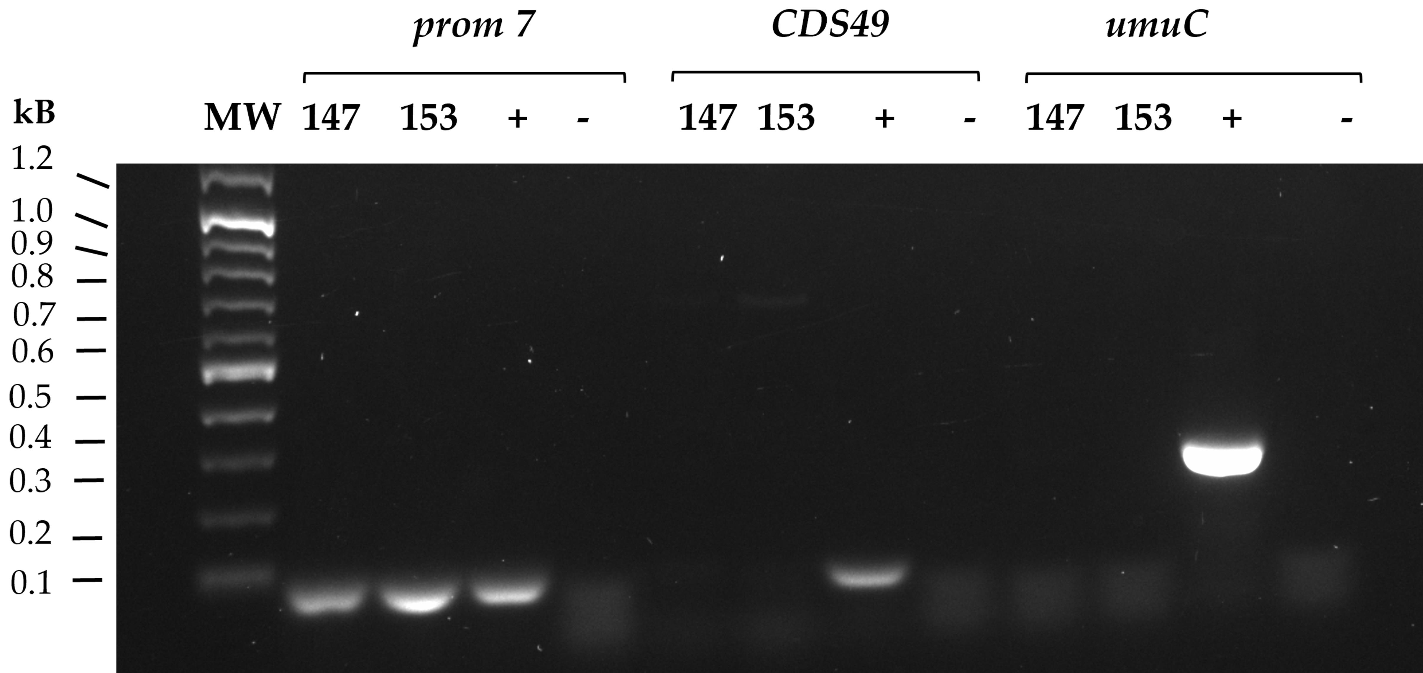
Figure 5. PCR screening of bacterial DNA to detect pMEGA loss. To prove deletion of the pMEGA plasmid, the screening amplified three different fragments: i) the prom7 fragment (80 bp), located on chromosome I, ii) the CDS49 fragment (100 bp), included in the pMEGA plasmid, and iii) the umuC (259 bp) fragment, included both in pMEGA and in the inserted suicide vector. PhTAC125 bacterial DNA was used as a template. MW = Lambda DNA EcoRI/HindIII ladder; 147 and 153: clone numbers 147 and 153 selected after 32 h induction of the PTasRNA interference plate; (+) positive control using extracted genomic DNA of PhTAC125 KrPL insPolV; (-) negative controls performed without DNA template were set up for all three PCRs.
8. Once the mutants are verified, inoculate the colony from the replica plate corresponding to the KrPL2 clone in 3 mL of TYP media.
9. Incubate at 15 °C for 24 h.
Note: It is recommended to prepare a glycerol stock by collecting 750 μL of saturated culture and mixing it with 250 μL of 80% glycerol. Store at -80 °C.
C3. Culture propagation to eliminate the pB40-79-PTasRNArepB
To remove the pB40-79-PTasRNArepB vector used for pMEGA curing (in steps C1 and C2), the KrPL2 strain, still harboring the PTasRNArepB vector, is subjected to culture propagation in the defined medium GG.
1. Dilute the culture to 0.1 OD600 in 3 mL of GG medium supplemented with Schatz salts.
2. Incubate at 15 °C for 24 h.
Note: During this period, the culture undergoes approximately five generations.
3. Collect cells corresponding to 0.1 OD600 and perform serial dilution up to 1:104.
4. Spread 100 μL of the dilution onto non-selective TYP agar plates.
5. Incubate at 15 °C for 3 days.
6. Replicate the grown colonies on TYP agar plates in the presence and absence of 100 μg/mL of ampicillin.
7. Incubate at 15 °C for 24 h.
Note: Culture propagation promotes the isolation of clones spontaneously cured of the pB40-79-PTasRNArepB plasmid used for pMEGA depletion. Colonies that have lost the plasmid will not grow in the presence of ampicillin. If no ampicillin-sensitive colonies are obtained, the propagation step (steps C3.1–7) should be repeated until ampicillin-sensitive clones are detected.
8. Pick the ampicillin-sensitive colonies and inoculate in 3 mL of TYP medium.
9. Incubate at 15 °C for 24 h.
10. Using 500 μL of overnight culture, extract plasmid DNA using the Monarch® Plasmid Miniprep kit following the manufacturer’s instructions.
Note: Prepare a glycerol stock by collecting 750 μL of saturated culture and mixing it with 250 μL of 80% glycerol. Store at -80 °C.
11. Load the extracted plasmid DNA on a 0.8% w/v agarose gel and run electrophoresis to verify the absence of the vector.
Note: Load on agarose gel 100 ng of pB40-79-PTasRNArepB vector to use as a positive control.
12. To further confirm plasmid loss, perform PCR screening using Taq DNA polymerase according to the manufacturer’s instructions and the primers listed in Table 1 (XhoI-asRNArepB fw, PstI-asRNArepB rv). Apply the following PCR program:
a. Initial denaturation, 95 °C, 30 s.
b. 30 cycles of denaturation, 95 °C, 15 s; annealing at 56 °C for 15 s, and an extension at 68 °C for 10 s.
c. Final extension, 68 °C, 5 min.
Note: Use 1 ng of pB40-79-PTasRNArepB vector as PCR positive control (expected amplicon: 112 nt).
13. Load 10 μL of each PCR on a 1.2% w/v agarose gel and run electrophoresis to verify plasmid loss.
Note: Ampicillin-sensitive clones will show no PCR amplification compared to the positive control.
Validation of protocol
This protocol has been used and validated in the following research article:
• Severino et al. [15]. Engineering the Marine Pseudoalteromonas haloplanktis TAC125 via the pMEGA Plasmid Targeted Curing Using PTasRNA Technology. Microorganisms. (Figure 2, Supplemental figures 1–3 and 5, and Supplementary tables 1–2).
Supplementary information
The following supporting information can be downloaded here:
1. Dataset S1. Homology Region of umuC gene (PSHA_p00030)
2. Dataset S2. FASTA sequence of the PSHA_p00057 gene with upstream 5′ UTR region
3. Dataset S3. FASTA sequence of the antisense RNA (asRNA) targeting PSHA_p00057
4. Dataset S4. FASTA sequence of the pB40-79-PTasRNArepB
5. Figure S1. RNAFold web server interface
Acknowledgments
Conceptualization, A.S., C.L., M.L.T.; Investigation, A.S., C.L.; Writing—Original Draft, A.S.; Writing—Review & Editing, A.S., C.L., M.C., E.P., M.L.T.; Funding acquisition, M.L.T.; Supervision, C.L., M.L.T.
During manuscript preparation, OpenAI assistants were used for grammatical refinement and lexical clarity. All content was subsequently reviewed, edited, and validated by the authors, who take full responsibility for the final version of the manuscript. Icons used in the graphical overview were obtained from BioRender (biorender.com) and adapted for this work.
This research was funded by the Italian Ministry of University and Research (MUR) within the PRIN 2022 project “Optimization of human CDKL5 recombinant production in the Antarctic bacterium Pseudoalteromonas haloplanktis TAC125: a multiparametric approach towards the exploitation of the enzyme replacement therapy for the CDKL5 Deficiency Disorder (ERT4CDD)” (Grant No. 2022J5X82R) to M.L.T.; the University of Pennsylvania Orphan Disease Center on behalf of the Loulou Foundation “Assessment of the therapeutic potential of TATk-hCDKL5 isoform 1 and 2 produced in recombinant Antarctic Pseudoalteromonas haloplanktis TAC125 (pilot award no. CDKL5-23-103-1) to M.L.T.; the National Center 5 “National Biodiversity Future Center” (code CN00000033), “Biodiversità”, financed by (PNRR)- Missione 4, Componente 2 “Dalla Ricerca all’Impresa” Investimento 1.4 “Potenziamento strutture di ricerca e creazione di campioni nazionali di R&S su alcune Key Enabling Technologies” UE—Next Generation EU, which funded the salary of C.L.; by the Regione Campania (Fondo di Rotazione ex L. 183/1987- Avvio delle misure organizzative per la "Promozione di progetti di ricerca, sviluppo sperimentale e innovazione collaborativi nel campo delle malattie rare") project "Verso la terapia proteica sostitutiva per il trattamento della sindrome da deficienza di CDKL5: produzione, caratterizzazione e somministrazione" (Intervento n. 26); and by the Italian Parents’ Association “La fabbrica dei sogni 2—New developments for Rett syndrome” (to A.S. M.C., C.L. and M.L.T.).
This protocol was used in [15].
Competing interests
The authors declare no conflicts of interest.
References
- Lauritsen, I., Kim, S. H., Porse, A. and Nørholm, M. H. H. (2018). Standardized Cloning and Curing of Plasmids. Methods Mol Biol. 1772: 469–476. https://doi.org/10.1007/978-1-4939-7795-6_28
- Trevors, J. (1986). Plasmid curing in bacteria. FEMS Microbiol Lett. 32: 149–157. https://doi.org/10.1111/j.1574-6968.1986.tb01189.x
- Rodríguez-Beltrán, J., DelaFuente, J., León-Sampedro, R., MacLean, R. C. and San Millán, Á. (2021). Beyond horizontal gene transfer: the role of plasmids in bacterial evolution. Nat Rev Microbiol. 19(6): 347–359. https://doi.org/10.1038/s41579-020-00497-1
- Buckner, M. M. C., Ciusa, M. L. and Piddock, L. J. V. (2018). Strategies to combat antimicrobial resistance: anti-plasmid and plasmid curing. FEMS Microbiol Rev. 42(6): 781–804. https://doi.org/10.1093/femsre/fuy031
- Tolmasky, M. E., Actis, L. A., Welch, T. J. and Crosa, J. H. (2007). Plasmids. In Methods for General and Molecular Microbiology (pp. 709–734). John Wiley & Sons, Ltd. https://doi.org/10.1128/9781555817497.ch30
- Calvanese, M., Balestra, C., Colarusso, A., Lauro, C., Riccardi, C., Fondi, M., Parrilli, E. and Tutino, M. L. (2023). Development of high-copy number plasmids in Pseudoalteromonas haloplanktis TAC125. Appl Microbiol Biotechnol. 107(7–8): 2469–2481. https://doi.org/10.1007/s00253-023-12448-w
- Calvanese, M., Colarusso, A., Lauro, C., Parrilli, E. and Tutino, M. L. (2022). Soluble Recombinant Protein Production in Pseudoalteromonas haloplanktis TAC125: The Case Study of the Full-Length Human CDKL5 Protein. In E. Garcia Fruitós and A. Arís Giralt (Eds.), Insoluble Proteins (Vol. 2406, pp. 219–232). New York, NY: Springer US. https://doi.org/10.1007/978-1-0716-1859-2_13
- Colarusso, A., Lauro, C., Calvanese, M., Parrilli, E. and Tutino, M. L. (2020). Improvement of Pseudoalteromonas haloplanktis TAC125 as a Cell Factory: IPTG-Inducible Plasmid Construction and Strain Engineering. Microorganisms. 8(10): Article 10. https://doi.org/10.3390/microorganisms8101466
- Colarusso, A., Lauro, C., Calvanese, M., Parrilli, E. and Tutino, M. L. (2022). Active human full-length CDKL5 produced in the Antarctic bacterium Pseudoalteromonas haloplanktis TAC125. Microb Cell Fact. 21: 211. https://doi.org/10.1186/s12934-022-01939-6
- Lauro, C., Colarusso, A., Calvanese, M., Parrilli, E. and Tutino, M. L. (2022). Conditional gene silencing in the Antarctic bacterium Pseudoalteromonas haloplanktis TAC125. Res Microbiol. 173: 103939. https://doi.org/10.1016/j.resmic.2022.103939
- Parrilli, E., Tedesco, P., Fondi, M., Tutino, M. L., Lo Giudice, A., de Pascale, D. and Fani, R. (2021). The art of adapting to extreme environments: The model system Pseudoalteromonas. Phys Life Rev. 36: 137–161. https://doi.org/10.1016/j.plrev.2019.04.003
- Hall, J. P. J., Botelho, J., Cazares, A. and Baltrus, D. A. (2022). What makes a megaplasmid? Phil Trans R Soc B. 377(1842): Article 1842. https://doi.org/10.1098/rstb.2020.0472
- Médigue, C., Krin, E., Pascal, G., Barbe, V., Bernsel, A., Bertin, P. N., Cheung, F., Cruveiller, S., D'Amico, S., Duilio, A., et al. (2005). Coping with cold: The genome of the versatile marine Antarctica bacterium Pseudoalteromonas haloplanktis TAC125. Genome Res. 15(10): 1325–1335. https://doi.org/10.1101/gr.4126905
- Qi, W., Colarusso, A., Olombrada, M., Parrilli, E., Patrignani, A., Tutino, M. L. and Toll-Riera, M. (2019). New insights on Pseudoalteromonas haloplanktis TAC125 genome organization and benchmarks of genome assembly applications using next and third generation sequencing technologies. Sci Rep. 9(1): Article 1. https://doi.org/10.1038/s41598-019-52832-z
- Severino, A., Lauro, C., Calvanese, M., Riccardi, C., Colarusso, A., Fondi, M., Parrilli, E. and Tutino, M. L. (2025). Engineering the Marine Pseudoalteromonas haloplanktis TAC125 via the pMEGA Plasmid Targeted Curing Using PTasRNA Technology. Microorganisms. 13(2): 324. https://doi.org/10.3390/microorganisms13020324
- Giuliani, M., Parrilli, E., Pezzella, C., Rippa, V., Duilio, A., Marino, G. and Tutino, M. L. (2012). A Novel Strategy for the Construction of Genomic Mutants of the Antarctic Bacterium Pseudoalteromonas haloplanktis TAC125. In A. Lorence (Ed.), Recombinant Gene Expression (Vol. 824, pp. 219–233). Totowa, NJ: Humana Press. https://doi.org/10.1007/978-1-61779-433-9_11
- Sambrook, J., Fritsch, E. F. and Maniatis, T. (1989). Molecular cloning: a laboratory manual. Molecular cloning: a laboratory manual., Ed. 2, Article Ed. 2. Retrieved from https://www.cabdirect.org/cabdirect/abstract/19901616061
- Tutino, M., Duilio, A., Parrilli, E., Remaut, E., Sannia, G. and Marino, G. (2001). A novel replication element from an Antarctic plasmid as a tool for the expression of proteins at low temperature. Extremophiles. 5(4): 257–264. https://doi.org/10.1007/s007920100203
- Calvanese, M., D’Angelo, C., Lauro, C., Tutino, M. L. and Parrilli, E. (2024). Recombinant protein production in Pseudoalteromonas haloplanktis TAC125 biofilm. Biofilm. 7: 100179. https://doi.org/10.1016/j.bioflm.2024.100179
- Duilio, A., Madonna, S., Tutino, M. L., Pirozzi, M., Sannia, G. and Marino, G. (2004). Promoters from a cold-adapted bacterium: definition of a consensus motif and molecular characterization of UP regulative elements. Extremophiles. 8(2): 125–132. https://doi.org/10.1007/s00792-003-0371-2
- Lorenz, R., Bernhart, S. H., Höner zu Siederdissen, C., Tafer, H., Flamm, C., Stadler, P. F. and Hofacker, I. L. (2011). ViennaRNA Package 2.0. Algorithms Mol Biol. 6(1): 26. https://doi.org/10.1186/1748-7188-6-26
Article Information
Publication history
Received: Feb 12, 2026
Accepted: Apr 3, 2026
Available online: Apr 17, 2026
Published: May 20, 2026
Copyright
© 2026 The Author(s); This is an open access article under the CC BY-NC license (https://creativecommons.org/licenses/by-nc/4.0/).
How to cite
Severino, A., Lauro, C., Calvanese, M., Parrilli, E. and Tutino, M. L. (2026). Plasmid Curing of Pseudoalteromonas haloplanktis TAC125 Using Homologous Recombination and PTasRNA Gene Silencing. Bio-protocol 16(10): e5687. DOI: 10.21769/BioProtoc.5687.

 Download Citation in RIS Format
Category
Microbiology > Microbial genetics > Plasmid
Molecular Biology > RNA > RNA interference
Microbiology > Microbial genetics > Recombination
Do you have any questions about this protocol?
Post your question to gather feedback from the community. We will also invite the authors of this article to respond.
 Post a Question


