


- Protocols

- Articles and Issues
- For Authors
- About
- Become a Reviewer


Assessing Mitochondrial Respiratory Complex-Associated Function From Previously Frozen Mouse Placental Tissue

(*Contributed equally to this work, §Technical contact: podinict@mcmaster.ca) Published: Vol 16, Iss 9, May 5, 2026 DOI: 10.21769/BioProtoc.5667 Views: 442
Reviewed by: Migla MiskinyteAnonymous reviewer(s)
 PDF
PDF  Ask a question
Advertisement



Protocol Collections
Comprehensive collections of detailed, peer-reviewed protocols focusing on specific topics




Related protocols
Abstract
The placenta is a metabolically active organ whose mitochondrial activity is tightly linked to fetal growth, oxygenation, and nutrient transport, mediating fetal susceptibility to environmental exposures. Accordingly, aberrant mitochondrial function has been implicated in the progression of placental dysfunction. However, existing respirometry platforms require primarily fresh or cryopreserved placental tissue and offer limited throughput, rendering these platforms impractical in the context of large-scale placental dissections. Here, we describe and validate a Seahorse XF approach for measuring mitochondrial respiration in previously frozen placentae, enabling the functional interrogation of placental mitochondria in prenatal studies. Our protocol fundamentally relies on the restoration of matrix substrates that are depleted due to increased mitochondrial membrane permeability following freeze-thaw cycles. We provide a strategy to assess complex I and II-associated respiration adapted for the Seahorse XFe24 Analyzer and further demonstrate comparable oxygen consumption readouts between fresh and frozen placentae. We further demonstrate distinct differences in the magnitude of oxygen consumption between fresh and frozen placentae in the absence of exogenous NADH. Taken together, we present a simplified and convenient protocol for the assessment of respiratory enzyme complex-associated respiration from archived placental tissue.
Key features
• This protocol is suitable for use with previously frozen mouse placental tissue.
• Streamlined protocol for complex-associated respirometry assessments following large-scale placental dissections.
• Respirometry data may be acquired in <4 hours.
Keywords: Placenta Mitochondria
Respirometry
Metabolism
Electron transport-chain enzyme activity
Respiration
Bioenergetics
Oxygen consumption
Fresh tissue
Frozen tissue
Mitochondria
Respirometry
Metabolism
Electron transport-chain enzyme activity
Respiration
Bioenergetics
Oxygen consumption
Fresh tissue
Frozen tissue
Graphical overview
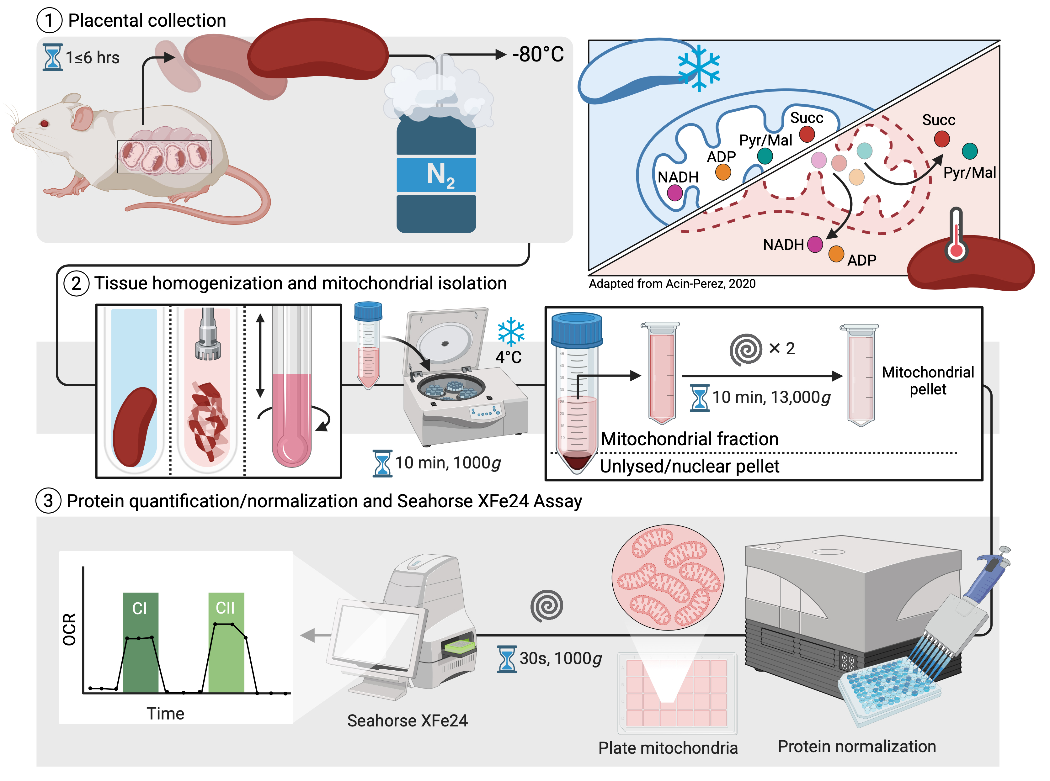
Overview of workflow for respirometry in previously frozen mouse placental tissue
Background
Placentation encompasses several key physiological and cellular stages, spanning from implantation to trophoblast differentiation [1]. This transient organ facilitates nutrient, gas, and waste exchange, while selectively transporting substances across the maternal–fetal interface and producing key pregnancy and fetal growth hormones [2,3]. As such, the energy-intensive demands of this multifunctional organ incur a significant metabolic cost sustained by its rich mitochondrial content [4]. Placental mitochondria undergo morphological and functional changes as villous cytotrophoblasts differentiate into syncytiotrophoblasts [5–7]. In addition, mitochondrial content and respiration have been shown to increase throughout pregnancy in mouse [8] and human placentae [9], respectively. Moreover, placental oxygen consumption and energy expenditure increase dramatically as pregnancy progresses, with up to 70% of O2 being consumed from the uterine circulation [4,10–12], suggesting a reliance on mitochondrial respiration for fetal growth. Placental mitochondrial dysfunction has increasingly been implicated in the etiology of common pregnancy disorders, including pre-eclampsia (PE) [13,14], fetal growth restriction (FGR) [15], and gestational diabetes mellitus (GDM) [15–18]. During oxidative phosphorylation (OXPHOS), electron leakage at complexes I, II, and III gives rise to mitochondrial reactive oxygen species (mtROS). Aberrant mtROS production, a hallmark of mitochondrial dysfunction, has been demonstrated in response to placental hypoxia and environmental insults [14]. Thus, mitochondrial respiratory chain enzyme complex–specific respiration is a critical parameter to consider when evaluating downstream mtROS production and oxidative pressure, as defects in electron transport may precede broader placental dysfunction.
In reproductive biology, both fresh and frozen placental tissues have been used to assess mitochondrial respiration [9,19–24]. Although fresh tissue is generally preferred, the timing of gestational stages and the logistical challenges of collecting placentae, particularly in late pregnancy, make it challenging to measure fresh samples in a study-controlled manner. These practical constraints, including the need to sacrifice large numbers of animals, limit the feasibility of fresh-tissue observations. To overcome this, recent studies have increasingly turned to frozen placentae to evaluate mitochondrial respiration [25–28]. Freeze-thaw cycles can disrupt mitochondrial membranes and result in the leakage of matrix substrates such as NADH, thereby compromising coupled mitochondrial respiration (29). The respiratory chain complexes themselves remain structurally and functionally intact [28,30]. Accordingly, respiratory enzyme complex-linked oxygen consumption has been measured in various snap-frozen tissues by supplying exogenous substrates and inhibitors [30–34], an approach that has been successfully applied to placental tissues using both Seahorse assays [25] and high-resolution respirometry in cryopreserved biopsies (28).
Here, we present a detailed protocol for measuring mitochondrial respiration in frozen mouse placentae using the Seahorse XF24 Analyzer. We outline recommended starting amounts of mitochondrial protein and provide suggestions on selecting and modifying injection port combinations to target specific mitochondrial complexes.
Materials and reagents
Biological materials
1. 6–11-week-old male and female CD1 mice (Charles River Laboratories) (see General note 1)
Reagents
1. PRAErrane isoflurane (Baxter, catalog number: CA2L9108)
2. HyCloneTM Hank’s 1× balanced salt solution (1× HBSS) (Thermo Fisher, Cytiva, catalog number: SH3058802)
3. D-mannitol (BioShop, catalog number: MAN509)
4. Sucrose (BioShop, catalog number: SUC507)
5. Potassium phosphate monobasic (KH2PO4) (Sigma-Aldrich, catalog number: P5379-100G)
6. Magnesium chloride (MgCl2) (Sigma-Aldrich, catalog number: M8266-100G)
7. Bovine serum albumin, fraction V, fatty acid free (BSA) (GoldBio, catalog number: A-421-250)
8. N-2-Hyxdroxyethylpiperazine-N’-2-ethanesulfonic acid (HEPES) (BioShop, catalog number: HEP001.500)
9. Ethylene glycol bis (2-Aminoethyl Ether) N,N,N’,N’ tetraacetic acid (EGTA) (BioShop, catalog number: EGT101)
10. Potassium hydroxide (KOH) (Sigma-Aldrich, 221473-25G)
11. Reduced β-nicotinamide adenine dinucleotide, reduced dipotassium salt (NADH) (Sigma-Aldrich, catalog number: N4505-100MG)
12. cOmpleteTM, Mini, EDTA-free protease inhibitor cocktail (Sigma-Aldrich, catalog number: 4693159001)
13. PhosSTOPTM (Sigma-Aldrich, catalog number: 4906837001)
14. Rotenone (Sigma-Aldrich, catalog number: R8875-10G)
15. Sodium succinate dibasic hexahydrate (succinate) (Sigma-Aldrich, catalog number: S2378)
16. Adenosine 5-diphosphate, potassium salt (ADP) (Sigma-Aldrich, catalog number: 117105)
17. Antimycin A (Sigma-Aldrich, catalog number: A8674-25MG)
18. Cytochrome c (Sigma-Aldrich, catalog number: C3131-10MG)
19. Carbonyl cyanide p-trifluoro-methoxyphenyl hydrazone, uncoupling agent (FCCP) (Sigma-Aldrich, catalog number: C2920)
20. Sodium pyruvate (Sigma-Aldrich, catalog number: P2256-100G)
21. L-malic acid (Sigma-Aldrich, catalog number: M1000)
22. Oligomycin (Sigma-Aldrich, catalog number: O4876)
23. Bradford reagent (Abcam, catalog number: ab119216)
24. InvitrogenTM UltraPureTM distilled water (Thermo Fisher Scientific, catalog number: 10977015)
25. Ethyl alcohol, pure (Sigma-Aldrich, catalog number: 459836)
26. Dimethyl sulfoxide (DMSO) (Sigma-Aldrich, catalog number: D8418-50ML)
27. Anti-Tom20 (D8T4N) rabbit monoclonal antibody (Cell Signaling Tech., catalog number: 42406)
28. Anti-Gapdh (14C10) rabbit monoclonal antibody (Cell Signaling Tech., catalog number: 2118)
29. Seahorse XF calibrant solution (included in Seahorse XFe24 FluxPak) (Agilent, catalog number: 102340-100)
Solutions
1. HEPES stock (1 M) (see Recipes)
2. EGTA stock (100 mM) (see Recipes)
3. Mitochondrial Assay Solution (MAS) (see Recipes)
4. NADH stock (75 mM) (see Recipes)
5. ADP stock (500 mM) (see Recipes)
6. Malate stock (400 mM) (see Recipes)
7. Pyruvate stock (2 M) (see Recipes)
8. Succinate stock (1 M) (see Recipes)
9. Antimycin A stock (5 mM) (see Recipes)
10. Oligomycin stock (10 mM) (see Recipes)
11. FCCP stock (10 mM) (see Recipes)
12. Cytochrome c stock (10 mg/mL) (see Recipes)
13. MAS buffer with cytochrome c (20 μg/mL) (see Recipes)
14. Port reagents for complex I and II assessment (see Recipes)
Recipes (see General note 2)
1. HEPES stock (1 M)
Dissolve 23.83 g of HEPES in 80 mL of ddH2O in a glass beaker. Mix on a magnetic stirrer and adjust pH to 7.4 using 5 M KOH. Adjust volume to 100 mL with ddH2O. Store at 2–8 °C.
2. EGTA stock (100 mM)
Dissolve 1.9 g of EGTA powder in 20 mL of sterile water. Adjust pH to 11 with KOH to allow for dissolution. Once dissolved, adjust pH to 8 with HCl and add sterile water to make a final volume of 50 mL. Store at 2–8 °C for <3 months.
3. Mitochondrial assay solution (MAS)
| Reagent | Final concentration | Quantity or volume for 50 mL of MAS buffer |
|---|---|---|
| Mannitol | 220 mM | 2.00 g |
| Sucrose | 70 mM | 1.20 g |
| KH2PO4 | 10 mM | 68 mg |
| MgCl2 | 5 mM | 23.8 mg |
| Fatty acid-free (FAF) BSA | 0.1% (w/v) | 50 mg |
| HEPES | 2 mM | 100 μL |
| EGTA | 1 mM | 200 μL |
Dissolve all reagents in 40 mL of ddH2O and adjust pH to 7.4 with KOH. Adjust volume to 50 mL and pH to 7.4, filter sterilize, and store at 2–8 °C for <2 weeks.
4. NADH stock (75 mM)
Dissolve 111.8 mg of NADH in 2 mL of sterile water. Aliquot to avoid freeze-thawing and store at -20 °C for <3 months.
5. ADP stock (500 mM)
Dissolve 1 g of ADP in 2.3 mL of sterile water and carefully neutralize with ~900 μL of 5 M KOH. Add 243.96 mg of MgCl2 and continue stirring until the white precipitate dissolves. Check and adjust pH to 7 with <50 μL of KOH (if necessary). Adjust volume with sterile water up to 4 mL. Aliquot and store at -80 °C to avoid freeze-thawing.
6. Malate stock (400 mM)
Dissolve 268.2 mg of L-malic acid in 3 mL of sterile water. Neutralize the solution with ~900 μL of 5 M KOH by adding 100–200 μL incrementally and monitoring pH. Adjust the final volume to 5 mL with sterile water. Aliquot and store at -20 °C.
Note: As this reaction is exothermic, it is recommended to dissolve L-malic acid in a volumetric glass flask or a closed 15-mL Falcon tube to minimize volume loss due to evaporation.
7. Pyruvate stock (2 M)
Dissolve 44 mg of sodium pyruvate in 200 μL of sterile water. Make the pyruvate stock fresh immediately prior to making the port reagents.
8. Succinate stock (1 M)
Dissolve 1.3505 g of succinate in 3 mL of sterile water. Check pH and adjust to 7 with ~65 μL of 1 M HCl. Adjust the volume to 5 mL with sterile water. Aliquot and store at -20 °C.
9. Antimycin A stock (5 mM)
Dissolve 5.4 mg of antimycin A in 2 mL of absolute ethanol. Aliquot and store at -20 °C.
10. Oligomycin stock (10 mM)
Dissolve 5 mg of oligomycin by adding 632.11 μL of DMSO into the glass vial. Mix well. Aliquot and store at -20 °C.
11. FCCP stock (10 mM)
Dissolve 2.54 mg of FCCP in 1 mL of absolute ethanol. Aliquot and store at -20 °C.
12. Cytochrome c stock (10 mg/mL)
Dissolve 10 mg of cytochrome c in 1 mL of sterile water. Aliquot to avoid freeze-thawing and store at -20 °C for <3 months.
13. MAS buffer with cytochrome c (20 μg/mL)
Add 10 μL of 10 mg/mL cytochrome c stock solution to 4990 μL of MAS warmed to room temperature. Make immediately prior to use in step D6.
14. Port reagents for complex I and II assessment
| Port | Compound | Stock concentration | Working concentration in the well | Volume of stock added in 2 mL of MAS buffer |
|---|---|---|---|---|
| A | NADH | 75 mM | 1 mM | 80 μL |
| A | ADP | 500 mM | 4 mM | 40 μL |
| A | Pyruvate | 2 M | 20 mM | 30 μL |
| A | Malate | 400 mM | 2 mM | 15 μL |
| B | Rotenone | 1 mM | 2 μM | 16 μL |
| C | Succinate | 1 M | 10 mM | 100 μL |
| D | Antimycin A | 5 mM | 4 μM | 10 μL |
There are four ports (labeled A–D) where substrates and inhibitors may be loaded for sequential injections in the assay. Port reagents are prepared at 9× of working concentration in 2 mL of MAS and loaded at 50 μL per port. Make stock concentration of reagents and store at -20 °C in one-time use aliquots. Do not reuse freeze-thawed aliquots. These working concentrations are adjusted to a starting volume of 100 μL of mitochondrial-enriched fraction loaded in the well. However, volumes can be changed based on the starting material.
Laboratory supplies
1. VWR® #5 precision tweezers (Avantor, catalog number: 89259-986)
2. Liquid nitrogen
3. Corning® polypropylene cryogenic vials (Thermo Fisher Scientific, catalog number: 09-761-71)
4. Seahorse XFe24 FluxPak (culture plates, sensor cartridge, calibrant) (Agilent, catalog number: 102340-100)
5. Falcon® 14-mL round-bottom high clarity PP test tube (Corning, catalog number: 352059)
6. PYREX® 5-mL Potter-Elvehjem tissue grinder with PTFE pestle (Corning, catalog number: 7725T-5)
7. Falcon® 15-mL high clarity PP conical centrifuge tube (Corning, catalog number: 352096)
8. 2-mL microtubes (Millipore Sigma, Corning, catalog number: AXYMCT200CS)
9. Corning® 96-well clear flat bottom UV-transparent microplate (Corning, catalog number: 3635)
10. FisherbrandTM disposable pipette basins (Thermo Fisher Scientific, catalog number: 13-601-508)
Equipment
1. Type 37900 culture incubator, 37 °C (Marshall Scientific, Thermolyne, model: 137925, catalog number: T37900)
2. Polytron® PT 1300 D handheld disperser (Kinematica, catalog number: 11010032)
3. EppendorfTM centrifuge 5810 R (4 °C) (Thermo Fisher Scientific, catalog number: 05-400-61)
4. Thermo Scientific IEC CL30 centrifuge (Marshall Scientific, catalog number: TSCL30)
5. Seahorse XFe24 Analyzer (Agilent, catalog number/model: S7801B)
6. TECAN Infinite 200 PRO (Tecan, catalog number/model: 490016-20BL)
Software and datasets
1. Seahorse Wave Desktop Software (Agilent, version 2.6.1), unlimited license with instrument
2. Seahorse Wave Controller Software (Agilent, version 2.2.1), unlimited license with instrument
Procedure
A. Mouse placental tissue harvest (estimated time: 1 h)
[On embryonic day 18.5 (E18.5)] (see General note 3)
1. Anesthetize pregnant mouse with isoflurane overdose (isoflurane vaporizer set to 5% and oxygen to 1–2 L/min) and cervically dislocate to sacrifice.
Note: For a detailed, visual guide on opening the abdominal cavity and exteriorizing the uterine horns from pregnant dams, we suggest following a previously established protocol where this procedure is illustrated [35].
2. Perform a caesarean-section by carefully cutting the abdomen down the midline to expose the uterus.
3. Retrieve the uterine horn from the abdomen and dissect at both the ovarian and cervical attachment sites. Place the uterine horn in a Petri dish containing ice-cold 1× HBSS on ice.
4. Separate feto-placental units prior to dissecting the placentae from each fetus.
5. Remove the umbilical cord, yolk sac, and metrial gland tissue from each placenta using #5 precision tweezers.
6. Immediately place the placenta into a cryovial and snap freeze in liquid N2 for future frozen placental tissue respirometry assessment or proceed without snap freezing for fresh placental tissue respirometry assessment.
7. Fresh tissues should be immediately suspended in 3 mL ice-cold MAS in 14 mL round-bottom Falcon tubes and kept on ice for <30 min until homogenization.
Note: In this protocol, fresh (n = 3) and frozen placentae (n = 3) were extracted in <20 min to avoid potential tissue degradation (total of 6 placentae). Tissue collections (fresh, frozen, fresh, etc.) were staggered to ensure that all placentae were treated equally and to ensure consistency during tissue extraction. However, we cannot recommend a minimum amount of time between the fresh tissue extraction and homogenization.
8. Store all snap-frozen placentae at -80 °C.
B. Preparation of the Seahorse cartridge (estimated time: 1 h)
(Day before assay)
1. Open the Seahorse XFe24 Flux Assay kit containing the utility plate, hydro booster (pink), sensor cartridge (green), and cartridge lid.
2. Set aside the sensor cartridge carefully and add 1 mL of Seahorse XF calibrant solution to each well of the utility plate.
3. Place the hydro booster on the utility plate and lower the sensor cartridge to submerge it into solution.
4. Incubate in a non-CO2, 37 °C incubator for at least 4 h or overnight.
5. Prepare stock solutions of all drugs and reagents listed in the Recipes section.
C. Mouse placental homogenization (estimated time: 1 h)
(Day of assay)
1. Cool a sterile metal tray and tweezers with liquid N2 prior to handling previously frozen placental tissue. Weigh and record the placental weights of both fresh and frozen placentae (see General note 4).
2. Immediately place weighed placental tissues into 3 mL of ice-cold MAS (containing protease and phosphatase inhibitors and FAF BSA) in 14 mL round-bottom Falcon tubes.
3. Sterilize the drill bit of the Polytron PT 1300 D mechanical homogenizer with 70% ethanol and ddH2O before use and set it to 15,000 rpm.
4. Submerge the grinder and homogenize each placental sample by moving the sample up and down for 5 s.
5. Transfer 3 mL of tissue homogenates into pre-cooled 5 mL glass-Teflon Dounce homogenizers on ice. Further homogenize the tissue lysate by performing 20 slow, twisting strokes per sample.
Critical: Keep an aliquot (50 μL) of whole tissue homogenate for normalization purposes (see Section G).
6. Perform the Bradford assay (according to instructions in Section G) to determine protein concentrations corresponding to whole tissue lysates for all samples.
7. Transfer 3 mL homogenates into pre-cooled 15 mL conical Falcon tubes and centrifuge at 1,000× g for 10 min at 4 °C to pellet unlysed, whole-cell, and nuclear debris. Discard the pellet containing unlysed cells and nuclear protein or snap-freeze pellet for nuclear protein extraction.
Pause point: Proceed to complete Section E during this centrifugation step.
D. Mouse placental mitochondrial-enriched fraction isolation (estimated time: 2 h)
1. Collect the resulting supernatant (<3 mL) after centrifugation at 1,000× g as the placental homogenate sample and transfer equal volumes of 1.2 mL of supernatant into two pre-cooled 1.5 mL Eppendorf tubes per sample.
2. Centrifuge at 13,000× g for 10 min at 4 °C to pellet placental mitochondria. Discard the rest of the supernatant or snap freeze and store at -80 °C for cytosolic protein extraction.
Pause point: Proceed to complete Section F during this centrifugation step. The calibration requires 15–20 min after loading port reagents. To minimize time between mitochondrial isolation and assay initialization, it is important to stagger sample preparation and instrument calibration accordingly.
3. After removing ~700 μL of supernatant from one tube and all of the supernatant from the other tube, combine pellets from each 1.5 mL Eppendorf tube into one tube using 500 μL of pre-existing MAS. Add 1 mL of fresh, ice-cold MAS.
4. Repeat centrifugation at 13,000× g for 10 min. Discard supernatant.
5. Resuspend the resulting mitochondrial pellet in adjusted volumes by normalizing to equal whole homogenate protein concentrations, such that the lowest concentration sample derived from step C6 will be resuspended with 200 μL of MAS buffer. Adjust the rest of the samples accordingly, where samples with higher whole homogenate protein concentration will correspond with slightly higher volumes of resuspended pellet (see Section G). This volume will then be sufficient to load samples in duplicates and thereby quantify mitochondrial function based on equal WTL amounts.
Notes:
1. Our data suggest that loading between 40 and 60 μg of enriched mitochondrial protein into the Seahorse XF24 wells will yield optimal oxygen consumption values.
2. Wherever possible, all experimental groups should be included on a single plate to avoid plate-to-plate variability. If samples need to be run on multiple plates, ensure the normalization factor is consistent between plates, where the protein concentration of the whole homogenate per 200 μL is the same across plates.
6. Once resuspended (>200 μL), plate mitochondrial-enriched fractions with equal volumes of 100 μL directly into each well of an XF24 microplate with warm MAS (supplemented with 20 μg/mL of cytochrome c) (see Recipes). MEFs are plated in duplicates, per each biological replicate, with 100 μL of MEF per well per replicate.
7. Proceed to spin the microplate in a plate centrifuge at 1,000× g for 2 min with disabled brakes.
8. Remove the lid and place the plate onto the tray of the Seahorse XF24 Analyzer and start the run.
E. Seahorse XF assay template and instrument setup
1. Open the Wave Controller software.
2. Create a new protocol.
3. Under Group Definitions, click on Add Injection Strategy to define your injection sequences. Label the ports according to the protocol.
a. For Injection Strategy 1 (complex I and II protocol), label ports accordingly: Port A, NADH; Port B, Rotenone; Port C, Succinate; Port D, Antimycin A.
b. For Injection Strategy 2 (MST protocol without NADH), label ports accordingly: Port A, ADP; Port B, Oligomycin; Port C, FCCP; Port D, Antimycin A.
Optional: For ports containing multiple compounds, you may choose to define all compounds in each port.
4. Proceed to the Plate Map tab and click on Add to create new treatment groups. Re-label each group name and color coordinate to correspond with your treatment groups. Select designated wells on the plate map to associate those wells with a treatment group.
5. To finish setting up the treatment group, select MAS in the dropdown for Assay Media, select Perm in the dropdown for Pretreatments, and select Experimental in the dropdown for Cell Type. Repeat this for all groups.
6. Under Instrument Protocol, define the mix and measure cycles as described in Table 1 below.
Table 1. Mix and measure parameters
| Step/Port | Cycle # | Mix time (min) | Wait time (min) | Measure time (min) |
|---|---|---|---|---|
| 1. Basal | 3 | 00:40 | 00:40 | 3:00 |
| 2. Port A | 3 | 00:40 | 00:40 | 3:00 |
| 3. Port B | 3 | 00:40 | 00:40 | 3:00 |
| 4. Port C | 3 | 00:40 | 00:40 | 3:00 |
| 5. Port D | 3 | 00:40 | 00:40 | 3:00 |
F. Preparation and loading of port reagents
1. Prepare working solutions of port reagents (see Recipes).
Critical: Protect light-sensitive compounds (i.e., FCCP, Rotenone, Antimycin A) from light using foil and minimize light exposure.
2. Retrieve the hydrated cartridge plate from the non-CO2 incubator.
3. For the complex I and II assessment protocol, load 50 μL of each substrate or inhibitor into the appropriate cartridge port according to the injection sequence: Port A, ADP, NADH, Pyruvate, Malate; Port B, Rotenone; Port C, Succinate; Port D, Antimycin A. Ensure all ports are filled with port solutions, including those associated with blank wells containing MAS buffer without any mitochondrial enriched fraction.
Critical: Invert or flick port reagent solutions and refrain from vortexing to minimize the formation of air bubbles.
4. For the mitochondrial stress test (MST) protocol in the absence of NADH, load 50 μL of each substrate or inhibitor into the appropriate cartridge port according to the injection sequence: Port A – ADP, Pyruvate, Malate; Port B – Oligomycin; Port C – FCCP; Port D – Antimycin A. Ensure all ports are filled with port solutions, including those associated with blank wells.
Critical: It is important to avoid air bubbles during loading as they may interfere with the pressure required for the injection, block capillary action inside the sensor cartridge, and lead to improper mixing in the well. These issues could result in variability and compromise reproducibility. Instead, dispense slowly against the side of the wall of the port when pipetting.
5. Remove the XF hydro booster (pink) and place the loaded cartridge (green) back into the utility plate containing the Seahorse XF calibrant.
Critical: Gently lower the cartridge into the utility plate to avoid disturbing the port reagents.
6. Insert the loaded cartridge and utility plate into the Seahorse XF Analyzer.
7. Proceed with the plate calibration in the Cartridge Request window. This calibration will require 15–20 min to complete.
G. Normalization
To compare mitochondrial respiration between different groups and conditions, protein concentrations of whole homogenate (25) or protein concentrations of the cytosolic fraction [30] are used to normalize mitochondrial respiration values. This would then provide mitochondrial respiration values relative to the whole cell/tissue amount. This is the standard method of normalizing respiration values and has been used in previous works, listed in the Validation of Protocol section.
1. To normalize the assay, proceed with a Bradford protein determination assay of the whole homogenate or supernatant fraction before starting the last centrifugation spin (see General note 5).
a. Prepare six BSA standards of 0, 5, 25, 50, 125, and 250 μg/mL in sterile water.
b. Prepare 1/50 or 1/100 dilutions of each unknown mitochondrial sample in sterile water.
c. Prepare a 1:4 Bradford reagent solution with sterile water and warm to room temperature.
d. Load 10 μL/well of standards in duplicate wells and 10 μL/well of each unknown sample in triplicate wells into a 96-well microplate.
e. Add 200 μL/well of Bradford reagent to all wells using a multichannel pipette and sterile reservoir.
f. Incubate for 5 min at 37 °C.
g. Read the absorbance at 595 nm on a microplate reader.
h. Use protein concentrations of whole homogenate or cytosolic fraction to adjust the volumes used to resuspend mitochondrial-enriched fractions. We set the lowest yield to a resuspension volume of 200 μL, which is adequate to load the sample in duplicates at 100 μL/well. All the other samples with greater concentrations via Bradford assay will have their resuspension volumes adjusted accordingly, while maintaining equal volumes of MEFs loaded, at 100 μL/well (see General note 6).
Data analysis
Raw oxygen consumption rate (OCR) data were exported directly from the Seahorse XF software for downstream analysis. To calculate complex I–driven respiration, we identified the peak OCR value following sequential addition of NADH, ADP, pyruvate, and malate, and subtracted the minimal rate obtained immediately after rotenone inhibition. Complex II–driven respiration was computed analogously: the peak OCR following succinate injection was measured, and the lowest OCR after antimycin A injection was subtracted to derive succinate-supported respiratory capacity.
Each biological replicate was plated in technical duplicates, with an acceptable coefficient of variation (CV) threshold of ≤10% between paired wells. If the CV exceeds 10%, we recommend excluding both technical wells from downstream analysis for that biological replicate. If more than one biological replicate per experimental group fails this criterion within a single assay run, we recommend repeating the experiment.
For experiments run across multiple plates, we strongly advise inclusion of a plate loading control—a pooled placental mitochondrial sample derived from several tissues, aliquoted and loaded identically into the same wells on every plate. This allows tracking of inter-plate variability and enables correction when comparing datasets across independent runs. This strategy is consistent with plate-normalization approaches used in high-throughput mitochondrial studies, including those described by Mosharov and colleagues [36].
Validation of protocol
Our protocol provides a reliable and accessible method for assessing mitochondrial respiration from previously frozen mouse placental tissue. Using gentle homogenization and centrifugation (Figure 1A), this protocol yields heavily enriched mitochondrial fractions (Figure 1B, C) that maintain electron transport chain (ETC) integrity despite freeze-thaw exposure. Consistent with prior reports [30], mitochondrial respiration is negligible in the absence of NADH-supplemented assay buffer when using frozen samples (Figure 1D, E). However, replenishment of NADH and the use of defined substrate–inhibitor combinations restored measurable OCR, yielding modest differences compared with freshly isolated mitochondria across a range of input amounts (Figure 1F, G). Lastly, we also provide a practical guide for XF24 assay optimization with ~40 μg of mitochondrial protein per well, producing consistent and interpretable respiratory profiles in frozen mouse placental tissues (Figure 1H, I). Overall, this workflow has been employed in multiple tissues and pathological contexts, including aging models and complex I–related mutations.
In placental tissue, Beetch et al. [25] (previously frozen mouse placenta, Figure 2 from the referenced article) applied a similar approach to demonstrate reduced complex I–dependent respiration in placenta-specific mTOR knockout models, further suggesting the sensitivity and applicability of this method. Overall, this streamlined ~4-h protocol enables reliable measurement of mitochondrial respiration in frozen placental tissue and provides a practical alternative when fresh sample collection is not feasible.
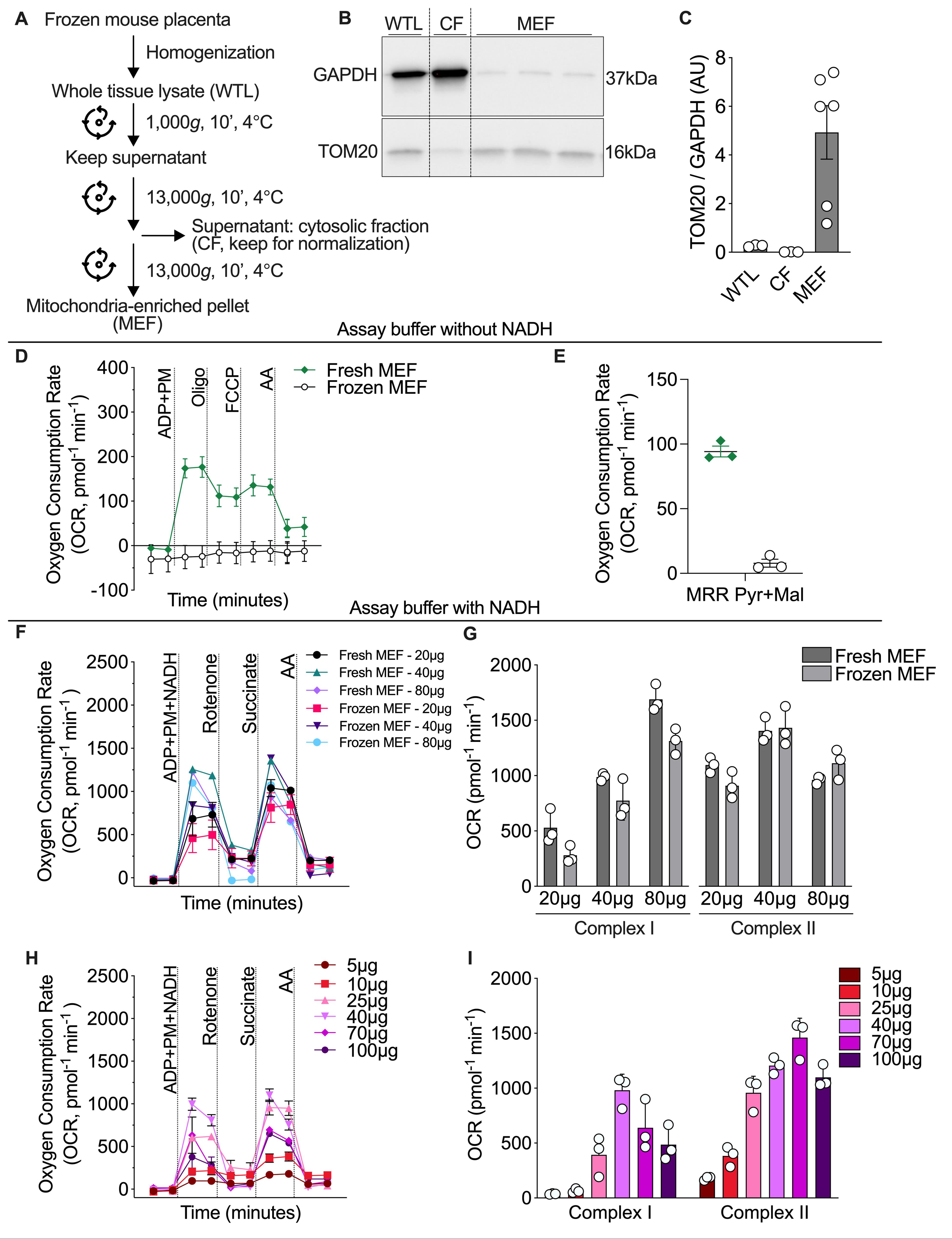
Figure 1. Previously frozen placental mitochondria exhibit comparable complex I- and II-associated respiration to fresh placental mitochondria. (A) Schematic of the workflow used to obtain whole-tissue lysate (WTL), cytosolic (CF), and mitochondrial-enriched fractions (MEF) from previously frozen mouse placental tissue. (B) Representative immunoblots of TOM20 and GAPDH in WTL, CF, and MEF. (C) Quantification of TOM20/GAPDH protein expression in WTL (n = 3), CF (n = 3), and MEF (n = 6), represented in arbitrary units (AU). (D) Representative oxygen consumption tracings in fresh-MEF and frozen-MEF samples following the sequential injection of ADP + pyruvate + malate (ADP + PM), oligomycin (oligo), FCCP, and antimycin A (AA) in the absence of NADH. (E) Oxygen consumption rate (OCR) depicting PM-dependent maximal respiration rate (MRR) in fresh-MEF (n = 3) and frozen-MEF (n = 3) without NADH, in pmol per min. (F) Representative oxygen consumption tracings for complex I- and II-associated respiration corresponding to 20, 40, and 80 μg loading amounts from fresh-MEF and frozen-MEF following the sequential addition of NADH + ADP + pyruvate + malate (ADP + PM + NADH), rotenone, succinate, and antimycin A (AA). (G) Quantification of complex I- and II-associated OCR in 20, 40, and 80 μg of fresh-MEF (n = 3) and frozen-MEF (n = 3), in pmol per min. (H) Representative oxygen consumption tracings for complex I- and II- associated respiration corresponding to 5, 10, 25, 40, 70, and 100 μg loading amounts from frozen-MEF (n = 3), in pmol per min. (I) Quantification of complex I- and II-associated OCR corresponding to 5–100 μg in frozen-MEF (n = 3), in pmol per min. Data are presented as the mean ± SEM.
General notes and troubleshooting
General notes
1. While the present study was only conducted in CD1 mice, this protocol may be performed on placentae from other species of mice and rats as well as on human placental tissue.
2. Unless otherwise indicated, aliquoted stocks stored at -20 °C or -80 °C can typically be used for up to ~6 months when protected from repeated freeze-thaw cycles. Labile reagents (e.g., NADH, pyruvate, cytochrome c) should either be prepared fresh or replaced within 3 months if aliquoted and stored at -20 °C. Stability under these storage conditions was not formally assessed in this study.
3. Our protocol utilizes mouse placental tissue collected at embryonic day 18.5 (E18.5); however, our protocol is flexible and suitable for placentae collected at any gestational timepoint. It should be noted that earlier gestational timepoints will have smaller placentae and a different distribution of placental zones and mitochondrial content, and it is therefore recommended to conduct a mitochondrial protein loading curve.
4. Provided that whole placental weights across experimental groups were within 30 mg, whole placentae were homogenized to preserve the representation of all placental zones. However, if this range is exceeded, placentae may be sectioned by a central sagittal cut and further divided into quarter sections, and tissue mass may then be roughly matched across samples. This method of sectioning is preferable to minimize over- or under-representation of placental zones. Partial sampling should be avoided, as this may bias mitochondrial measurements toward zone-specific populations [4]. Some treatments and/or exposures may result in zone-specific, as opposed to global, alterations in placental mitochondrial content and/or function. Therefore, it may be advantageous to perform respirometry in isolated placental zones.
5. Here, we provide empirical evidence of respiration values of mitochondrial-enriched fractions per given protein amounts loaded (Figures 1G, I). When comparing different groups and conditions, our protocol includes up-front normalization based on the whole protein homogenate. We recommend first quantifying whole-tissue homogenate protein concentrations using a Bradford assay and subsequently adjusting all mitochondrial-enriched fractions to equal volumes and equal whole homogenate concentrations before plate loading. This method of normalization captures respiration changes that are reflective of and relative to alterations in mitochondrial mass that may result from a given treatment or exposure. As a result, each well receives equal volumes of mitochondrial-enriched fraction derived from equal amounts of whole tissue homogenate, removing the need for additional post hoc normalization once the run is complete.
6. As an example, if whole-tissue lysates 1, 2, and 3 correspond to protein readouts of 2327, 3000, and 3546 μg/mL as determined by the Bradford assay, then the corresponding mitochondrial pellets 1, 2, and 3 are to be resuspended in 200, 257, and 304 μL, respectively. This allows for the loading of 100 μL of MEFs per well in duplicate wells.
Troubleshooting
Problem 1: Low respiration rates (overall low OCR).
Possible cause: Insufficient mitochondrial protein loading in the well.
Solution: Optimize the total mitochondrial protein loaded per well by performing a protein loading curve (see Figure 1I) that yields robust basal and substrate-dependent respiration without signal saturation.
Problem 2: Blunted responses to substrates or incomplete inhibition of OCR following inhibitor addition.
Possible cause: Ineffective substrate and inhibitor concentrations.
Solution: Assuming the total amount of mitochondrial protein in the well has been optimized (e.g., ~50 μg), we recommend increasing the concentration of substrates and/or inhibitors to increase their efficacy against the optimized protein amount.
Acknowledgments
Conceptualization, T.P., D.X.; Investigation, T.P., D.X., C.M.; Writing—Original Draft, T.P., D.X.; Writing—Review & Editing, T.P., D.X., C.M., J.N., S.R.; Funding acquisition, T.P., S.R.; Supervision, J.N., S.R.
This work was supported by NSERC Discovery funding (RGPIN-2020–06739) to S.R. and NSERC PGS-D funding to T.P.
The following figures were created in BioRender. Podinic, T. (2026) https://BioRender.com/6cvak3l. We further acknowledge several previously published works from which our protocol was adapted [30,31].
Competing interests
The authors declare no conflicts of interest.
Ethical considerations
All animal work was approved by the McMaster University Animal Research Ethics Board under Animal Utilization Protocol number 21-06-16.
References
- Cindrova-Davies, T. and Sferruzzi-Perri, A. N. (2022). Human placental development and function. Semin Cell Dev Biol. 131: 66–77. https://doi.org/10.1016/j.semcdb.2022.03.039
- Zeldovich, V. B., Clausen, C. H., Bradford, E., Fletcher, D. A., Maltepe, E., Robbins, J. R. and Bakardjiev, A. I. (2013). Placental Syncytium Forms a Biophysical Barrier against Pathogen Invasion. PLoS Pathog. 9(12): e1003821. https://doi.org/10.1371/journal.ppat.1003821
- Costa, M. A. (2016). The endocrine function of human placenta: an overview. Reprod Biomed Online. 32(1): 14–43. https://doi.org/10.1016/j.rbmo.2015.10.005
- Sferruzzi-Perri, A. N., Higgins, J. S., Vaughan, O. R., Murray, A. J. and Fowden, A. L. (2019). Placental mitochondria adapt developmentally and in response to hypoxia to support fetal growth. Proc Natl Acad Sci USA. 116(5): 1621–1626. https://doi.org/10.1073/pnas.1816056116
- Fisher, J. J., McKeating, D. R., Cuffe, J. S., Bianco-Miotto, T., Holland, O. J. and Perkins, A. V. (2019). Proteomic Analysis of Placental Mitochondria Following Trophoblast Differentiation. Front Physiol. 10: e01536. https://doi.org/10.3389/fphys.2019.01536
- Fisher, J., McKeating, D., Pennell, E., Cuffe, J., Holland, O. and Perkins, A. (2019). Mitochondrial isolation, cryopreservation and preliminary biochemical characterisation from placental cytotrophoblast and syncytiotrophoblast. Placenta. 82: 1–4. https://doi.org/10.1016/j.placenta.2019.05.004
- Podinić, T., MacAndrew, A. and Raha, S. (2023). Trophoblast Syncytialization: A Metabolic Crossroads. Results Probl Cell Differ. 71: 101–125. https://doi.org/10.1007/978-3-031-37936-9_6
- Bartho, L. A., Fisher, J. J., Walton, S. L., Perkins, A. V. and Cuffe, J. S. M. (2022). The effect of gestational age on mitochondrial properties of the mouse placenta. Reproduction and Fertility 3(1): 19–29. https://doi.org/10.1530/raf-21-0064
- Holland, O. J., Hickey, A. J., Alvsaker, A., Moran, S., Hedges, C., Chamley, L. W. and Perkins, A. V. (2017). Changes in mitochondrial respiration in the human placenta over gestation. Placenta. 57: 102–112. https://doi.org/10.1016/j.placenta.2017.06.011
- Fowden, A. L., Forhead, A. J., White, K. L. and Taylor, P. M. (2000). Equine uteroplacental metabolism at mid- and late gestation. Exp Physiol. 85(5): 539–545. https://doi.org/10.1017/s0958067000020674
- Jones, C. T. and Rolph, T. P. (1985). Metabolism during fetal life: a functional assessment of metabolic development. Physiol Rev. 65(2): 357–430. https://doi.org/10.1152/physrev.1985.65.2.357
- Carter, A. (2000). Placental Oxygen Consumption. Part I: In Vivo Studies—A Review. Placenta. 21: S31–S37. https://doi.org/10.1053/plac.1999.0513
- Hu, X. Q. and Zhang, L. (2022). Mitochondrial Dysfunction in the Pathogenesis of Preeclampsia. Curr Hypertens Rep. 24(6): 157–172. https://doi.org/10.1007/s11906-022-01184-7
- Holland, O. J., Cuffe, J. S. M., Dekker Nitert, M., Callaway, L., Kwan Cheung, K. A., Radenkovic, F. and Perkins, A. V. (2018). Placental mitochondrial adaptations in preeclampsia associated with progression to term delivery. Cell Death & Disease 9(12): e1038/s41419–018–1190–9. https://doi.org/10.1038/s41419-018-1190-9
- Voros, C., Stavros, S., Sapantzoglou, I., Mavrogianni, D., Daskalaki, M. A., Theodora, M., Antsaklis, P., Drakakis, P., Loutradis, D., Daskalakis, G., et al. (2025). The Role of Placental Mitochondrial Dysfunction in Adverse Perinatal Outcomes: A Systematic Review. J Clin Med. 14(11): 3838. https://doi.org/10.3390/jcm14113838
- Kadam, L., Chan, K., Tawater, E. and Myatt, L. (2025). Dysregulation of placental mitochondrial structure dynamics and clearance in maternal obesity and gestational diabetes. Placenta. 171: 140–149. https://doi.org/10.1016/j.placenta.2025.09.019
- Sobrevia, L., Valero, P., Grismaldo, A., Villalobos-Labra, R., Pardo, F., Subiabre, M., Armstrong, G., Toledo, F., Vega, S., Cornejo, M., et al. (2020). Mitochondrial dysfunction in the fetoplacental unit in gestational diabetes mellitus. Biochim Biophys Acta Mol Basis Dis. 1866(12): 165948. https://doi.org/10.1016/j.bbadis.2020.165948
- Valent, A. M., Choi, H., Kolahi, K. S. and Thornburg, K. L. (2021). Hyperglycemia and gestational diabetes suppress placental glycolysis and mitochondrial function and alter lipid processing. FASEB J. 35(3): e202000326rr. https://doi.org/10.1096/fj.202000326rr
- Mandò, C., De Palma, C., Stampalija, T., Anelli, G. M., Figus, M., Novielli, C., Parisi, F., Clementi, E., Ferrazzi, E., Cetin, I., et al. (2014). Placental mitochondrial content and function in intrauterine growth restriction and preeclampsia. Am J Physiol Endocrinol Metab. 306(4): E404–E413. https://doi.org/10.1152/ajpendo.00426.2013
- Tong, W., Allison, B. J., Brain, K. L., Patey, O. V., Niu, Y., Botting, K. J., Ford, S. G., Garrud, T. A., Wooding, P. F. B., Lyu, Q., et al. (2025). Placental mitochondrial metabolic adaptation maintains cellular energy balance in pregnancy complicated by gestational hypoxia. J Physiol. : e1113/jp287897. https://doi.org/10.1113/jp287897
- Salazar-Petres, E., Pereira-Carvalho, D., Lopez-Tello, J. and Sferruzzi-Perri, A. N. (2022). Placental structure, function, and mitochondrial phenotype relate to fetal size in each fetal sex in mice. Biol Reprod. 106(6): 1292–1311. https://doi.org/10.1093/biolre/ioac056
- Murray, A., Armstrong, J. and O'Brien, K. (2025). Placental mitochondria in high-altitude pregnancy: metabolic adaptations and a toolkit for respiratory assessment. Philos Trans R Soc Lond B Biol Sci. 380(1933): e0175. https://doi.org/10.1098/rstb.2024.0175
- Dalton-O’Reilly, J., Heazell, A. E. P., Greenwood, S., Dilworth, M. and Desforges, M. (2025). Sex-specific alterations in placental mitochondria, oxidative damage and apoptosis in mice of advanced maternal age. Reproduction. 170(4): e1530/rep–25–0160. https://doi.org/10.1530/rep-25-0160
- Miller, C. N., Dye, J. A., Henriquez, A. R., Stewart, E. J., Lavrich, K. S., Carswell, G. K., Ren, H., Freeborn, D. L., Snow, S. J., Schladweiler, M. C., et al. (2020). Ozone-induced fetal growth restriction in rats is associated with sexually dimorphic placental and fetal metabolic adaptation. Mol Metab. 42: 101094. https://doi.org/10.1016/j.molmet.2020.101094
- Beetch, M., Oribamise, E., Jo, S., Clifton, B., Larson, S., Hausmann, A., Wong, A., Akhaphong, B., Morgan, E., Alejandro, E. U., et al. (2025). Placental mTOR signalling links mitochondrial dysfunction, nutrient transport and neonatal beta cell perturbations in mice. Diabetologia. 68(12): 2823–2839. https://doi.org/10.1007/s00125-025-06542-z
- Yung, H. W., Colleoni, F., Dommett, E., Cindrova-Davies, T., Kingdom, J., Murray, A. J. and Burton, G. J. (2019). Noncanonical mitochondrial unfolded protein response impairs placental oxidative phosphorylation in early-onset preeclampsia. Proc Natl Acad Sci USA. 116(36): 18109–18118. https://doi.org/10.1073/pnas.1907548116
- Colleoni, F., Morash, A., Ashmore, T., Monk, M., Burton, G. and Murray, A. (2012). Cryopreservation of placental biopsies for mitochondrial respiratory analysis. Placenta. 33(2): 122–123. https://doi.org/10.1016/j.placenta.2011.11.019
- Giovarelli, M., Serati, A., Zecchini, S., Guelfi, F., Clementi, E. and Mandò, C. (2023). Cryopreserved placental biopsies maintain mitochondrial activity for high-resolution respirometry. Mol Med. 29(1): e1186/s10020–023–00645–2. https://doi.org/10.1186/s10020-023-00645-2
- Petrenko, A. (1992). A mechanism of latent cryoinjury and reparation of mitochondria. Cryobiology. 29(1): 144–152. https://doi.org/10.1016/0011-2240(92)90015-t
- Acin‐Perez, R., Benador, I. Y., Petcherski, A., Veliova, M., Benavides, G. A., Lagarrigue, S., Caudal, A., Vergnes, L., Murphy, A. N., Karamanlidis, G., et al. (2020). A novel approach to measure mitochondrial respiration in frozen biological samples. EMBO J. 39(13): e2019104073. https://doi.org/10.15252/embj.2019104073
- Osto, C., Benador, I. Y., Ngo, J., Liesa, M., Stiles, L., Acin‐Perez, R. and Shirihai, O. S. (2020). Measuring Mitochondrial Respiration in Previously Frozen Biological Samples. Curr Protoc Cell Biol. 89(1): e116. https://doi.org/10.1002/cpcb.116
- Vekaria, H. J., Kalimon, O. J., Prajapati, P., Velmurugan, G. V. and Sullivan, P. G. (2024). An efficient and high-throughput method for the evaluation of mitochondrial dysfunction in frozen brain samples after traumatic brain injury. Front Mol Biosci. 11: e1378536. https://doi.org/10.3389/fmolb.2024.1378536
- Sarver, D. C., Saqib, M., Chen, F. and Wong, G. W. (2024). Mitochondrial respiration atlas reveals differential changes in mitochondrial function across sex and age. eLife. 13: e4. https://doi.org/10.7554/elife.96926.4
- Yao, P. J., Munk, R., Gorospe, M. and Kapogiannis, D. (2023). Analysis of mitochondrial respiration and ATP synthase in frozen brain tissues. Heliyon. 9(3): e13888. https://doi.org/10.1016/j.heliyon.2023.e13888
- Pennington, K. A., Schlitt, J. M. and Schulz, L. C. (2012). Isolation of Primary Mouse Trophoblast Cells and Trophoblast Invasion Assay. J Visualized Exp. 59: e3791/3202. https://doi.org/10.3791/3202
- Mosharov, E. V., Rosenberg, A. M., Monzel, A. S., Osto, C. A., Stiles, L., Rosoklija, G. B., Dwork, A. J., Bindra, S., Junker, A., Zhang, Y., et al. (2025). A human brain map of mitochondrial respiratory capacity and diversity. Nature. 641(8063): 749–758. https://doi.org/10.1038/s41586-025-08740-6
Article Information
Publication history
Received: Jan 27, 2026
Accepted: Mar 18, 2026
Available online: Apr 9, 2026
Published: May 5, 2026
Copyright
© 2026 The Author(s); This is an open access article under the CC BY-NC license (https://creativecommons.org/licenses/by-nc/4.0/).
How to cite
Podinic, T., Xhuti, D., Monaco, C., Nederveen, J. P. and Raha, S. (2026). Assessing Mitochondrial Respiratory Complex-Associated Function From Previously Frozen Mouse Placental Tissue. Bio-protocol 16(9): e5667. DOI: 10.21769/BioProtoc.5667.

 Download Citation in RIS Format
Category
Cell Biology > Cell metabolism > Respirometry
Cell Biology > Cell-based analysis > Mitochondrial respiration
Systems Biology > Metabolomics > Tissue
Do you have any questions about this protocol?
Post your question to gather feedback from the community. We will also invite the authors of this article to respond.
 Post a Question


