


- Protocols

- Articles and Issues
- For Authors
- About
- Become a Reviewer


Preparing Adjuvanted Nanoliposomes for Applications Toward Recombinant Influenza Vaccine Development


Published: Vol 16, Iss 7, Apr 5, 2026 DOI: 10.21769/BioProtoc.5662 Views: 491
Reviewed by: Navnita DuttaChhuttan L MeenaAnonymous reviewer(s)
 PDF
PDF  Ask a question
Original research article
The authors used this protocol in:

Mar 2024
Advertisement



Protocol Collections
Comprehensive collections of detailed, peer-reviewed protocols focusing on specific topics




Abstract
Nanoparticle vaccines can provide advantages over traditional vaccine methodologies, including adjuvant delivery to enhance the effectiveness of recombinant antigens. Many approaches exist to formulate different vaccine nanoparticles, which are designed for different biomolecular cargos, adjuvant compositions, and disease targets. Here, a protocol is described to produce nanoliposomes whose surface is decorated with recombinant protein influenza antigens with monophosphoryl lipid A and QS-21 adjuvants incorporated into the lipid bilayer for protection against influenza infection. This protocol includes methods for producing adjuvanted liposomes and coupling with His-tagged antigens for surface decoration of the particle. This allows for a rapid methodology of producing immunogenic antigen-presenting liposomes that can be tailored to display a combination of influenza surface antigens.
Key features
• This protocol uses recombinant proteins, which can be adapted to the desired strain sequence.
• Production of this influenza vaccine formulation uses in vitro techniques, eliminating the need for virus growth in eggs.
• Additional lipid adjuvants can be included in the liposome production step to be incorporated into the surface membrane.
• Cobalt-porphyrin phospholipid provides a flexible platform for binding His-tag-bearing proteins.
Keywords: Influenza Nanoparticle vaccine
Nanoliposomes
Recombinant protein vaccine
Vaccine adjuvant
Nanoparticle vaccine
Nanoliposomes
Recombinant protein vaccine
Vaccine adjuvant
Graphical overview

Application of liposomes as a platform for multistrain influenza vaccines. Cobalt-porphyrin liposomes present multiple surface antigens through spontaneous associations between cobalt ions and histidine residues on recombinant proteins. Figure reproduced under Creative Commons license from [1].
Background
The rapid rate of mutation among the viruses in the Orthomyxoviridae family, commonly known as influenza, requires vaccines to be updated on an annual basis to effectively provide protection against seasonal epidemics. Furthermore, genetic transfers between zoonotic strains, such as avian influenza, with human competent strains pose a significant threat of spontaneously forming novel influenza strains, which could expand into a global pandemic with a high mortality rate [2]. The adaptability of influenza viruses is primarily due to two major surface proteins on the virion, hemagglutinin and neuraminidase, which serve to facilitate viral uptake into and release from host cells, respectively [3–5]. These antigens undergo continuous mutation and reassortment between antigenically distinct strains, resulting in annual epidemics [6]. To combat the ever-evolving landscape of influenza viruses, an epidemic response demands a robust vaccine, which can rapidly shift production to utilize newly emerging antigens, providing the most up-to-date protection possible. Traditional influenza vaccines produce whole viruses within chicken eggs, which are split with a detergent or attenuated for use in immunizations. These methods incur numerous challenges, including production bottlenecks based on egg supply, a time-intensive process of selecting and cultivating egg-adapted viruses, the possibility of egg-specific mutations replacing epitopes found in human-competent viruses, and anaphylaxis in egg-allergic individuals. Recombinant protein production would allow for faster production and greater quality control, yet the technique remains relatively underutilized due to the limited antigenicity of isolated surface proteins, requiring high doses of antigen to be effective [7]. Adjuvants provide a critical supportive component to recombinant vaccines by increasing the immune response to these antigens through the activation of immunologic signaling pathways and increased uptake into antigen-presenting cells [8,9]. The nanoliposomes described in this protocol provide a robust adjuvant and vehicle for recombinant vaccines. The combination of surface presentation of influenza antigens and the effect of lipid adjuvants has been shown to significantly increase protection against lethal influenza infections in preclinical animal studies on mice and ferrets; additionally, these liposomes have been shown to increase immunological responses against a wide range of hemagglutinin and neuraminidase variants, providing a straightforward admixture method for producing multiplexed vaccine particles [1,10]. The surface binding properties of these liposomes are facilitated by the cobalt-porphyrin phospholipids (CoPoP) embedded in the bilayer membrane, which bind to chains of histidine residues called His-tags, which are routinely added to recombinant proteins for purification (Graphical overview). This platform is compatible with a wide array of antigenic proteins, including other viral infections such as SARS-CoV-19 [11,12], surface proteins of the malaria parasite [13,14], and cancer markers [15,16]. This protocol describes the process for generating CoPoP, formulating ~100 nm diameter adjuvanted liposomes with CoPoP-bearing membranes, and incubating these liposomes with recombinant influenza antigens to produce immunogenic vaccine liposomes for preclinical testing.
Materials and reagents
Biological materials
1. Cobalt porphyrin-phospholipid (CoPoP) (POP Biotechnologies, Inc.)
2. Recombinant influenza surface antigens bearing 6×-His-tag (purchased or produced)
3. Cholesterol (Evonik, catalog number: 00078928)
4. 1,2-dioleoyl-sn-glycero-3-phosphocholine (DOPC) (CordenPharma, catalog number: LP-R4-070)
5. Monophosphoryl hexa-acyl lipid A, 3-deacyl (3D6A-PHAD) (Avanti, catalog number: 699855)
6. QS-21 (Quillaja Saponaria derivative) (Desert King)
7. Anti-mouse IgG with HRP conjugate (Thermo Fisher, catalog number: 62-6520)
8. Chicken whole blood with Alsevers (Pel-Freez, catalog number: 33133-1)
9. Receptor destroying enzyme (RDE), lyophilized powder (Sigma-Aldrich, catalog number: C8772-1VL)
Reagents
1. Hydrochloric acid (HCl) (Millipore Sigma, catalog number: 320331)
2. Phosphate-buffered saline, pH 7.4 (1× PBS and 10× PBS) (Sigma-Aldrich, catalog number: P3813)
3. 200-proof ethanol, pure research grade (EtOH) (Sigma-Aldrich, catalog number: 459844)
4. Sodium hydroxide (NaOH) (Sigma-Aldrich, catalog number: 795429)
5. Deionized water, lab grade (Thermo Fisher, catalog number: A57775)
6. 5× ELISA coating buffer (Bio-Rad, catalog number: BUF030C)
7. SuperBlockTM blocking buffer (Thermo Fisher, catalog number: 37515)
8. Bovine serum albumin (BSA), heat shock fraction, pH 7, >98% (Sigma-Aldrich, catalog number: A9647)
9. Tween 20 washing buffer, 1% in 10× PBS (Thermo Fisher, catalog number: J63314.K3)
10. 1-StepTM Ultra TMB-blotting substrate solution (Thermo Fisher, catalog number: 37574)
Solutions
1. CPQ liposomes (see Recipes)
Recipes
1. CPQ liposomes
| Reagent | Amount | Molar weight (g/mol) | Final concentration (mg/mL, mM) |
|---|---|---|---|
| CoPoP | 12.5 mg | 1068.5 | 2.5 mg/mL, 2.34 mM |
| Cholesterol | 62.5 mg | 386.65 | 12.5 mg/mL, 32.3 mM |
| DOPC | 250.0 mg | 734.04 | 50.0 mg/mL, 68 mM |
| 3D6A-PHAD | 5.0 mg | 1763.47 | 1.0 mg/mL, 0.57 mM |
| QS-21* | 5.0 mg | 1990.1 | 1.0 mg/mL, 0.5 mM |
| PBS | 4.0 mL | 10 mM phosphate, pH 7.4 | |
| Ethanol | 1.0 mL | ||
| Total | 5 mL |
*QS-21 is added to the liposomes following ethanol removal.
CP and CA can be made by omitting QS-21 and QS-21/3D6A-PHAD, respectively.
Laboratory supplies
1. Snap-top microcentrifuge tubes, 0.6 mL sterile (Thermo Fisher, catalog number: 3449)
2. Snap-top microcentrifuge tubes, 1.5 mL sterile (Thermo Fisher, catalog number: 3451)
3. 0.2 μm syringe filter (Foxx Life Sciences, catalog number: 379-2215-OEM)
4. 15 mL centrifuge tube (Thermo Scientific, catalog number: 339651)
5. 20 mL glass vials (DWK Life Sciences, catalog number: 986532)
6. 5 mL syringe (Becton, Dickinson & Co., catalog number: 309646)
7. 50 mL centrifuge tube (Thermo Scientific, catalog number: 339653)
8. Serological pipettes, various volumes (VWR, catalog number: 75816)
9. Micropipette tips, various sizes (VWR, catalog number: 76322)
10. 96-well flat-bottom plate (clear) (Greiner Bio-one, catalog number: 655801)
11. 96-well V-bottom plate (clear) (Greiner Bio-One, catalog number: 651261)
12. Clear flat-bottom Immuno 96-well plates, MaxiSorp coating (Thermo Fisher, catalog number: 439454)
13. Aluminum foil (Reynold’s wrap)
14. Dialysis clips (Spectrum Chemical, catalog number: 888-11584-PK)
15. Dialysis tubing 12,000–14,000 MWCO (Fisherbrand, catalog number: 21-152-16)
16. Cuvettes (BrandTech, catalog number: 759075D)
17. Parafilm (Amcor, catalog number: PM-996)
18. Becton-Dickinson ultra-fine insulin syringe, 0.3 mL, 31G × 8 mm (BD, catalog number: 320440)
19. Microtainer® capillary blood collector, gold, serum separator tube (Fisher Scientific, catalog number: 02-675-185)
Equipment
1. Pipette controller (e.g., VWR, catalog number: 612-7179)
2. Single-channel 0.5–10 μL micropipette (VWR, catalog number: 89079-962)
3. Single-channel 2–20 μL micropipette (VWR, catalog number: 89079-964)
4. Single-channel 20–200 μL micropipette (VWR, catalog number: 89079-970)
5. Single-channel 100–1,000 μL micropipette (VWR, catalog number: 89079-974)
6. 1 L graduated cylinder (Eisco, catalog number: CH0344O)
7. 0.08 μm extruder filter (Cytiva, catalog number: 10419306)
8. 0.1 μm extruder filter (Cytiva, catalog number: 10419506)
9. 0.2 μm extruder filter (Cytiva, catalog number: 10417006)
10. 10 mL Lipex extruder (Evonik, catalog number: 002108)
11. 1 L glass beaker (Eisco, catalog number: CH0126KPK6)
12. 2 L laboratory glass bottle (VWR, catalog number: 10754-822)
13. Analytical balance (Mettler Toledo, catalog number: XSR104)
14. Antistatic spatulas (VWR, catalog number: 80081-194)
15. Bath sonicator (Branson, catalog number: CPX2800H)
16. Heated circulating bath (VWR, catalog number: 1130A)
17. Hot water bath (IKA, catalog number: HB10S098)
18. Magnetic stir bar, 20 and 30 mm length (VWR, catalog numbers: 442-4523 and 442-4525)
19. Microcentrifuge (Thermo Scientific, catalog number: 75002447)
20. Compressed nitrogen gas (Praxair, catalog number: AR LC160-230)
21. Plate reader (abs/fluorescence) (Tecan, catalog number: F129013)
22. Serological pipetter (Drummond Scientific)
23. Stir plate (VWR, catalog number: 97042-714)
24. Vortexer (VWR, catalog number: 58816-121)
25. Dynamic Light Scattering (DLS) machine (BeNano)
26. UV-VIS spectrophotometer (Perkin Elmer LAMBDA 365+)
27. Incubating microplate shaker (e.g., Fisher Scientific, catalog number: 02-217-760)
28. Class II biosafety cabinet (e.g., Labconco Type A2 model)
Software and datasets
1. PerkinElmer UV WinLab software
2. BeNano particle solutions software
Procedure
A. Liposome production
1. Lipid mixture
a. Determine the desired adjuvant formulation for the liposome application. See Table 1 for lipid and adjuvant ratios to produce various types of binding liposomes.
Table 1. Cobalt-porphyrin phospholipids (CoPoP) liposome formulations
| Formulation | Composition (weight ratio) |
|---|---|
| No adjuvant (CA) | DOPC:Chol:CoPoP = 20:5:1 |
| CoPoP/PHAD (CP) | DOPC:Chol:CoPoP:PHAD = 20:5:1:0.4 |
| CoPoP/PHAD/QS-21 (CPQ) | DOPC:Chol:CoPoP:PHAD:QS21 = 20:5:1:0.4:0.4 |
b. Remove lipids from the -20 °C freezer, allow them to reach room temperature, weigh the appropriate lipid quantities, and combine them into a single 20 mL glass vial on the analytical balance. See Recipe 1 for the target weight for each lipid.
Note: Lipid weights may be scaled as needed according to the formulation and volume of liposomes.
c. Add 1 mL of ethanol preheated to 45 °C to the vial containing lipids.
Note: If large precipitates are observed after the addition of ethanol, the solution should be sonicated to break up large aggregates.
d. Heat lipids in ethanol to 45 °C in the water bath for 5 min. Sonicate briefly as needed to completely dissolve lipids.
Note: Use an electric tea kettle to heat water to 45 °C before adding it to the sonicator bath.
e. Add 4 mL of preheated 1× PBS at 55 °C to the ethanol lipid mixture.
f. Sonicate the suspension for 20–30 s to break up potential aggregates.
g. Incubate in a 55 °C water bath for 5 min, then sonicate for 20–30 s prior to extrusion.
2. Extrusion
a. Preheat the circulating water bath for the extruder to 55 °C.
b. Prepare the extruder for use (Figure 1).
c. Check that the unit is clean, then install the stacked polycarbonate filter membranes. The 0.08 μm filters should be placed first at the bottom, followed by 0.1 μm and then 0.2 μm at the top.
d. Connect the extruder to a nitrogen gas tank and set the pressure to 300 psi.
e. Fill the extruder with 5 mL of PBS.
f. Pressurize the extruder to remove the PBS in the extruder.
g. Retain at least 80 µL of pre-extruded liposome samples for later analysis.
h. Depressurize the extruder and fill the barrel with the liposome suspension.
i. Pressurize the extruder to extrude the sample and collect it into the 20 mL starting vial.
j. Repeat steps A2h–i for 10 total passes. On the final pass, collect the sample in a clean 20 mL glass vial.
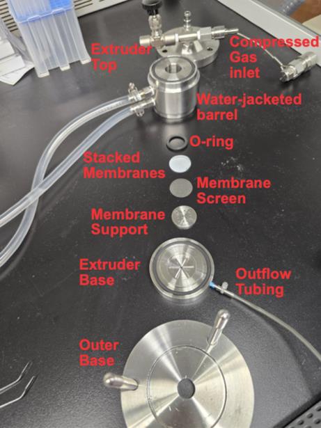
Figure 1. Expanded view of nitrogen extruder. The model displayed is a Lipex 10 mL; components in other models may differ.
3. Dialysis
a. Add 1 L of 1× PBS buffer at 4 °C into a clean beaker with a magnetic stir bar.
b. Cut an appropriate length of dialysis tubing (12,000–14,000 MWCO) and seal with a clip at one end. Add to the beaker to hydrate for 5 min.
c. Add liposome suspension to the open-end pipette and seal the pouch with a clip.
d. Cover the beaker with a layer of aluminum foil.
e. Dialyze the sample against 1 L of 1× PBS overnight. Exchange buffer following overnight incubation.
Note: If an overnight incubation is not desired, then buffer exchange may be performed at 4–6 h intervals.
f. Perform buffer exchange by emptying and refilling the stirring beaker with a second liter of 1× PBS for 4–6 h.
Note: For convenience, 2 L of 1× PBS can be prepared, with 1 L reserved at 4 °C in anticipation of the buffer exchange.
g. After dialysis, transfer liposomes to a clean 20 mL glass vial and then store the liposomes at 4 °C.
4. Sterile filtration
a. Filter the liposomes through a 0.2 μm syringe filter into a 20 mL glass vial.
Note: This work can be done in a biosafety cabinet to minimize potential contamination. Additionally, vials may be autoclaved to further reduce contaminant risk.
b. Cap the autoclaved vial and store at 4 °C until needed.
B. Characterization of liposomes
1. UV–Vis quantification of CP liposomes
a. Use the pre-filtered CP liposome suspension to prepare a standard curve and perform measurements using a UV–Vis spectrophotometer.
b. Dilute pre-filtered CP liposomes in ethanol. Specifically, mix 40 µL of sample with 1,960 µL of ethanol and mix thoroughly using a pipette for a 50× dilution standard. Prepare and measure also 100×, 200×, and 400× dilutions of the pre-filtered CP liposome suspension to generate a standard curve.
c. For the sterile CP liposome sample, mix 40 µL of sample with 1,960 µL of ethanol and transfer to a 2 mL cuvette for measurement.
d. In PerkinElmer UV WinLab (UV–Vis software), set the wavelength scan range to 300–700 nm and record the absorbance peak at 645 nm.
e. Generate a standard curve by plotting absorbance vs. dilution and fitting the data using a linear equation. Then, interpolate the absorbance values of the samples using the standard curve to determine the concentration of the liposome samples.
2. Size measurements
a. Add 1 mL of 1× PBS to a disposable cuvette.
b. Add 5 μL of sample to the cuvette.
c. Mix using a 1 mL micropipette, with an effort to avoid introducing bubbles or dust particles. This should achieve a 1:200 sample to diluent ratio for measurement.
d. Measure size and polydispersity of sample using BeNano particle solutions software. Typical average diameters range from 90 to 120 nm, with a polydispersity index (PDI) typically below 0.2 and no greater than 0.5.
C. Representative liposome applications
CoPoP facilitates antigen binding through the binding affinity between cobalt ions held within the porphyrin rings and His-tags appended to the membrane anchoring region of recombinant surface proteins. This affinity allows a wide range of recombinant proteins to be attached to the surface of liposomes via simple room-temperature admixture. For hemagglutinin (HA) proteins, the His-tag is included at the C-terminus of the primary sequence, while for neuraminidase (NA) proteins, the His-tag is present at the N-terminus. Because the proteins bind to liposome surfaces from solution rather than forming endogenously, artificial multimerization domains must be added to the recombinant protein sequences to achieve trimer and tetramer formation in HA and NA, respectively.
1. Antigen incubation
a. Thaw stock vial of His-tagged influenza antigen in a water bath at room temperature.
b. Generate an antigen working stock by diluting in sterile 1× PBS to a concentration of 80 μg/mL.
c. Combine equal volumes of 320 μg/mL CoPoP liposome stock with 80 μg/mL working stock of antigen to achieve a desired quantity of 1:4 antigen to CoPoP mass ratio. At this step, the incubation volume may be lower than the intended final volume.
Note: Using a 1:4 mass ratio ensures antigen coupling of most antigens to the CoPoP liposome. Using a lower ratio may result in some antigen remaining in solution.
d. Incubate antigen and liposome admixture at room temperature for 3 h.
e. Following the incubation period, add sterile 1× PBS to achieve the final desired volume of antigen–nanoliposome suspension for experimental use or preclinical administration.
2. Blood collection
a. Shave the quadriceps of the mouse, apply petroleum jelly to the exposed skin, locate the saphenous vein, and pierce with a needle or lancet.
b. Collect blood in microcentrifuge tubes containing serum separation gel.
c. Centrifuge whole blood at 2,000× g for 15 min at 4 °C.
d. Extract serum and aliquot into 0.6 mL microcentrifuge tubes at up to 50 μL per aliquot, store at 4 °C for use within 2 weeks, or snap-freeze and store at -80 °C indefinitely.
3. Vaccination strategy
a. For primary vaccination, prepare 50 μL per dose of antigen-liposome vaccine solution for mice, with 10% of the total volume as excess to account for loss from syringe loading. See Table 2 for typical antigen and CPQ composition per dose.
b. Load a 0.3 mL syringe with 50 μL of vaccine and safely set aside.
c. Gently immobilize the mouse using a grip technique or containment device.
d. Apply an antiseptic wipe or alcohol swab to the exposed skin to prevent infection of the injection site.
e. Inject the vaccine into the left quadriceps muscle.
f. When administering a booster dose, repeat steps C2–3 three weeks following the initial dose.
Table 2. Composition of typical vaccine doses
| Component | Stock concentration | Volume (per 50 µL dose) | Final concentration |
| His-tagged antigen | 1 mg/mL | 1.35 µL | 27.0 µg/mL |
| CoPoP | 320 µg/mL | 16.9 µL* | 108 µg/mL |
| QS21 | 128 µg/mL | * | 43.2 µg/mL |
| PHAD | 128 µg/mL | * | 43.2 µg/mL |
| PBS | n/a | 31.3 µL | n/a |
*Adjuvants added in a single volume of liposome suspension.
4. ELISA assay
a. Coat 96-well clear-bottom MaxiSorp plates with 100 μL per well of 1 μg/mL target antigen in ELISA coating buffer. If needed, adjust pH with NaOH or HCl.
Notes:
1. If using 5× ELISA coating buffer concentrate, dilute 5-fold in sterile 1× PBS; ideal pH should fall within the 7.8–8.2 range.
2. A coating buffer may be produced by mixing a 100 mM sodium bicarbonate buffer, with the pH adjusted to within the 9.4–9.6 range.
b. Incubate plates overnight (>18 h) at 4 °C.
c. Empty plates by aspiration or inversion and rinse wells with 100 μL of 10× PBS with 0.1% Tween 20 (PBST).
d. Add 100 μL of blocking buffer to each well and incubate at room temperature for 1–2 h with constant shaking provided by a microplate shaker.
Note: A commercial blocking buffer could be used, or one could be created by dissolving 2% (w/v) BSA in PBST.
e. Perform serial dilution of serum in 1% (w/v) BSA in PBST (Figure 2).
Note: To perform serial dilution, add 100 μL of 1% BSA in PBST to wells in rows B–H of a 96-well plate, and 125 μL of 1% BSA in PBST to row A. To this row, add 1.25 μL of sample to wells, achieving an initial concentration of 1:100 sample in diluent. Then, add 25 μL of this initial dilution to each corresponding well in each column of row B using a multichannel pipette, mixing contents by repeated pipetting. This achieves a dilution factor of 1:5 from the initial dilution, or a 1:500 overall sample dilution. Repeat this process for rows C–G. In row H, sample inclusion can be omitted to provide a negative control and baseline measurement.

Figure 2. ELISA plate layout example. Samples are assayed in triplicate, allowing four samples to be assessed on each plate. The colorimetric assay becomes dark blue when horseradish peroxidase (HRP) reacts with 3,3',5,5'-tetramethylbenzidine (TMB), and yellow when the reaction is halted with an acid.
f. Incubate the serial dilutions of the sample in an antigen-coated plate at room temperature for 1 h with constant shaking.
g. Empty plates and rinse three times with 100–200 μL of PBST.
h. Add 100 μL of 1 μg/mL secondary antibody with HRP conjugate and incubate for 30 min at room temperature with shaking.
i. Empty plates and rinse 5 times with PBST.
Note: Miniscule amounts of secondary antibody can lead to colorimetric results; additional washing ensures all unbound secondary antibody is removed.
j. Add 100 μL of TMB substrate to each well and allow 5–10 min for the reagent to undergo colorimetric reaction.
k. Stop the reaction by adding 100 μL of 1 M acid solution, such as HCl.
l. Read the absorbance at 450 nm in a plate reader. Analyze data using appropriate software.
5. Hemagglutinin inhibition (HAI) assay
Note: This assay is only applicable to vaccine studies involving the HA antigen and will not measure inhibition of NA or other antigens.
a. Incubate 20 μL of each serum sample with 60 μL of RDE and incubate in a 37 °C water bath overnight, ideally between 18 and 22 h.
Notes:
1. To reconstitute lyophilized RDE, add 5 mL of sterile water and aliquot. Store frozen for long-term stability.
2. It is important to seal the plate to prevent fluid from the water bath from entering, as well as reducing the loss of moisture to evaporation.
b. Deactivate RDE by incubating samples in a water bath at 56 °C for 30 min.
c. Isolate red blood cells (RBCs) by filtering 1 mL of whole chicken blood through sterile 2 × 2 gauze into a 15 mL conical tube, followed by 10 mL of 1× PBS. Discard the filter and invert to mix. Centrifuge at 200× g for 5 min at 4 °C for three cycles. Remove the supernatant and resuspend the RBC pellet with 10 mL of sterile PBS each time.
d. Find the appropriate viral dilution titer by performing 1:2 serial dilutions of the virus, starting with a 1:10 dilution (5 μL from virus stock plus 45 μL of 1× PBS). Observe and record the last well in which hemagglutination occurs.
e. Based on the dilution factor found in the prior step, create a virus working stock by diluting the original virus stock in sterile 1× PBS.
f. Allow samples to cool to room temperature and add 20 μL of sterile 1× PBS to achieve a final volume of 100 μL, a 1:5 dilution of the original sample.
Note: Because some volume can be lost during incubation due to evaporation, it may be necessary to adjust the volume of PBS added to achieve a final volume of 100 μL.
g. Load V-bottom 96-well plates by adding 25 μL of 1× PBS to rows B–H and 50 μL of the appropriate RDE II-treated serum to row A1–A10 wells. Each sample can be tested in pairs of adjacent columns for accuracy. To well A11, add 50 μL of the viral antigen solution to serve as a viral activity positive control, and to well A12, add 1× PBS to serve as the negative control.
Note: When performing this assay with a new virus for the first time, a standard curve should be established to determine the hemagglutinating units (HAU) of the stock virus solution.
h. Perform a 1:2 serial dilution by transferring 25 μL of sample from row A to row B and repeating this process between each subsequent row, such that the sample dilution ranges from 1:5 in row A to 1:640 in row H.
Note: After RBC suspension is added to the wells, the final virus dilution factors will range from 1:10 to 1:1,280.
i. For wells in columns 1–10, add 25 μL of virus working stock. In columns 11–12, add 25 μL of 1× PBS to the control samples.
j. Incubate samples at room temperature for 30 min.
k. Create a 0.5%–1% suspension of RBCs in 1× PBS by combining 125 μL of the RBC pellet with 25 mL of sterile 1× PBS.
l. Add 50 μL of RBC suspension to each well of the plate.
m. Incubate samples at room temperature for an additional 30 min.
n. Record hemagglutination inhibition results. The HAI titer corresponds to the last well that does not form an RBC pellet, indicating HA activity and thus viral potency. See Figure 3 for an example of HAI plate reading.
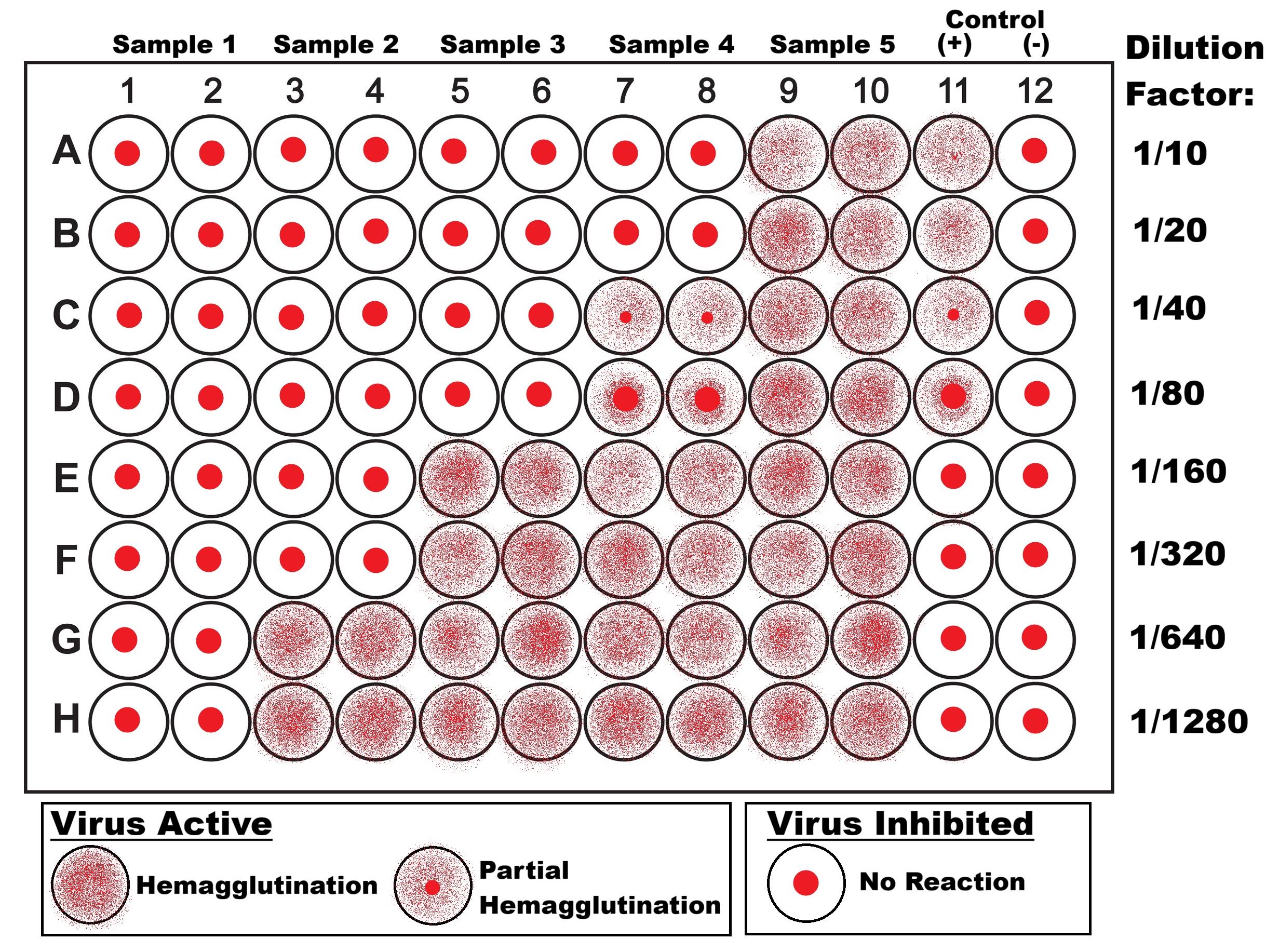
Figure 3. Hemagglutinin inhibition (HAI) plate layout example. Samples in the diagram are arranged with the highest titer on the left and the lowest on the right, depicting HAI titers of >1,280, 320, 80, 20, and <10, respectively.
6. Influenza challenge
a. Three weeks after the booster vaccination, as described above, prepare the appropriate virus inoculum by diluting a quickly thawed virus stock aliquot and sonicating for 45 s in a sonicating water bath. Then, place on ice. Dilute virus appropriately with ice-cold 1× PBS with 100 µg/mL penicillin and streptomycin solution (P&S) by performing serial dilutions (Table 3).
Table 3. Dilution strategy for influenza virus
| Initial stock | Dilution 1 | Dilution 2 | |
|---|---|---|---|
| Amount of PBS+P&S | - | 900 μL | 2,125 μL |
| Amount from previous dilution | 100 μL | 100 μL | 375 μL |
| Dilution factor | Undiluted | 1.0 × 10⁻¹ | 1.5 × 10⁻² |
| Putative titer (PFU/mL) | 1.6 × 10⁸ | 1.6 × 10⁷ | 2.4 × 10⁶ |
| Inoculum (PFU/50 μL) | 8.0 × 10⁶ | 8.0 × 10⁵ | 1.2 × 10⁵ |
b. In an induction chamber exposure chamber, anesthetize mice with 3.0% vaporized isoflurane in 100% oxygen at a flow rate of 0.5–1.0 L/min.
c. Inoculate mice with virus inoculum via dropwise application of 50 μL with a 20–200 μL micropipette to the nares of the mouse, alternating between left and right nares with each drop (~5 μL), and ensuring inoculum is inhaled upon each drop.
d. Following inoculation, place mice in a cage with a microisolator top.
Caution: While mouse-to-mouse and mouse-to-human transmission of airborne mouse-adapted influenza has not been observed to date, an abundance of caution should be observed to prevent unnecessary exposures.
e. Monitor mice for 14 days following inoculation to assess body weight, clinical scoring of observed symptoms (e.g., lethargy, hunched posture, absence of grooming, etc.), and other biometric data as desired. Mice that lose >25% of the initial body weight should be euthanized.
f. After 14 days, euthanize mice and collect blood, bronchioalveolar lavage, lung tissue, and spleen for histological analysis.
Data analysis
For calculating IgG titers by ELISA, it is recommended to use at least three replicates per sample group. Once absorbance data has been collected via a plate reader, the antibody titer can be calculated by identifying the absorbance values within the sample column immediately above and below the threshold value. Threshold values can be determined in various ways; if an ELISA kit is used, a threshold value may be provided; otherwise, an approximate threshold value of 2× mean negative control absorbance can provide a simple delineation between positive and negative results. Using the dilution and absorbance values corresponding to the threshold, the ELISA titer can be calculated with the following formula:
Xtiter = Xhigh - (Xhigh - Xlow)((Th - Alow)/(Ahigh - Alow))
Xtiter = ELISA titer
Xhigh = Higher dilution factor (e.g., 106-fold dilution from original sample)
Xlow = Lower dilution factor (e.g., 105-fold dilution from original sample)
Th = Threshold value
Ahigh = Absorbance corresponding to the higher dilution factor (<Th)
Alow = Absorbance corresponding to the lower dilution factor (>Th)
Validation of protocol
This protocol (or parts of it) has been used and validated in the following research article(s):
• Lovell et al. [17]. Porphysome nanovesicles generated by porphyrin bilayers for use as multimodal biophotonic contrast agents. Nature Materials.
• Shao et. al. [18]. Functionalization of cobalt porphyrin–phospholipid bilayers with his-tagged ligands and antigens. Nature Chemistry.
• Sia et. al. [10]. A liposome-displayed hemagglutinin vaccine platform protects mice and ferrets from heterologous influenza virus challenge. Proceedings of the National Academy of Sciences.
• Sia et. al. [1]. Adjuvanted nanoliposomes displaying six hemagglutinins and neuraminidases as an influenza virus vaccine. Cell Reports Medicine.
• Sia [19]. Respiratory Vaccination with Hemagglutinin Nanoliposomes Protects Mice from Homologous and Heterologous Strains of Influenza Virus. Journal of Virology.
General notes and troubleshooting
General notes
A. Storage
1. Lipids should be stored in aliquots at -20 °C under argon or nitrogen overlay to achieve optimal long-term storage.
2. Once CoPoP liposomes are formed, they should not be frozen but can be stored at 4 °C.
3. Working liposome stocks may be stored in microcentrifuge tubes at a CoPoP concentration of 320 μg/mL.
B. Safety considerations
1. Most influenza strains, especially mouse-adapted strains, require BSL-2/ABSL-2 containment protocols, with PPE including standard nitrile gloves, eye protection, and a lab coat. Any aerosol-producing procedures (i.e., virus inoculum preparation, mouse infection, tissue harvest, and processing) should be conducted in a biosafety cabinet. Pathogenic influenza strains (i.e., highly pathogenic avian influenza H5N1) require BSL-3/ABSL-3 containment.
2. When working with laboratory animals, PPE should include a disposable gown, medical exam gloves, and eye protection. N95 masks can further reduce the risk of exposure to aerosols, which may cause sensitization and the development of allergies in researchers.
3. Infected animals should be housed within cages equipped with a microisolator lid. While mouse-adapted influenza strains have not been observed to infect humans or other animals, exposures should be limited wherever possible.
4. Surfaces and workspaces that may have been in contact with materials containing viruses or blood and other bodily tissues should be decontaminated with at minimum 70% ethanol solution and thoroughly wiped down with a disposable towel.
5. Containers used for blood or viral solutions should be washed with bleach before being disposed of in appropriate biohazardous waste bags or containers.
6. Acids and bases used for pH adjustment or ELISA should be kept separated to prevent exothermic neutralization reactions and stored in appropriately labeled cabinets with secondary containment.
Troubleshooting
| Problem | Possible cause | Solution |
| Low or no ELISA absorbance | Low protein binding | Increase the pH of the coating buffer with NaOH; aim for pH 9 |
| Incorrect plate | Ensure plates have MaxiSorp or a similar protein-binding coating | |
| Excessive ELISA absorbance | Insufficient plate blocking | Increase blocking buffer volume and/or incubation time; review product information if using preformulated blocking buffer |
| Insufficient plate washing | Increase the number of wash cycles with PBST between steps | |
| Hemagglutination is not observed in the samples | Excessive RDE | Reduce the RDE component during sample incubation, using PBS to compensate for the total volume |
| Insufficient virus activity | Ensure the virus warms to at least room temperature before use; perform a titering curve and decrease the dilution factor of the test virus | |
| Excessive hemagglutination observed | Contaminated or expired whole blood | Replace blood for use in HAI with a fresh lot |
| High viral concentration | Perform a titering curve and increase the dilution factor of the test virus |
Acknowledgments
The protocol for liposome production is based on a prior report [17], with influenza study protocols used in [10]. This study was funded, in part, by NIH grants 1R41 AI149954, 1R44 AI181479, and 5R01 HL151498. This material is also the result of work supported with resources and the use of facilities at the Veterans Administration Western New York Healthcare System (VAWNYHS), Buffalo, NY. The contents of this manuscript do not represent the views of the Department of Veterans Affairs or the United States Government. Experimental protocols involving mice, including immunizations and subsequent challenge studies, were reviewed and approved by the University at Buffalo and VAWNYHS IACUC and Biosafety Committees.
Competing interests
J.F.L. and W.-C.H. hold interest in POP Biotechnologies.
Ethical considerations
This protocol contains the use of animal subjects, with adjuvanted liposome treatment of mice approved by the IACUC of Buffalo, NY.
References
- Sia, Z. R., Roy, J., Huang, W. C., Song, Y., Zhou, S., Luo, Y., Li, Q., Arpin, D., Kutscher, H. L., Ortega, J., et al. (2024). Adjuvanted nanoliposomes displaying six hemagglutinins and neuraminidases as an influenza virus vaccine. Cell Rep Med. 5(3): 101433. https://doi.org/10.1016/j.xcrm.2024.101433
- Harrington, W. N., Kackos, C. M. and Webby, R. J. (2021). The evolution and future of influenza pandemic preparedness. Exp Mol Med. 53(5): 737–749. https://doi.org/10.1038/s12276-021-00603-0
- Skehel, J. J. and Waterfield, M. D. (1975). Studies on the primary structure of the influenza virus hemagglutinin. Proc Natl Acad Sci USA. 72(1): 93–97. https://doi.org/10.1073/pnas.72.1.93
- Skehel, J. J. and Wiley, D. C. (2000). Receptor Binding and Membrane Fusion in Virus Entry: The Influenza Hemagglutinin. Annu Rev Biochem. 69(1): 531–569. https://doi.org/10.1146/annurev.biochem.69.1.531
- Gamblin, S. J. and Skehel, J. J. (2010). Influenza Hemagglutinin and Neuraminidase Membrane Glycoproteins. J Biol Chem. 285(37): 28403–28409. https://doi.org/10.1074/jbc.r110.129809
- Nelson, M. I., Viboud, C., Simonsen, L., Bennett, R. T., Griesemer, S. B., St. George, K., Taylor, J., Spiro, D. J., Sengamalay, N. A., Ghedin, E., et al. (2008). Multiple Reassortment Events in the Evolutionary History of H1N1 Influenza A Virus Since 1918. PLoS Pathog. 4(2): e1000012. https://doi.org/10.1371/journal.ppat.1000012
- Wong, S. S. and Webby, R. J. (2013). Traditional and New Influenza Vaccines. Clin Microbiol Rev. 26(3): 476–492. https://doi.org/10.1128/cmr.00097-12
- Tregoning, J. S., Russell, R. F. and Kinnear, E. (2018). Adjuvanted influenza vaccines. Hum Vaccin Immunother. 14(3): 550–564. https://pmc.ncbi.nlm.nih.gov/articles/PMC5861793/
- Del Giudice, G., Rappuoli, R. and Didierlaurent, A. M. (2018). Correlates of adjuvanticity: A review on adjuvants in licensed vaccines. Semin Immunol. 39: 14–21. https://doi.org/10.1016/j.smim.2018.05.001
- Sia, Z. R., He, X., Zhang, A., Ang, J. C., Shao, S., Seffouh, A., Huang, W. C., D’Agostino, M. R., Teimouri Dereshgi, A., Suryaprakash, S., et al. (2021). A liposome-displayed hemagglutinin vaccine platform protects mice and ferrets from heterologous influenza virus challenge. Proc Natl Acad Sci USA. 118(22): e2025759118. https://doi.org/10.1073/pnas.2025759118
- Huang, W., Zhou, S., He, X., Chiem, K., Mabrouk, M. T., Nissly, R. H., Bird, I. M., Strauss, M., Sambhara, S., Ortega, J., et al. (2020). SARS‐CoV‐2 RBD Neutralizing Antibody Induction is Enhanced by Particulate Vaccination. Adv Mater. 32(50): e202005637. https://doi.org/10.1002/adma.202005637
- Lovell, J. F., Baik, Y. O., Choi, S. K., Lee, C., Lee, J. Y., Miura, K., Huang, W. C., Park, Y. S., Woo, S. J., Seo, S. H., et al. (2022). Interim analysis from a phase 2 randomized trial of EuCorVac-19: a recombinant protein SARS-CoV-2 RBD nanoliposome vaccine. BMC Med. 20(1): 462. https://doi.org/10.1186/s12916-022-02661-1
- Song, Y., Deng, B., Long, C. A., Li, Q., Diakite, M., Huang, W. C., Zhou, S., Luo, Y., Ivanochko, D., MacGill, R. S., et al. (2025). Priming or Boosting Plasmodium falciparum Transmission-Blocking Responses With Recombinant Vaccines or Gametocyte Extract. J Infect Dis. 232(6): 1418–1426. https://doi.org/10.1093/infdis/jiaf318
- Huang, W. C., Deng, B., Lin, C., Carter, K. A., Geng, J., Razi, A., He, X., Chitgupi, U., Federizon, J., Sun, B., et al. (2018). A malaria vaccine adjuvant based on recombinant antigen binding to liposomes. Nat Nanotechnol. 13(12): 1174–1181. https://doi.org/10.1038/s41565-018-0271-3
- Luo, Y., Li, Q., Zhou, S., Oh, H., Jablonski, J., Song, Y., Su, Y., Wu, Y., Zhu, H., Ortega, J., et al. (2026). Lipid nanoparticles that co-deliver poly(I:C) and short peptide antigens elicit anti-tumor responses with vaccination. Biomaterials. 327: 123754. https://doi.org/10.1016/j.biomaterials.2025.123754
- Luo, Y., Zhou, S., Song, Y., Huang, W. C., Wilding, G. E., Jablonski, J., Quinn, B. and Lovell, J. F. (2025). Iterative selection of lipid nanoparticle vaccine adjuvants for rapid elicitation of tumoricidal CD8+ T cells. Bioact Mater. 48: 189–199. https://doi.org/10.1016/j.bioactmat.2025.01.028
- Lovell, J. F., Jin, C. S., Huynh, E., Jin, H., Kim, C., Rubinstein, J. L., Chan, W. C. W., Cao, W., Wang, L. V., Zheng, G., et al. (2011). Porphysome nanovesicles generated by porphyrin bilayers for use as multimodal biophotonic contrast agents. Nat Mater. 10(4): 324–332. https://doi.org/10.1038/nmat2986
- Shao, S., Geng, J., Ah Yi, H., Gogia, S., Neelamegham, S., Jacobs, A. and Lovell, J. F. (2015). Functionalization of cobalt porphyrin–phospholipid bilayers with his-tagged ligands and antigens. Nat Chem. 7(5): 438–446. https://doi.org/10.1038/nchem.2236
- Sia, Z. R., Chiem, K., Huang, W. C., Seffouh, A., Teimouri Dereshgi, A., Hogan, T., Ortega, J., Davidson, B. A., Martinez-Sobrido, L., Lovell, J. F., et al. (2022). Respiratory Vaccination with Hemagglutinin Nanoliposomes Protects Mice from Homologous and Heterologous Strains of Influenza Virus. J Virol. 96(19): e01006–22. https://doi.org/10.1128/jvi.01006-22
Article Information
Publication history
Received: Jan 13, 2026
Accepted: Mar 3, 2026
Available online: Mar 26, 2026
Published: Apr 5, 2026
Copyright
© 2026 The Author(s); This is an open access article under the CC BY-NC license (https://creativecommons.org/licenses/by-nc/4.0/).
How to cite
Sia, Z. R., Huang, W., Willadsen, M., Kutscher, H. L., Lovell, J. F. and Davidson, B. A. (2026). Preparing Adjuvanted Nanoliposomes for Applications Toward Recombinant Influenza Vaccine Development. Bio-protocol 16(7): e5662. DOI: 10.21769/BioProtoc.5662.

 Download Citation in RIS Format
Category
Immunology > Mucosal immunology > Vaccine adjuvant
Biological Engineering > Biomedical engineering > Drug Delivery
Biophysics > Bioengineering > Nanomaterials
Do you have any questions about this protocol?
Post your question to gather feedback from the community. We will also invite the authors of this article to respond.
 Post a Question


