- Protocols

- Articles and Issues
- For Authors
- About
- Become a Reviewer

Simple Induction and Detection of Anthocyanins in Arabidopsis thaliana: A Tool for Mutant Screening and Metabolic Analysis
Published: Vol 16, Iss 5, Mar 5, 2026 DOI: 10.21769/BioProtoc.5628 Views: 143
Reviewed by: Shweta PanchalSujata KumariJose Roberto Torres
Advertisement


Protocol Collections
Comprehensive collections of detailed, peer-reviewed protocols focusing on specific topics


Related protocols
Abstract
Anthocyanins are specialized flavonoid pigments that play critical roles in plant coloration, photoprotection, and responses to environmental stress. Arabidopsis thaliana serves as a valuable genetic model for dissecting anthocyanin biosynthesis and regulatory networks. Conventional methods for anthocyanin quantification, such as crude spectrophotometric assays, often compromise pigment integrity, yield inconsistent results, and provide limited information on compound composition. Here, we describe a simple, reproducible, and high-fidelity protocol for the induction, extraction, quantification, and chromatographic profiling of anthocyanins in Arabidopsis thaliana seedlings. The workflow employs well-defined anthocyanin-inductive conditions (AIC), methanol/formic acid extraction, lyophilization for dry-weight normalization, and dual quantification via spectrophotometry and High-performance liquid chromatography with diode-array detection (HPLC-DAD) analysis. This protocol enables accurate comparison between wild-type and mutant genotypes, facilitating both mutant screening and metabolic pathway analysis. The approach minimizes pigment degradation, enhances reproducibility across replicates, and offers a robust tool for research in plant metabolism, stress physiology, and flavonoid biochemistry.
Key features
• This protocol establishes well-defined anthocyanin-inductive conditions (AIC) using sucrose and continuous light, enabling reproducible pigment accumulation in Arabidopsis thaliana seedlings.
• This protocol employs methanol/formic acid extraction and lyophilization to maintain anthocyanin stability and minimize degradation during sample processing.
• This protocol integrates spectrophotometric OD532 normalization with HPLC-DAD profiling for quantification of total anthocyanins and characterization of individual anthocyanin species.
• This protocol is suitable for mutant screening, metabolic pathway analysis, and stress-response studies in Arabidopsis thaliana.
Keywords: Anthocyanin-inductive conditions (AIC)Graphical overview
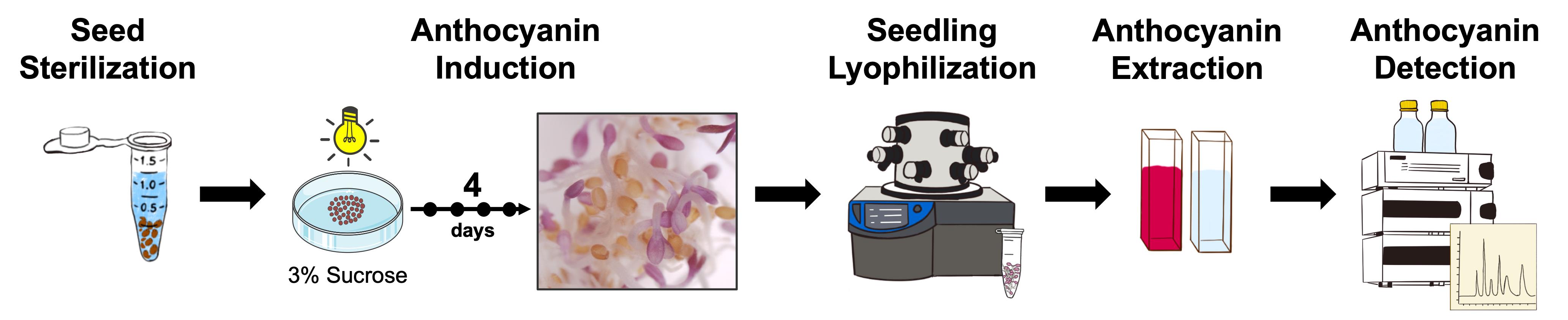
Graphical overview of anthocyanin induction, extraction, quantification, and HPLC-based detection in Arabidopsis thaliana. The schematic summarizes the complete workflow, including seed sterilization, anthocyanin induction under anthocyanin-inductive conditions (AIC), pigment extraction from lyophilized seedlings, spectrophotometric quantification, and high-performance liquid chromatography with diode-array detection (HPLC-DAD)-based detection of anthocyanins.
Background
Anthocyanins (Figure 1) are a class of water-soluble flavonoid pigments responsible for the red, purple, and blue coloration observed in many plant tissues [1,2]. Beyond their visual functions in attracting pollinators and serving as stress indicators [3,4], anthocyanins perform physiological roles by scavenging reactive oxygen species (ROS) and enhancing tolerance to abiotic stresses such as high light, nutrient deficiency, and low temperature [5,6]. Arabidopsis thaliana has become a widely used model system for investigating anthocyanin biosynthesis and regulation, owing to its fully sequenced genome, genetic tractability, and availability of numerous pigment-related mutants [7–9]. Its responsiveness to sucrose supplementation and continuous light exposure makes it particularly suitable for anthocyanin induction under controlled anthocyanin-inductive conditions (AIC) [10–14].
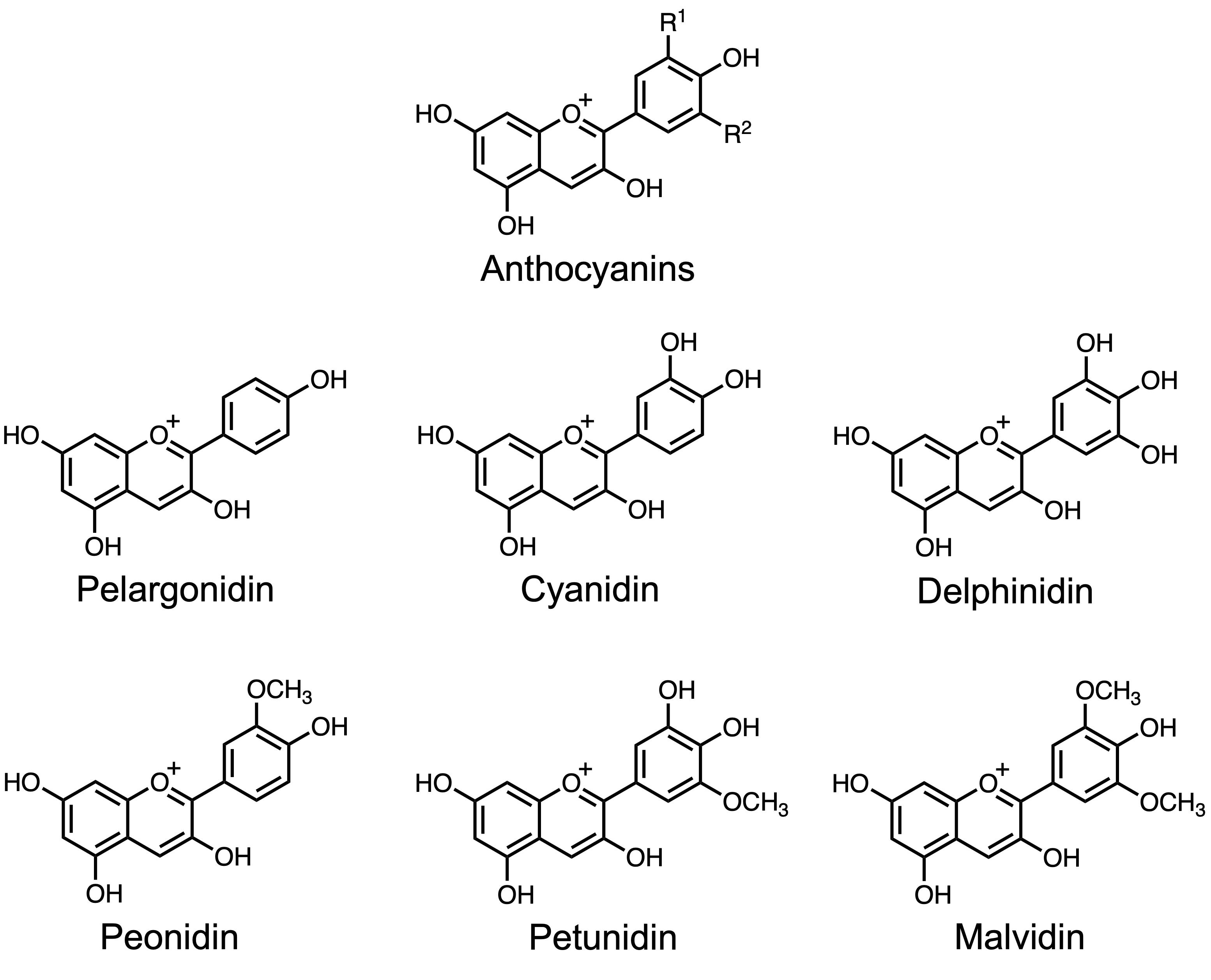
Figure 1. Chemical structures of representative anthocyanins. The basic anthocyanidin backbone consists of a flavylium cation structure with variable hydroxylation and methoxylation patterns that define specific anthocyanidins, such as pelargonidin, cyanidin, delphinidin, peonidin, petunidin, and malvidin. R1, R2: -H, -OH, -OCH3.
Traditional quantification of anthocyanins in Arabidopsis thaliana typically involves spectrophotometric assays of crude extracts, often based on absorbance at 530 nm with corrections for chlorophyll interference [15,16]. While these assays are rapid and cost-effective, they lack specificity in distinguishing individual anthocyanin species and provide limited resolution for comparative analyses among genotypes or treatments. In contrast, chromatographic techniques such as high-performance liquid chromatography (HPLC), coupled with diode array detection (DAD) or mass spectrometry [1], enable precise separation, identification, and quantification of individual anthocyanin compounds [12,17,18]. Several previously reported extraction methods rely on harsh conditions such as strong acid hydrolysis, which may result in modification of glycosylated forms [19,20].
The protocol presented here integrates optimized steps for anthocyanin induction, extraction, quantification, and chromatographic characterization in Arabidopsis thaliana. By combining a mild methanol/formic acid extraction system with lyophilization and normalization by dry weight, the method enhances pigment recovery, stability, and reproducibility. The inclusion of the chalcone isomerase mutant tt5–2 [9,21] alongside the wild-type Col-0 provides a robust system for comparing anthocyanin-deficient and anthocyanin-producing phenotypes, facilitating both genetic and metabolic analyses. Beyond Arabidopsis, this protocol can be adapted and optimized for other plant systems in which anthocyanin accumulation is environmentally or developmentally regulated, including crop and ornamental species. The protocol’s precision and reproducibility make it suitable for metabolic engineering and synthetic biology applications, where anthocyanins are targeted for enhancement of nutritional value, stress resilience, or natural pigment production in engineered plants and cell cultures.
Compared with existing methods, this protocol defines reproducible anthocyanin-inductive conditions (AIC), standardizes extraction buffers compatible with HPLC analysis, and integrates dual quantification approaches, spectrophotometric and chromatographic, for comprehensive pigment profiling. Beyond its utility in basic plant physiology, this approach is broadly applicable to studies of anthocyanin biosynthesis, stress physiology, metabolic engineering for pigment production, and functional genomics of the flavonoid pathway in Arabidopsis thaliana and other plant species [10,12,17,18].
Materials and reagents
Biological materials
1. Arabidopsis thaliana Col-0 seeds [Arabidopsis Biological Resource Center (ABRC), stock number: CS70000]
2. Arabidopsis thaliana tt5-2 seeds (ABRC, stock number: CS2105574)
Reagents
1. Ethanol (MiliporeSigma, catalog number: 64-17-5)
2. Bleach (Clorox, catalog number: USA001229)
3. Triton X-100 (VWR, catalog number: 9002-93-01)
4. Sucrose (FisherScientific, catalog number: 57-50-1)
5. Methanol (MeOH) (FisherScientific, catalog number: 67-56-1)
6. Formic acid (FisherScientific, catalog number: 270480010)
7. Acetonitrile (FisherScientific, catalog number: 75-05-08)
Solutions
1. 70% ethanol (see Recipes)
2. 30% bleach (see Recipes)
3. 0.1% Triton X-100 (see Recipes)
4. Sterilizing solution (see Recipes)
5. 3% sucrose (see Recipes)
6. Extraction buffer (see Recipes)
7. HPLC running buffer A (see Recipes)
8. HPLC running buffer B (see Recipes)
Recipes
1. 70% ethanol (500 mL)
| Reagent | Final concentration | Quantity or volume |
|---|---|---|
| Ethanol | 70% (v/v) | 350 mL |
| dH2O | n/a | 150 mL |
| Total | n/a | 500 mL |
Store at room temperature (RT).
2. 30% bleach (500 mL)
| Reagent | Final concentration | Quantity or volume |
|---|---|---|
| Commercial bleach | 30% (v/v) | 150 mL |
| dH2O | n/a | 350 mL |
| Total | n/a | 500 mL |
Store at RT.
3. 0.1% Triton X-100 (250 mL)
| Reagent | Final concentration | Quantity or volume |
|---|---|---|
| Triton X-100 | 0.1% (v/v) | 0.25 mL |
| dH2O | n/a | 249.75 mL |
| Total | n/a | 250 mL |
Store at RT.
4. Sterilizing solution (50 mL)
| Reagent | Final concentration | Quantity or volume |
|---|---|---|
| Commercial bleach | 27% (v/v) | 45 mL |
| Triton X-100 | 0.01% (v/v) | 5 mL |
| Total | n/a | 50 mL |
Prepare before use.
5. 3% sucrose (500 mL)
| Reagent | Final concentration | Quantity or volume |
|---|---|---|
| Sucrose | 3% (w/v) | 30 g |
| dH2O | n/a | 500 mL |
| Total | n/a | 500 mL |
Filter the solution using a sterile bottle top filter inside a fume hood. Store at RT.
6. Extraction buffer (50 mL)
| Reagent | Final concentration | Quantity or volume |
|---|---|---|
| MeOH | 80% (v/v) | 40 mL |
| Formic acid | 3% (v/v) | 1.5 mL |
| dH2O | n/a | 8.5 mL |
| Total | n/a | 50 mL |
Store at RT.
7. HPLC running buffer A (1 L)
| Reagent | Final concentration | Quantity or volume |
|---|---|---|
| Formic acid | 0.1% (v/v) | 1 mL |
| dH2O | n/a | 999 mL |
| Total | n/a | 1,000 mL |
Store at RT.
8. HPLC running buffer B (1 L)
| Reagent | Final concentration | Quantity or volume |
|---|---|---|
| Formic acid | 0.1% (v/v) | 1 mL |
| Acetonitrile | n/a | 999 mL |
| Total | n/a | 1,000 mL |
Store at RT.
Laboratory supplies
1. Pipette tips 20–200 μL and 100–1,000 μL (FisherScientific, catalog numbers: 94052320, 94052410)
2. Falcon tubes 15 and 50 mL (FALCON Corning Products, catalog numbers: 352196, 352098)
3. 1.7 mL graduated microcentrifuge tubes (VWR, catalog number: 10025-716)
4. 60 × 15 mm Petri dish (VWR, catalog number: 77589-162)
5. 500 mL bottle top filter (FisherScientific, catalog number: FB12566510)
6. Parafilm (Bemis, catalog number: PM-992)
7. Weighing paper (VWR, catalog number: 12578-121)
8. HPLC vial (FisherScientific, catalog number: C4000-12)
9. HPLC vial insert (MUHWA Scientific, catalog number: X003K4XYWZ)
10. HPLC screw cup (Alwsci technologies, catalog number: C0002896)
11. Symmetry C18 column, 100 Å, 3.5 µm, 4.6 mm × 150 mm (Waters Corporation, catalog number: WAT200632)
12. Semi-micro cuvette (MiliporeSigma, catalog number: BR759015)
Equipment
1. Pipetteman pipettes (Gilson, catalog number: FD10006)
2. Graduated cylinder (PYREX, catalog number: 3042)
3. CorningTM PYREXTM reusable media storage bottles, Pyrex (FisherScientific, catalog number: 10-462-711)
4. Philips 30W 36in T12 cool white fluorescent tube (Bulbs.com, SKU: 272427)
5. Digital orbital shaker (ONiLAB, SKU: SK-O180-S)
6. Spectrometer (LI-COR Biosciences, model: LI-180)
7. Stainless steel forceps (MiliporeSigma, catalog number: Z168793)
8. Drawing pin (Amazon, catalog number: DH05101901)
9. -80 °C ultra-Low freezer (PHCbi, catalog number: MDF-DU502VH)
10. FreeZone Benchtop Freeze Dryer (Labconcom, catalog number: 700201000)
11. Vortex (Scientific Industries, catalog number: SI-0236)
12. Microcentrifuge (VWR, catalog number: VWRI521-2656)
13. HPLC system (Agilent Technologies, model: Agilent 1260)
14. Euromex NexiusZoom Series high-precision zoom binocular stereomicroscope (AmScope, catalog number: NZ1902-P)
15. Camera (AmScope, catalog number: MU1803)
16. Fine balance (FisherScientific, catalog number: 02-035SD)
17. Branson UltrasonicsTM CPX8800H ultrasonic cleaning bath (FisherScientific, catalog number: 15-336-124)
18. 0.22 mm filter (VWR, catalog number: 76478-992)
19. Digital timer (VWR, catalog number: 609-0204)
20. UV-1600PC scanning spectrophotometer (VWR, catalog number: 10037-436)
Software and datasets
1. AmScope version 4.12.24446.20240114 (Irvine, California)
2. Agilent OpenLab Control Panel, version 3.5.0 (Santa Clara, California)
3. Agilent OpenLab Acquisition, Build 2.6.0.691 (Santa Clara, California)
4. Agilent OpenLab Data Analysis, Build 2.206.0.780 (Santa Clara, California)
5. GraphPad Prism 10.1.1 (San Diego, California)
Procedure
A. Sterilization of Arabidopsis thaliana seeds
Note: Perform steps A2–9 under a sterile fume hood at RT.
1. Aliquot ~1,000 (~20 mg) Arabidopsis thaliana seeds (Col-0 and tt5-2) into 1.7 mL microcentrifuge tubes (Figure 2A).
2. Add 1 mL of 70% ethanol into the tube and invert every 10 s for 1 min.
3. Carefully remove the ethanol from the tube using a sterile pipette, minimizing seed removal.
4. Add 1 mL of sterilizing solution into the tube and invert every 30 s for 15 min.
5. Remove the sterilizing solution from the tube using a sterile pipette.
6. Add 1 mL of autoclaved water into the tube and invert every 30 s for 1 min.
7. Carefully remove the water from the tube using a sterile pipette.
8. Repeat steps A6–7 two more times.
9. Add 1 mL of autoclaved water into the tube (Figure 2B).
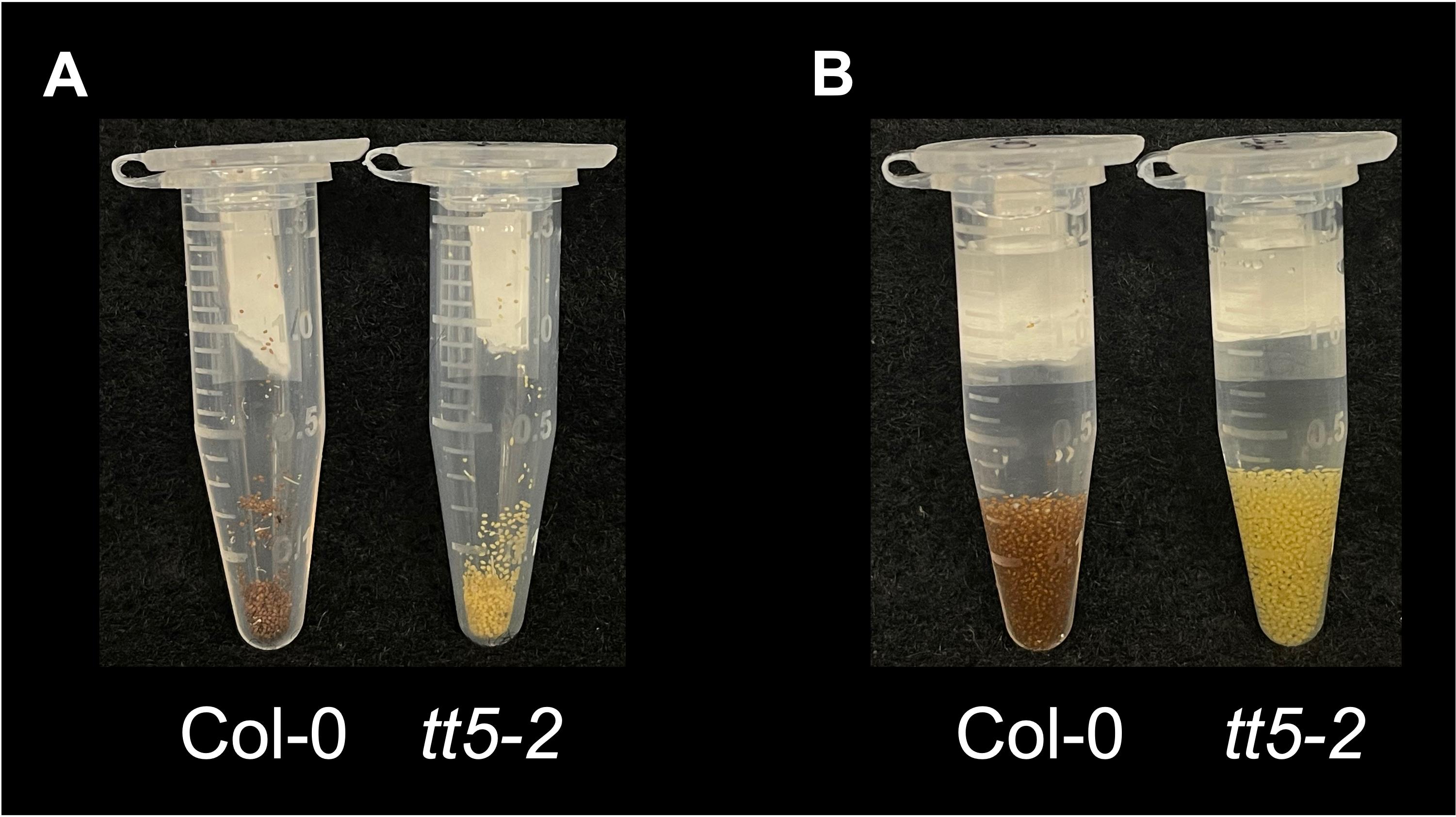
Figure 2. Sterilization of Arabidopsis thaliana seeds. (A) Representative image showing aliquoted Arabidopsis thaliana wild-type (Col-0) and anthocyanin-deficient mutant (tt5-2) seeds prior to sterilization. (B) Seeds following completion of the sterilization process with a fully hydrated appearance.
B. Anthocyanin induction under anthocyanin inductive conditions (AIC)
Note: Perform steps B1–3 under a sterile fume hood at RT.
1. Prepare three 60 × 15 mm Petri dishes with 10 mL of 3% sucrose solution per tube of sterilized seeds.
2. Transfer ~300 sterilized seeds (1/3 of the total amount) per Petri dish (Figure 3A, D).
3. Label the lids and seal the Petri dishes with parafilm.
4. (Critical) Place the Petri dishes on an orbital shaker set at 70 rpm, under 24-h consistent light with intensity of ~80 μmol cm-2 s-1, at a temperature of 21–22 °C.
5. Incubate the seeds under AIC for 4 days (Figure 3B, E). Anthocyanin accumulation is not observed in control samples grown in H2O (Figure 3C, F).
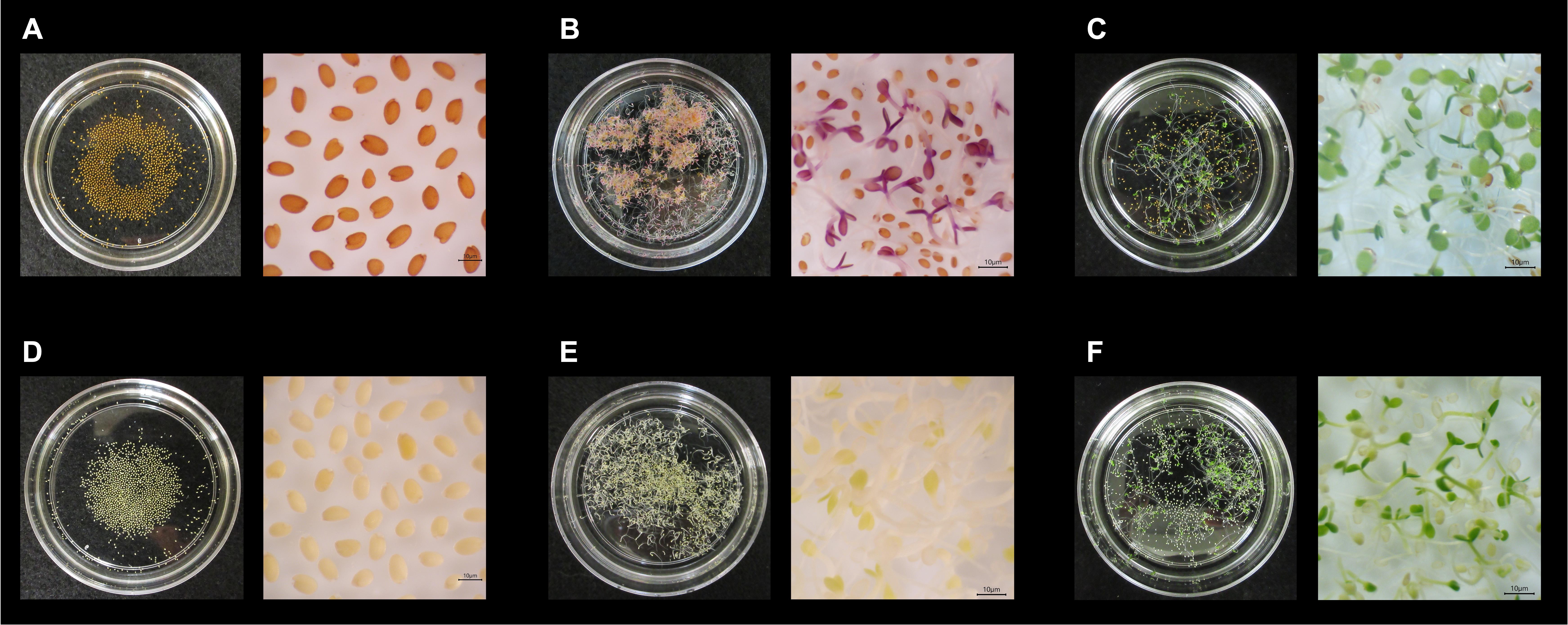
Figure 3. Anthocyanin induction in Arabidopsis thaliana seedlings under anthocyanin inductive conditions (AIC). (A) Arabidopsis thaliana wild-type (Col-0) seeds before anthocyanin induction, showing beige coloration on 3% sucrose medium. (B) Col-0 seedlings after 4 days under AIC, displaying strong purple pigmentation on hypocotyls and cotyledons. (C) Col-0 seedlings grown under control conditions (H2O only) for 4 days, showing green seedlings without detectable anthocyanin accumulation. (D) tt5-2 mutant seeds before induction, exhibiting light yellow coloration. (E) tt5-2 seedlings after 4 days under AIC, showing a lack of purple pigmentation due to disruption in the chalcone isomerase (TT5) gene, confirming the anthocyanin-deficient phenotype. (F) tt5-2 seedlings grown under control conditions (H2O only), also lacking anthocyanin pigmentation.
C. Anthocyanin extraction and quantification from lyophilized Arabidopsis thaliana seedlings
1. Take the seedlings from the Petri dishes using forceps. Gently remove the excess moisture by dabbing the seedlings with a paper towel.
2. Place the seedlings into a 1.7 mL tube and create a small vent hole in the cap of the tube using a drawing pin.
3. Keep the tubes in -80 °C for at least 1 h.
4. Turn on and monitor the freeze-dryer until it reaches -70 °C and 0.2–0.4 mbar; then, begin the freeze-drying cycle with the samples inside for ~16 h.
5. Weigh the lyophilized seedlings using a fine balance and record the dry weight.
6. Transfer the lyophilized seedlings into new 1.7 mL tubes.
Pause point: Lyophilized samples can be stored at RT for up to 1 month before extraction.
7. Add extraction buffer into the tubes at a ratio of 20 μL of extraction buffer per milligram of dry weight.
8. Soak the seedlings with extraction buffer at RT for ~16 h.
9. Vortex for 10 s and centrifuge at 13,000× g for 5 min at RT.
10. Collect the supernatant with extracted anthocyanins into new 1.7 mL tubes (Figure 4).
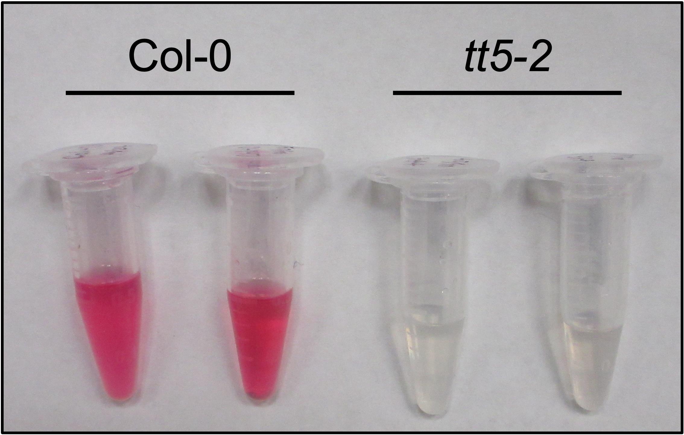
Figure 4. Extraction of anthocyanins from Arabidopsis thaliana seedlings. Representative image showing extracted anthocyanins from lyophilized Arabidopsis thaliana wild-type (Col-0) and tt5-2 mutant seedlings following methanol/formic acid extraction. Col-0 samples display a strong red coloration in the supernatant, indicative of high anthocyanin accumulation, whereas tt5-2 mutant extracts remain colorless.
Pause point: Extracted samples can be stored at -20 °C for up to two weeks before further analysis.
11. Add 100 μL of extraction buffer to the semi-micro cuvette, insert it into the spectrophotometer, and set it to measure OD at 532 nm. Click Blank.
12. Transfer 100 μL of extracted samples from step C10 into new semi-micro cuvettes and evaluate total anthocyanins by measuring OD at 532 nm. Normalize to the dry weight of tissue from step C5 and express as OD532/mg of dry weight (Figure 5).
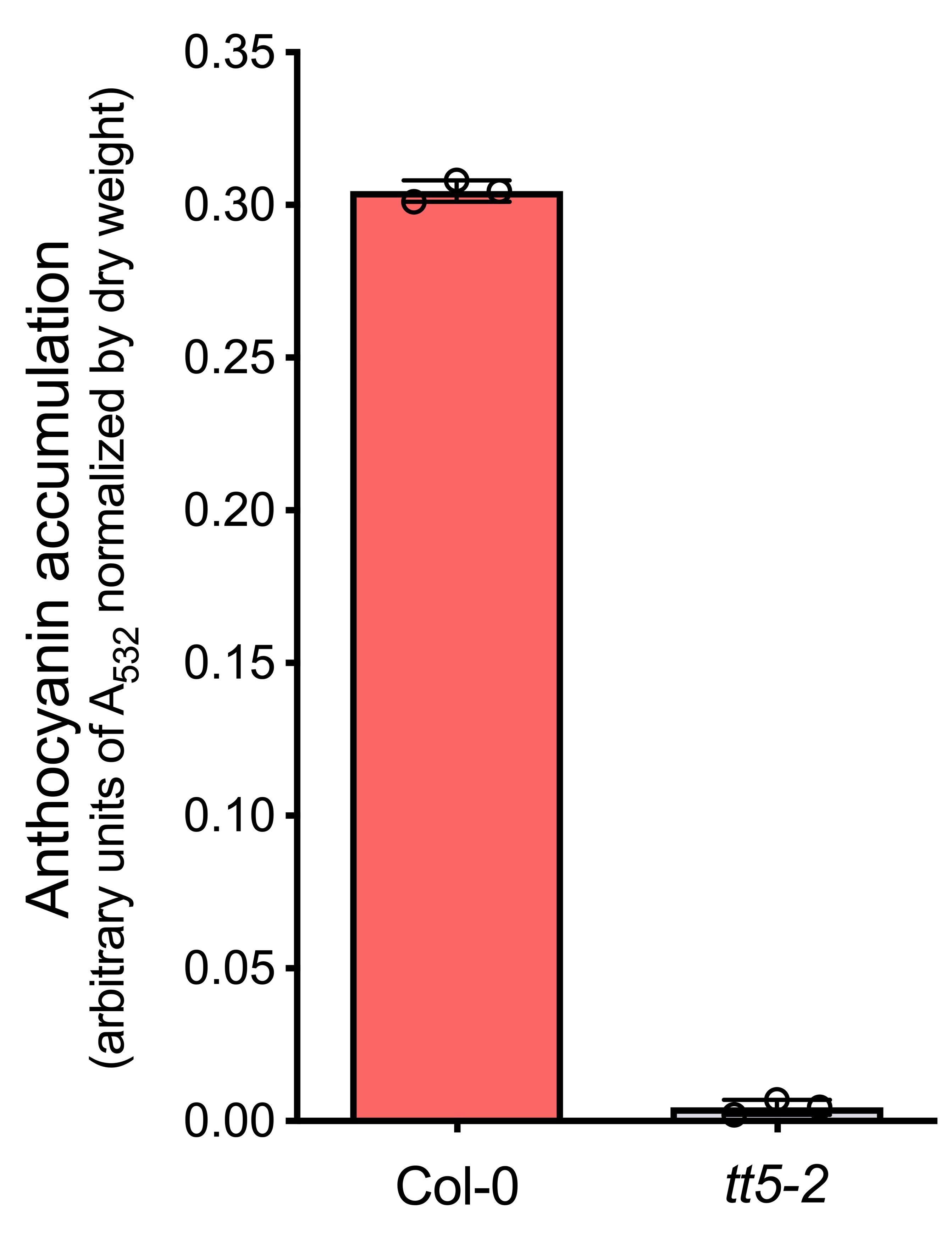
Figure 5. Quantification of anthocyanin accumulation in Arabidopsis thaliana seedlings. Spectrophotometric measurement of total anthocyanins extracted from Col-0 and tt5-2 seedlings. Absorbance at 532 nm was normalized to the dry weight. Bar graph shows a significant increase in anthocyanin content in Col-0 compared to the tt5-2 mutant, consistent with the visual pigment difference observed in Figure 4. Error bars represent the standard deviation from biological triplicates.
D. Detection of anthocyanins by high-performance liquid chromatography (HPLC)
1. Sample preparation
a. Apply extracted anthocyanins from step C10 to a 0.22 mm filter and collect the filtered samples.
b. Transfer 60 μL of filtered samples into HPLC vial inserts within HPLC vials.
c. Screw the HPLC cups.
d. Label the vials and flick them to make sure there are no air bubbles at the bottom of the inserts.
2. HPLC setup
a. Double-click the Agilent software Control Panel.
b. Set up 1260 Infinity II LC System using a Symmetry C18 column in the column compartment. Ensure the column is installed in the correct orientation. Tighten the connectors, but do not overtighten, as this may damage the column tip.
c. Click Instrument, select Project, and click Launch to open the software Acquisition.
d. Under the Home tab, select Method to set up the method parameters with a flow rate of 0.5 mL/min at a column temperature of 35 °C.
e. Create a 30-min gradient running method with HPLC running buffer A (H2O + 0.1% formic acid) and buffer B (acetonitrile + 0.1% formic acid). Use the following gradient running conditions: 0 min, 95% A and 5% B; 20 min, 75% A and 25% B; 22 min, 20% A and 80% B; 22.1 min, 5% A and 95% B; 25 min, 5% A and 95% B; 25.1 min, 95% A and 5% B; and 30 min, 95% A and 5% B. Set up diode array detector (DAD) signals to detect 532 nm. Save as method Anthocyanins.
f. Fill in the information in the sequence table by clicking Sequence1: include the vials (e.g., 1, 2, …), acquisition method (“Anthocyanins” from step D2e), sample amount (“20” as 20 µL), and data file and sample file names (e.g., blank, sample 1, sample 2, ….).
g. Equilibrate the system by running 95% A and 5% B at a flow rate of 0.3 mL/min for at least 30 min.
3. Sample running: Place the sample vials from step D1 in the autosampler following the corresponding vial numbers from step D2f. Click Run to run the samples.
4. Data processing and result analysis
a. Create a folder on your computer to save the result data.
b. Open the software Control Panel and select the corresponding project.
c. Click Start Data Analysis to open the software Data Analysis.
d. Select the dataset from the Data selection list and confirm that the dataset is correct.
e. Double-click the dataset to load data.
f. Select the GC/LC Quantitative method and click Link and process.
g. Under Data Processing > Signals, select Sig = 532.
h. Click Processing Method > Find Tools on the top menu. Select Post Processing Plugins to open the plugin configuration window. Choose CSV Export to enable automatic data output in CSV format. Click Browse and select the folder you created in step D4a as the output path. Click Save Method on the top menu.
i. Click Reprocess all to apply the method to all files. Open the output folder and confirm that files were generated successfully.
j. Create anthocyanin profiles by Prism using the exported CSV files (Figure 6).
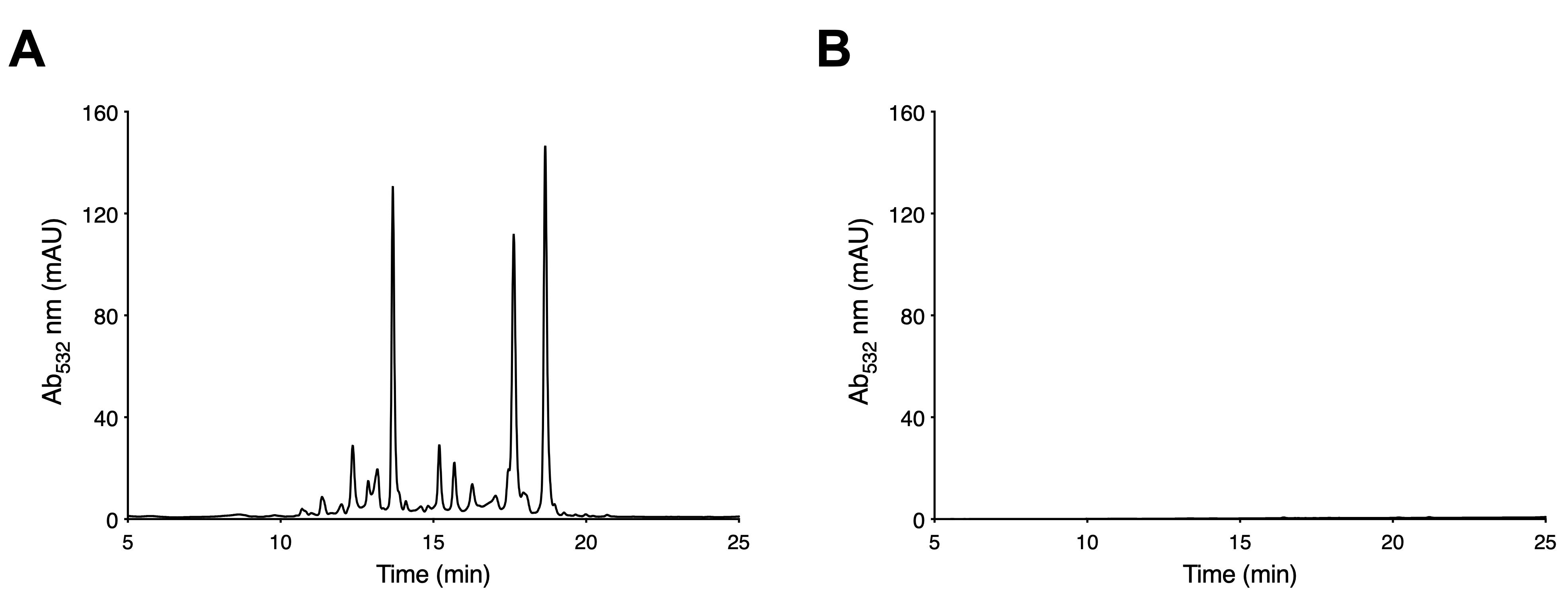
Figure 6. High-performance liquid chromatography (HPLC) chromatographic profiles of anthocyanin extracts from Arabidopsis thaliana seedlings under anthocyanin-inductive conditions (AIC). Representative HPLC chromatograms recorded at 532 nm. (A) The Col-0 sample exhibits distinct peaks corresponding to anthocyanin compounds, confirming successful pigment extraction and separation. (B) The tt5-2 mutant profile lacks detectable peaks, reflecting anthocyanin deficiency.
Validation of protocol
This protocol or parts of it has been used and validated in the following research articles:
Jiang et al. [22] Flavonoid pathway intermediates implicate UVR8 in functions beyond canonical UV-B signaling. Nature Communications (Figures 5 and S9).
Jiang et al. [17] Synergy between the anthocyanin and RDR6/SGS3/DCL4 siRNA pathways expose hidden features of Arabidopsis carbon metabolism. Nature Communications (Figures 1–3, 5, S3, S6, S7, S9, S10, and S13).
Jiang et al. [18] Diversity of genetic lesions characterizes new Arabidopsis flavonoid pigment mutant alleles from T-DNA collections. Plant Science (Figure 2).
General notes and troubleshooting
General notes
1. Anthocyanin accumulation is highly sensitive to environmental variables such as light intensity, temperature, and sucrose concentration. To ensure reproducibility, maintain stable growth conditions with continuous light at 21–22 °C. Using seedlings of uniform developmental stages and batch-prepared reagents will minimize variability across biological replicates.
2. Anthocyanins are prone to degradation under alkaline or high-temperature conditions. To preserve pigment integrity, extraction and handling should occur under acidic solvent and at room temperature. Avoid repeated freeze-thaw cycles by aliquoting extracts prior to storage at -20 °C or below.
3. Although this protocol utilizes UV-Vis spectrophotometry and HPLC-DAD, it can be readily adapted to liquid chromatography–mass spectrometry (LC–MS) or liquid chromatography–tandem mass spectrometry (LC–MS/MS) for structural identification of individual anthocyanins.
4. The sensitivity of spectrophotometric measurement may not resolve low-abundance or minor anthocyanin species. For genetically modified lines or stress conditions that yield weak pigmentation, chromatographic or mass spectrometric methods are preferred. Ensure complete lyophilization before weighing samples, as residual moisture can distort dry-weight normalization.
5. Anthocyanin profiles can vary among genotypes and environmental conditions. To improve inter-experimental comparability, report extraction volumes normalized to the sample dry weight and provide detailed information on induction parameters (light duration, sucrose concentration, and temperature).
Troubleshooting
Problem 1: Microbial contamination during seed sterilization or induction.
Possible cause: Incomplete sterilization of seeds, forceps, or working surfaces.
Solutions: Perform all seed handling steps under a sterile fume hood disinfected with 70% ethanol. Sterilize forceps by dipping in 30% bleach between samples. Prepare sterilizing solutions freshly before each use to maintain effectiveness.
Problem 2: Weak or inconsistent anthocyanin induction.
Possible cause: Fluctuations in light intensity, temperature, or sucrose concentration during incubation.
Solutions: Maintain continuous illumination (~80 μmol cm-2 s-1) and a constant temperature of 21–22 °C. Verify the accuracy of the 3% sucrose solution and ensure the orbital shaker speed is consistent (50–70 rpm).
Problem 3: Low anthocyanin yield after extraction.
Possible causes: Incomplete extraction or insufficient extraction duration.
Solutions: Ensure lyophilized samples are fully immersed in the extraction buffer. Extend incubation up to ~20 h if necessary, vortex thoroughly before centrifugation, and avoid sample clumping to ensure uniform extraction.
Problem 4: High background absorbance or unstable OD532 readings.
Possible causes: Cuvette contamination, incorrect blanking, or solvent residue.
Solutions: Rinse cuvettes with ethanol before each use. Re-blank the spectrophotometer using fresh extraction buffer. Avoid introducing air bubbles when loading samples and ensure consistent optical path length across measurements.
Problem 5: Inconsistent HPLC peak resolution or retention times.
Possible causes: Column contamination, incomplete system equilibration, or inaccurate gradient settings.
Solutions: Confirm correct gradient programming and equilibrate the system with 95% buffer A and 5% buffer B at 0.3 mL/min for at least 30 min before analysis. Regularly replace solvent filters and degas buffers to prevent air interference.
Problem 6: Degradation or discoloration of extracts during storage.
Possible causes: Repeated freeze-thaw cycles or prolonged exposure to elevated temperatures.
Solutions: Aliquot extracts into amber microtubes and store at -20 °C or lower. Avoid unnecessary thawing and handle samples at room temperature to preserve pigment stability. For long-term storage, consider vacuum-drying and re-dissolving before analysis.
Problem 7: Air bubbles in HPLC vials leading to injection errors.
Possible causes: Improper sample transfer, capping, or vial insert placement.
Solutions: Gently tap or flick capped vials to release trapped air bubbles before loading into the autosampler. Ensure vial inserts are properly seated and that caps are securely tightened to prevent leakage or misalignment during injection.
Acknowledgments
Conceptualization: E.G. and N.J.; Investigation: Y.H., S.I., and N.J.; Writing—Original Draft: Y.H., S.I., and N.J.; Writing—Review & Editing: Y.H., E.G., and N.J.; Funding Acquisition: N.J.; Supervision: N.J. Funding support from the USDA-NIFA Hatch Multistate project HAW05053-R and University of Hawaii at Mānoa Undergraduate Research Opportunities Program (UROP) is gratefully acknowledged. This protocol has been described and validated in the original research papers by Jiang et al. [17,18]. We appreciate the technical assistance and support provided by Xiu Yuan, Qing Li, and Zhiyan Du.
Competing interests
The authors declare no competing interests.
References
- Williams, C. A. and Grayer, R. J. (2004). Anthocyanins and other flavonoids. Nat Prod Rep. 21(4): 539. https://doi.org/10.1039/b311404j
- Grotewold, E. (2006). The Genetics and Biochemistry of Floral Pigments. Annu Rev Plant Biol. 57(1): 761–780. https://doi.org/10.1146/annurev.arplant.57.032905.105248
- Chalker‐Scott, L. (1999). Environmental Significance of Anthocyanins in Plant Stress Responses. Photochem Photobiol. 70(1): 1–9. https://doi.org/10.1111/j.1751-1097.1999.tb01944.x
- Narbona, E., del Valle, J. C., Arista, M., Buide, M. L. and Ortiz, P. L. (2021). Major Flower Pigments Originate Different Colour Signals to Pollinators. Front Ecol Evol. 9: e743850. https://doi.org/10.3389/fevo.2021.743850
- Naing, A. H. and Kim, C. K. (2021). Abiotic stress‐induced anthocyanins in plants: Their role in tolerance to abiotic stresses. Physiol Plant. 172(3): 1711–1723. https://doi.org/10.1111/ppl.13373
- Cerqueira, J. V. A., de Andrade, M. T., Rafael, D. D., Zhu, F., Martins, S. V. C., Nunes-Nesi, A., Benedito, V., Fernie, A. R. and Zsögön, A. (2023). Anthocyanins and reactive oxygen species: a team of rivals regulating plant development? Plant Mol Biol. 112: 213–223. https://doi.org/10.1007/s11103-023-01362-4
- Peer, W. A., Brown, D. E., Tague, B. W., Muday, G. K., Taiz, L. and Murphy, A. S. (2001). Flavonoid Accumulation Patterns of Transparent Testa Mutants of Arabidopsis. Plant Physiol. 126(2): 536–548. https://doi.org/10.1104/pp.126.2.536
- Shirley, B. W., Kubasek, W. L., Storz, G., Bruggemann, E., Koornneef, M., Ausubel, F. M. and Goodman, H. M. (1995). Analysis of Arabidopsis mutants deficient in flavonoid biosynthesis. Plant J. 8(5): 659–671. https://doi.org/10.1046/j.1365-313x.1995.08050659.x
- Appelhagen, I., Thiedig, K., Nordholt, N., Schmidt, N., Huep, G., Sagasser, M. and Weisshaar, B. (2014). Update on transparent testa mutants from Arabidopsis thaliana: characterisation of new alleles from an isogenic collection. Planta. 240(5): 955–970. https://doi.org/10.1007/s00425-014-2088-0
- Pourcel, L., Irani, N. G., Lu, Y., Riedl, K., Schwartz, S. and Grotewold, E. (2010). The Formation of Anthocyanic Vacuolar Inclusions in Arabidopsis thaliana and Implications for the Sequestration of Anthocyanin Pigments. Mol Plant. 3(1): 78–90. https://doi.org/10.1093/mp/ssp071
- Pourcel, L., Irani, N. G., Koo, A. J. K., Bohorquez‐Restrepo, A., Howe, G. A. and Grotewold, E. (2013). A chemical complementation approach reveals genes and interactions of flavonoids with other pathways. Plant J. 74(3): 383–397. https://doi.org/10.1111/tpj.12129
- Kovinich, N., Kayanja, G., Chanoca, A., Riedl, K., Otegui, M. S. and Grotewold, E. (2014). Not all anthocyanins are born equal: distinct patterns induced by stress in Arabidopsis. Planta. 240(5): 931–940. https://doi.org/10.1007/s00425-014-2079-1
- Chanoca, A., Kovinich, N., Burkel, B., Stecha, S., Bohorquez-Restrepo, A., Ueda, T., Eliceiri, K. W., Grotewold, E. and Otegui, M. S. (2015). Anthocyanin Vacuolar Inclusions Form by a Microautophagy Mechanism. Plant Cell. 27(9): 2545–2559. https://doi.org/10.1105/tpc.15.00589
- Chanoca, A., Burkel, B., Kovinich, N., Grotewold, E., Eliceiri, K. W. and Otegui, M. S. (2016). Using fluorescence lifetime microscopy to study the subcellular localization of anthocyanins. Plant J. 88(5): 895–903. https://doi.org/10.1111/tpj.13297
- Nakata, M., Mitsuda, N., Herde, M., Koo, A. J., Moreno, J. E., Suzuki, K., Howe, G. A. and Ohme-Takagi, M. (2013). A bHLH-Type Transcription Factor, ABA-INDUCIBLE BHLH-TYPE TRANSCRIPTION FACTOR/JA-ASSOCIATED MYC2-LIKE1, Acts as a Repressor to Negatively Regulate Jasmonate Signaling in Arabidopsis. Plant Cell. 25(5): 1641–1656. https://doi.org/10.1105/tpc.113.111112
- Nakata, M. and Ohme-Takagi, M. (2014). Quantification of Anthocyanin Content. Bio Protoc. 4(7): e1098. https://doi.org/10.21769/bioprotoc.1098
- Jiang, N., Gutierrez-Diaz, A., Mukundi, E., Lee, Y. S., Meyers, B. C., Otegui, M. S. and Grotewold, E. (2020). Synergy between the anthocyanin and RDR6/SGS3/DCL4 siRNA pathways expose hidden features of Arabidopsis carbon metabolism. Nat Commun. 11(1): e1038/s41467–020–16289–3. https://doi.org/10.1038/s41467-020-16289-3
- Jiang, N., Lee, Y. S., Mukundi, E., Gomez-Cano, F., Rivero, L. and Grotewold, E. (2020). Diversity of genetic lesions characterizes new Arabidopsis flavonoid pigment mutant alleles from T-DNA collections. Plant Sci. 291: 110335. https://doi.org/10.1016/j.plantsci.2019.110335
- Zhang, Z., Kou, X., Fugal, K., McLaughlin, J. (2004). Comparison of HPLC methods for determination of anthocyanins and anthocyanidins in bilberry extracts. J Agric Food Chem. 52(4):688–91. https://doi.org/10.1021/jf034596w
- Silva, S., Costa, E. M., Calhau, C., Morais, R. M. and Pintado, M. E. (2015). Anthocyanin extraction from plant tissues: A review. Crit Rev Food Sci Nutr. 57(14): 3072–3083. https://doi.org/10.1080/10408398.2015.1087963
- Shirley, B. W., Hanley, S. and Goodman, H. M. (1992). Effects of ionizing radiation on a plant genome: analysis of two Arabidopsis transparent testa mutations. Plant Cell. 4(3): 333–347. https://doi.org/10.1105/tpc.4.3.333
- Jiang, N., García Navarrete, T., Lee, Y. S., Pawlowski, E. G., Park, S. and Grotewold, E. (2025). Flavonoid pathway intermediates implicate UVR8 in functions beyond canonical UV-B signaling. Nat Commun. 16(1): e1038/s41467–025–63010–3. https://doi.org/10.1038/s41467-025-63010-3
Article Information
Publication history
Received: Dec 17, 2025
Accepted: Feb 1, 2026
Available online: Feb 12, 2026
Published: Mar 5, 2026
Copyright
© 2026 The Author(s); This is an open access article under the CC BY-NC license (https://creativecommons.org/licenses/by-nc/4.0/).
How to cite
He, Y., Iha, S., Grotewold, E. and Jiang, N. (2026). Simple Induction and Detection of Anthocyanins in Arabidopsis thaliana: A Tool for Mutant Screening and Metabolic Analysis. Bio-protocol 16(5): e5628. DOI: 10.21769/BioProtoc.5628.
Category
Plant Science > Plant biochemistry > Metabolite
Biochemistry > Other compound > Small molecular
Do you have any questions about this protocol?
Post your question to gather feedback from the community. We will also invite the authors of this article to respond.