- Protocols

- Articles and Issues
- For Authors
- About
- Become a Reviewer

Non-Enzymatic Isolation of Cancer-Associated Fibroblasts From Human Prostate Tumor Explants
(*contributed equally to this work) Published: Vol 16, Iss 5, Mar 5, 2026 DOI: 10.21769/BioProtoc.5614 Views: 74
Reviewed by: Vinit SharmaMartin V KolevAnonymous reviewer(s)
Original research article
The authors used this protocol in:
Apr 2022

Protocol Collections
Comprehensive collections of detailed, peer-reviewed protocols focusing on specific topics


Related protocols
Abstract
Prostate carcinoma (PCa) progression is strongly influenced by the surrounding tumor microenvironment, where cancer-associated fibroblasts (CAFs) represent the most abundant and functionally relevant stromal population. Despite their importance, the lack of stable cell lines representing CAF phenotypes limits the study of stromal–tumor interactions. To address this limitation, we provide an optimized protocol for isolating CAFs from fresh human PCa biopsies based on a mechanical procedure exploiting the specific CAF ability to migrate out from the tumor explants. This approach preserves tissue architecture and maintains CAF viability and phenotype. The resulting ex vivo CAF cultures provide a suitable model to investigate CAF biology within the tumor microenvironment.
Key features
• The protocol provides an optimized workflow for isolating CAFs from prostate tumor explants by exploiting their selective outgrowth.
• It is an optimized, enzymatic-free procedure that minimizes fibroblast cell stress while preserving cell phenotypic features.
Keywords: Prostate cancerGraphical overview
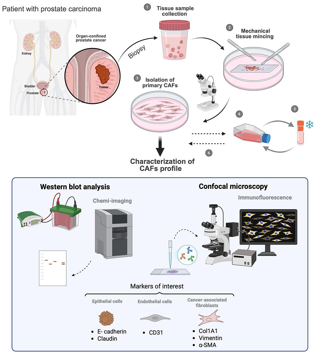
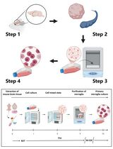
Workflow for cancer-associated fibroblasts (CAFs) isolation
Background
Prostate carcinoma (PCa) arises from the malignant transformation of epithelial cells residing within the prostatic acini. Although the genetic background of transformed cells is crucial for tumor initiation [1], increasing evidence indicates that PCa progression is strongly influenced by interactions with both resident and recruited cell populations of the surrounding microenvironment [2]. Under physiological conditions, the prostate stroma is composed of several cell types, including fibroblasts, endothelial cells, and a small number of immune cells. When stromal homeostasis is disrupted by PCa onset, many of these cells become “educated” by cancer cells, acquiring pro-tumorigenic features. Among pro-tumoral stromal components, cancer-associated fibroblasts (CAFs) constitute the predominant population across different PCa stages [3]. Recently, distinct CAF phenotypic subtypes, including myofibroblastic CAFs (myCAFs) and/or inflammatory CAFs (iCAFs), have been identified according to their specific expression profiles and spatial localization/interactions within the tumor mass [4–6]. Briefly, the dynamic crosstalk with the neighboring tumor cells results in profound phenotypic, transcriptional, and functional alterations compared with the normal fibroblasts populating the non-cancerous areas. In particular, tumor-induced activation of normal fibroblasts into a myCAF population results in higher contractility, primarily supported by increased expression of the alpha-smooth muscle actin (α-SMA) [7] and a metabolic shift toward enhanced glycolysis [8]. These CAF features are crucial to establish a crosstalk with epithelial tumor cells, ultimately promoting enhanced cell invasion, immune evasion, and resistance to therapy insults, key processes driving metastatic progression of tumors, including PCa [9–11].
Given the key role of CAFs in supporting PCa progression, developing in vitro models that accurately recapitulate CAF-tumor interactions is essential for research. The absence of cell lines recapitulating the CAF phenotype represents a limitation in the study of CAF pathobiology. In this regard, the establishment of CAF cultures ex vivo is an actual need, as it provides a physiologically relevant model to study this crosstalk in vitro. Indeed, fibroblast standard isolation procedures are mainly based on enzymatic dissociation procedures, followed by flow cytometry cell sorting or magnetic bead–based isolation [12, 13]. However, while these procedures are likely proper for single-cell transcriptomics analysis, they show restricted applicability for cell culture, as they may alter the phenotype of the isolated cells due to the exposure to enzymatic cocktails and to prolonged maintenance in non-adherent conditions. For a proper isolation of CAFs for cell culture applications, we optimized a protocol for mechanically isolating myCAFs from minced human fresh PCa biopsies by exploiting the intrinsic motility of these cells toward a nutrient-rich environment. This approach has the advantage of preserving tissue architecture, maintaining CAF viability and intrinsic phenotypic characteristics, providing a more physiologically relevant model for in vitro studies. Here, we provide a step-by-step procedure to selectively separate activated fibroblasts (i.e., myCAFs) from fresh human PCa biopsies; we also describe validation methods to assess the purity of the resulting primary cultures, as well as the acquisition of phenotypic and metabolic CAF-specific markers compared to normal fibroblasts (e.g., derived from benign prostatic hyperplasia). Applying the protocol described here allows the preparation of ex vivo fibroblast populations suitable for studying the mechanisms underlying fibroblast activation and for elucidating their role in the tumor microenvironment, especially focusing on PCa. These primary CAFs can be co-cultured with tumor cells, allowing a more faithful recapitulation of tumor physiology in vitro.
Materials and reagents
Biological materials
1. Human surgical explants from patients affected by benign prostatic hyperplasia (BPH) for (not-activated) healthy prostate fibroblasts (HPF) isolation
2. Human surgical explants from patients subjected to surgical intervention for prostate cancer (Gleason Group > 3) for CAFs isolation
3. Human epithelial PCa cell line DU145 (RRID: CVCL 0105) obtained from ATCC and routinely tested for Mycoplasma contamination using the MycoAlert Mycoplasma Detection kit (Lonza, #LOLT07710)
4. Endothelial colony-forming cells (ECFCs) isolated from umbilical cord blood
Notes: DU145 cells and ECFCs can be replaced with other similar commercially available cell lines to be used as epithelial and endothelial controls, respectively.
Reagents
1. Physiological saline solution, NaCl, 0.9% (B. Braun, catalog number: 030902391)
2. Phenol-red high glucose Dulbecco’s modified Eagle’s medium (DMEM) (Euroclone, catalog number: ECB7501L)
3. Fetal bovine serum (FBS) (Euroclone, catalog number: ECS5000L)
4. L-Glutamine (Merck Sigma, catalog number: G7513-100 ML)
5. 10,000 units penicillin/10 mg streptomycin solution in 0.9% (Merck Sigma, catalog number: P0781-100 ML)
6. Kanamycin (Merck, catalog number: 246933-9)
7. Amphotericin B (Fungizone) (Euroclone, catalog number: ECM0009D)
8. Trypsin (Sigma-Aldrich, catalog number: T4049)
9. Dimethyl sulfoxide (DMSO) (Sigma-Aldrich, catalog number: 472301)
10. RIPA lysis buffer (Thermo Fisher Scientific, catalog number: 8990)
11. Protease inhibitors (Sigma-Aldrich, catalog number: P8340)
12. Phosphatase inhibitors (Sigma-Aldrich, catalog number: P0044)
13. BCA Protein Assay kit (Sigma-Aldrich, catalog number: 1003579336)
14. 4%–20% acrylamide precast SDS-PAGE gels (Bio-Rad, catalog numbers: 4568093 and 4568096)
15. PVDF membranes (Bio-Rad, catalog number: 1704157)
16. Phosphate buffered saline (PBS) (Euroclone, catalog number: ECB4004L)
17. Tween 20 (Sigma-Aldrich, catalog number: P1379)
18. Non-fat dry milk (Regilait Ecreme 750gr)
19. Primary antibodies (Table 1)
Table 1. Primary antibodies list
| Target | Catalog number | Dilution | Secondary antibodies |
|---|---|---|---|
| E-Cadherin | Cell Signaling Technology, 24E10 | 1:1,000 | Rabbit |
| Claudin | Cell Signaling Technology, D5H1C | 1:1,000 | Rabbit |
| CD31 | Cell Signaling Technology, 89C2 | 1:1,000 | Mouse |
| Col1a1 | Cell Signaling Technology, 72026 | 1:1,000 | Rabbit |
| Vimentin | Cell Signaling Technology, D21H3 | 1:1,000 | Rabbit |
| Alpha-SMA | Merck Sigma, SAB5700835 | 1:1,000 | Mouse |
| LDHA | Santa Cruz Biotechnology, D0220 | 1:1,000 | Mouse |
| MCT4 | Santa Cruz Biotechnology, 376140 | 1:1,000 | Mouse |
| HSP90 | Santa Cruz Biotechnology, sc-69703 | 1:1,000 | Mouse |
20. Secondary antibodies: Anti-rabbit HRP (Santa Cruz, catalog number: sc-2357) and anti-mouse HRP (Santa Cruz, catalog number: sc-516102)
21. ECL substrates: Clarity Western ECL (Bio-Rad, catalog number: 1705061) or Clarity Max ECL (Bio-Rad, catalog number: 1705062)
22. Formaldehyde (Sigma-Aldrich, catalog number: 252549-1L)
23. Methanol (Sigma-Aldrich, catalog number: 34860-2.5L)
24. DAPI (Thermo Fisher Scientific, catalog number: D3571)
25. Triton X-100 (Sigma-Aldrich, catalog number: T8787-250ML)
26. Alexa Fluor-488 rabbit secondary antibody (1:1,000) (Thermo Fisher Scientific, catalog number: A-11008)
27. α-SMA antibody for immunofluorescence analysis (1:100) (Abcam, catalog number: ab5694)
28. Glass mountant Pro-Long (Invitrogen, catalog number: P36980)
29. Horse serum (Euroclone, catalog number: ECS0091L)
Solutions
1. Starvation medium (see Recipes)
2. Complete medium (see Recipes)
3. Freezing medium (see Recipes)
4. Non-fat dry milk (see Recipes)
Recipes
1. Starvation medium
| Reagent | Final concentration | Quantity or volume |
|---|---|---|
| DMEM high glucose | - | 400 mL |
| Penicillin-streptomycin (stock 100×) | 2× | 10 mL |
| L-Glutamine (stock 100×) | 2 mM | 5 mL |
2. Complete medium
| Reagent | Final concentration | Quantity or volume |
|---|---|---|
| DMEM high glucose | - | 400 mL |
| FBS | 20% (v/v) | 100 mL |
| Penicillin-streptomycin (stock 100×) | 2× | 10 mL |
| L-Glutamine (stock 100×) | 2 mM | 5 mL |
| Kanamycin (stock 100×) | 100 μg/mL | 5 mL |
| Fungizone (Amphotericin B stock 100×) | 2.5 μg/mL | 5 mL |
3. Freezing medium
| Reagent | Final concentration | Quantity or volume |
|---|---|---|
| FBS | 90% (v/v) | 9 mL |
| DMSO | 10% (v/v) | 1 mL |
4. Non-fat dry milk
| Reagent | Final concentration | Quantity or volume |
|---|---|---|
| Non-fat dry milk | 5% (w/v) | 5 g |
| T-PBS (PBS 1× + Tween 20 1%) | - | 100 mL |
Laboratory supplies
1. Cell culture plates/dishes [Euroclone, catalog numbers: ET2100 (p100) and ET2060 (p60)]
2. Tubes [Euroclone, catalog numbers: ET5015B (15 mL) and ET5050B (50 mL)]
3. Microcentrifuge (Euroclone catalog number: ET3415)
4. Pipettes (micropipettes, serological pipettes) [Euroclone, catalog numbers: EPS02N (2 mL), EPS05N (5 mL), EPS10N (10 mL), and EPS25N (25 mL)]; pipette controller Primo® Mate (Euroclone, catalog number: ECP2000); micropipettes P10, P20, P100, P200, and P1000 (Gilson MyPipetman); micropipette tips [Gilson, catalog numbers: DF10ST (0.1–20 μL), DF200ST (2–200 μL), and DF1000ST (100–1,000 μL)]
5. Glass coverslips (BioSigma, catalog number: VBS636)
Equipment
1. Tweezers (DIMART, catalog number: DIM.2120/14) sterilized in an oven at 180 °C for 3–4 h
2. Sterile scalpel (Feather, catalog number: 530050)
3. Microscope slides (BioSigma, catalog number: VBS654), individually packed in aluminum and sterilized as above
4. Laminar flow hood (CELLBIO, model: MARS)
5. CO2 cell culture incubator (Thermo Scientific, model: Forma Direct Heat)
6. Refrigerated centrifuge (Sigma, model: 1-14K)
7. Inverted microscope (Leica Microsystem, model: DMi1)
8. SDS-PAGE gel tank (Bio-Rad, model: Mini-PROTEAN Tetra System) and power supply (Bio-Rad, model: PowerPAC Basic)
9. Trans-Blot Unit (Bio-Rad, model: Trans-Blot Turbo)
10. Confocal microscope (Leica Microsystems, model: TCS SP8)
11. ChemiDoc MP imaging system (Bio-Rad)
12. 4 °C fridge (Gorenje), -20 °C freezer (Gorenje), and -80 °C freezer
13. Liquid nitrogen tank
Software and datasets
1. LAS-AF image acquisition software (Leica Microsystems)
Procedure
A. Sample collection
1. Collect fresh tissue samples (0.5–1 cm) of PCa and BPH aseptically immediately after surgery.
2. Keep samples in sterile physiological saline solution or PBS in a 50 mL Falcon on ice and transfer to the laboratory. Caution: Tissue samples should be processed as soon as possible (within 1–2 h) to prevent degradation.
3. Maintain tumor and healthy tissues separately to avoid cross-contamination.
B. Tissue preparation
1. In a biological laminar flow hood, transfer the samples to a sterile culture dish (100 mm plate) using sterile tweezers.
2. Remove any fat or necrotic areas using sterile scalpels and tweezers.
3. Cut the tissue into small fragments (approximately 2–3 mm3) using sterile scalpels.
4. Transfer 8–10 tissue fragments to a new culture plate (100 mm plate) using sterile tweezers, spacing them approximately 0.5–1 cm apart along the center of the plate.
5. Carefully place a sterile glass slide over the tissue fragments. Gently press with sterile forceps to flatten them, ensuring good contact with the culture surface to retain a proper pressure that avoids fragment movement.
C. Primary culture
1. Gently add 15 mL of complete medium to cover the explants, ensuring they remain in place.
2. Incubate the cultures at 37 °C in a humidified atmosphere containing 5% CO2.
3. After 24–48 h, verify that the tissue pieces remain adherent.
4. Replace the medium every 2–3 days to remove debris and provide fresh nutrients.
D. Fibroblast outgrowth and subculture
1. Fibroblasts typically begin to migrate out from the tissue fragments within 7–21 days (Figure 1).
2. Once a sufficient cell outgrowth (70%–80% coverage of the glass slide) is empirically observed by 21 days, aspirate the culture medium and remove the tissue pieces using sterile tweezers. Any remaining tissue fragments should then be carefully aspirated with a pipette to ensure complete removal and minimize residual material.
3. Wash the cells with 5 mL of sterile PBS.
4. Detach adherent cells using 1 mL of 0.25% Trypsin–EDTA, neutralizing it with 5 mL of complete culture medium, and collect the cell suspension.
5. Centrifuge at 300× g for 5 min, discard the supernatant, and resuspend the pellet in fresh complete medium.
6. Seed fibroblasts into a new culture dish (60 mm plate) at the desired density (2 × 10⁵ cells/plate) and maintain under standard culture conditions, changing the medium every 2–3 days.
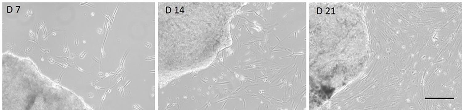
Figure 1. Fibroblast outgrowth from prostate carcinoma (PCa) tissue explant. Representative images showing migration and expansion of fibroblasts from the explant at days (D) 7, 14, and 21 (magnification 20×). Scale bar, 100 μm.
E. Cryopreservation
1. Detach fibroblasts at 70%–80% confluence (100 mm plate).
2. Aspirate the culture medium and wash with 5 mL of sterile PBS.
3. Add 1 mL of trypsin–EDTA, neutralize the enzyme with 5 mL of complete culture medium, and collect the cell suspension.
4. Count cells using a Burker chamber.
5. Centrifuge the cell suspension at 300× g for 5 min and discard the supernatant.
6. Resuspend the cell pellet in freezing medium.
7. Adjust the cell density to 1 × 10⁶ cells/mL and aliquot 1 mL into cryogenic vials.
8. Place the vials into a freezing container and transfer to a -80 °C freezer.
Caution: Minimize the time cells remain in freezing medium at room temperature.
9. After 24 h, transfer the vials to the liquid nitrogen tank (-196 °C) for long-term storage.
F. Thawing
1. Remove a vial containing frozen fibroblasts from liquid nitrogen storage.
2. Immediately place the vial in a 37 °C water bath.
3. As soon as the ice has melted, transfer the cell suspension into a 15 mL conical tube containing approximately 9 mL of complete medium to dilute the DMSO.
4. Mix gently and centrifuge at 300× g for 5 min at room temperature.
5. Carefully discard the supernatant.
6. Resuspend the cell pellet in 8 mL of fresh complete medium.
7. Transfer the entire suspension into the prepared 100 mm culture plate.
8. Leave the plate at 37 °C, 5% CO2.
9. After 24 h, replace the medium with fresh complete medium to remove residual DMSO and non-viable cells.
10. Change the medium every 2–3 days, monitoring cell attachment, morphology, and proliferation.
Data analysis
A. Western blot analysis characterization
After isolation, it is essential to assess any contamination of other cell types (e.g., epithelial or endothelial cells) rather than fibroblasts, as well as the specific activation of CAFs. Table 2 summarizes the main markers analyzed in this protocol for these purposes.
Table 2. Specific cell type markers
| Category | Marker | Description |
|---|---|---|
| Epithelial markers | E-Cadherin Claudin | Cell–cell adhesion molecule Tight junction protein |
| Endothelial markers | CD31(PECAM-1) | Adhesion molecule expressed on endothelial cells |
| Mesenchymal markers | Collagen Vimentin Alpha-SMA | Extracellular matrix structural protein Intermediate filament protein α-Smooth muscle actin; marker of activated fibroblasts/myofibroblasts/CAFs |
To examine the expression of these markers, perform western blot analysis as follows:
1. Place and grow cells to sub-confluence (60%–70%) in complete culture medium (2 × 10⁵ cells/60 mm plate).
2. The day after, incubate the cells with 3 mL of starvation medium for 24 h.
3. Lyse cells in 150 μL of ice-cold RIPA buffer supplemented with protease and phosphatase inhibitors.
4. Incubate on ice for 30 min with occasional mixing.
5. Centrifuge lysates at 3,000× g for 10 min at 4 °C to clarify.
6. Collect the supernatant without disturbing the pellet.
7. Determine protein concentration using the BCA Assay kit.
8. Prepare aliquots containing 5–25 μg of total protein per sample.
9. Load proteins on 4%–20% SDS-PAGE precast gels.
10. Run electrophoresis under standard conditions.
11. Transfer proteins to a PVDF membrane using the Trans-Blot Turbo Transfer Pack.
12. Block the membrane in PBS-T (0.1% Tween 20) containing 5% non-fat dry milk for 1 h at room temperature.
13. Incubate the membrane overnight at 4 °C with primary antibody (1:1,000 in PBS-T with 5% non-fat dry milk) (Table 1).
14. Wash the membrane 3 × 5 min with PBS-T.
15. Incubate with HRP-conjugated secondary antibodies (1:5,000 in PBS-T with 5% non-fat dry milk) for 1 h at room temperature.
16. Wash the membrane 3 × 5 min with PBS-T.
17. Apply Clarity ECL or Clarity Max ECL substrate. ECL substrates were prepared according to the indications provided by the manufacturer (1:1 peroxide:luminol/enhancer solution; 500 mL each).
18. Detect signals using the ChemiDoc MP Imaging System (Figure 2). Incubation time: 5 min; exposure time: 20–300 s.
For western blot analysis (Figure 2), samples were loaded once on a single 4%–20% Mini-PROTEAN® TGXTM precast protein gel. Each well was loaded with 15 μg of total protein for each sample. The PVDF membrane was cut as indicated by the dashed line. Red arrows indicate stripping and reprobing of the PVDF membrane section. Following the transfer of proteins on the PVDF membrane, we first analyzed the expression of E-cadherin, Vimentin, and Claudin proteins, as reported on the panel above. Following signal acquisition, membranes were washed twice with T-PBS and stripped with Euroclone StripABlot buffer (15 min in horizontal oscillation). Membranes were then washed again twice with T-PBS and immediately re-incubated with CD31 and alpha-smooth muscle actin (α-SMA) antibodies. The stripping and reprobing procedure was repeated the following day to allow the detection of the Hsp90 signal. Col1a1 expression was analyzed in a second moment on a separate gel. The outcomes of these analyses, illustrating contamination levels and cancer-associated fibroblast (CAF) activation, are presented in Figure 3.
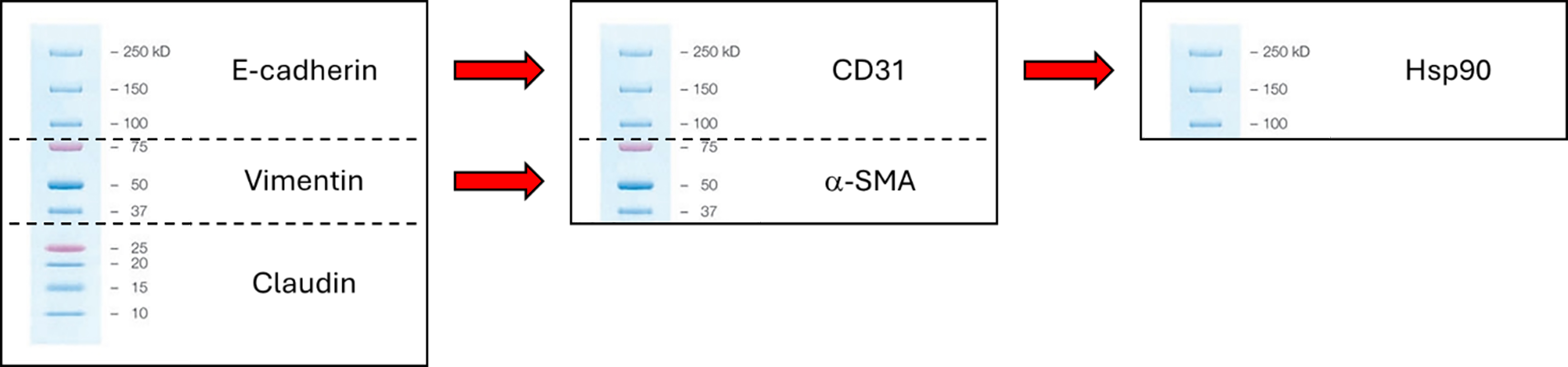
Figure 2. Experimental design of the WB analysis. Schematic overview of the experimental workflow used to assess protein expression levels by Western blot.
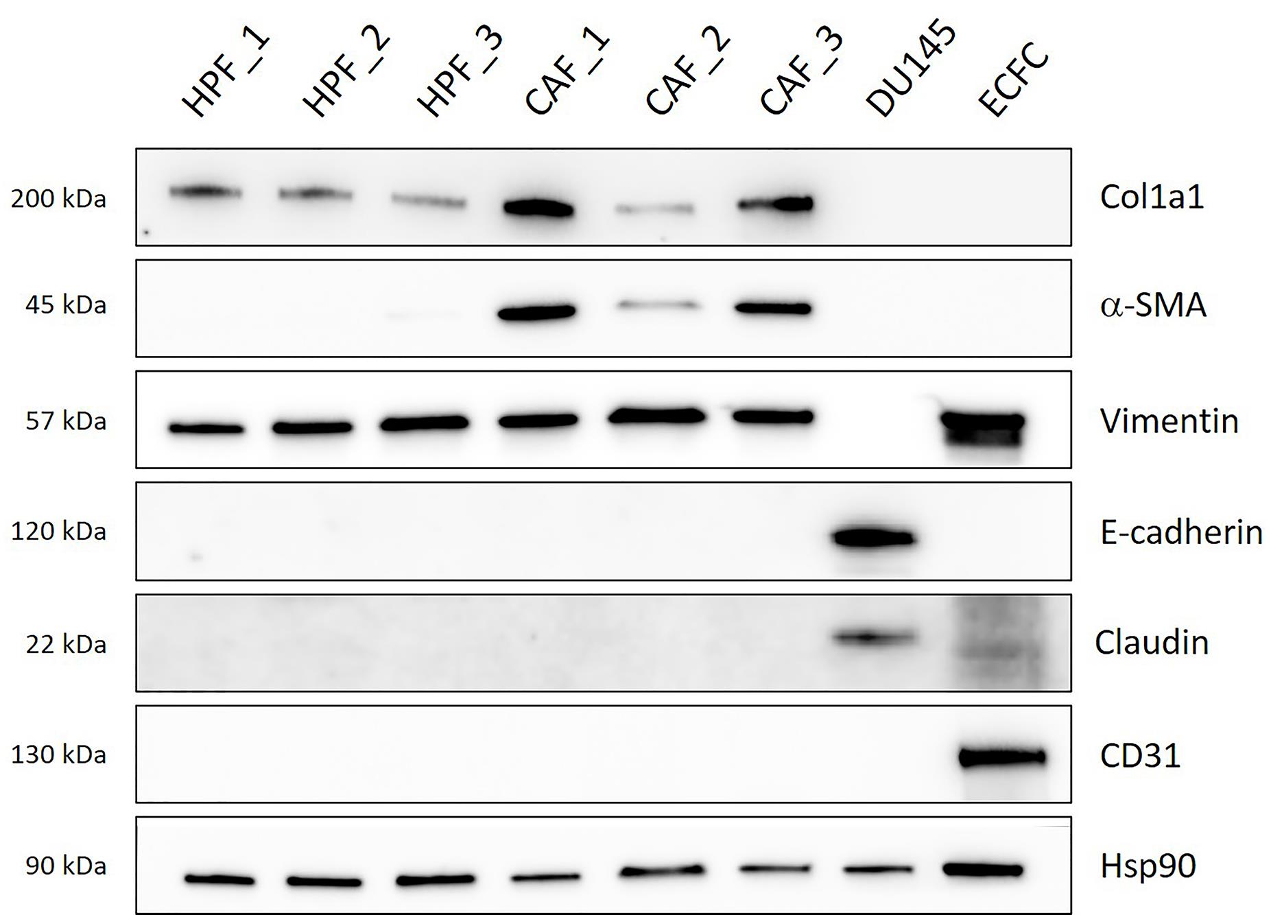
Figure 3. Western blot analysis of representative cell markers. The results indicate that collagen type I (col1a1) and vimentin are expressed in both healthy and cancer-associated fibroblasts (HPFs and CAFs), confirming their mesenchymal identity. Conversely, α-smooth muscle actin (α-SMA) is detected exclusively in CAFs, reflecting their activation compared to the HPFs. As expected, CD31 is exclusively expressed in endothelial cells (ECFCs), while E-cadherin and claudin are only detected in epithelial cells (DU145), in accordance with their role in epithelial junctions. HSP90 was used as a loading control to ensure equal protein content across samples. HPF and CAF were derived from three different explants. Note that α-SMA expression differs among the three CAF samples, reflecting inter-patient biological variability.
B. Immunofluorescence analysis
To confirm CAF activation, immunofluorescence analysis of α-SMA can be performed to highlight stress fibers associated with CAF activation (Figure 4). Perform immunofluorescence analysis as follows:
1. Seed HPF and CAF onto glass coverslips at 1–2 × 10⁵ cells per well of a 6-well plate.
2. Incubate with 3 mL of starvation medium for 24 h.
3. Fix cells with 1 mL of 4% formaldehyde for 10 min.
4. Permeabilize cells with 0.1% Triton X-100 for 15 min.
5. Block nonspecific binding using PBS-based buffer containing 2% horse serum and 5% BSA for 1 h at room temperature.
6. Incubate cells with primary antibodies overnight at 4 °C.
7. Wash cells 3 × 5 min with PBST (PBS+ 0.1% Tween 20).
8. Incubate with Alexa Fluor conjugate secondary antibodies (1:1,000) for 1 h at room temperature.
9. Counterstain cells with DAPI (300 ng/mL final concentration) for 10 min at room temperature.
10. Wash coverslips 2 × 30 s with PBS.
11. Rinse in ddH2O.
12. Mount samples for imaging using ProLong Glass antifade mountant.
13. For long-term storage, store samples at 4 °C protected from light.
14. Capture images using the TCS SP8 confocal microscope with LAS-AF software.
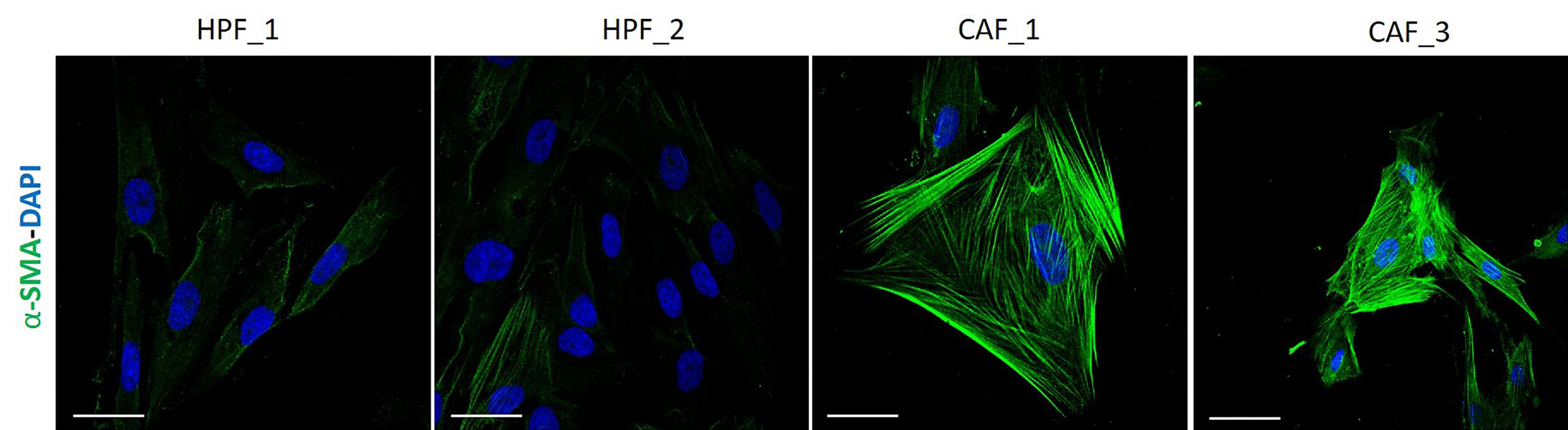
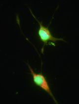
Figure 4. Immunofluorescence analysis of alpha-smooth muscle actin (α-SMA). Healthy and cancer-associated fibroblasts (HPFs and CAFs) were stained for α-SMA (green) and for nuclei (DAPI, blue). Representative confocal microscopy images reveal both enhanced and spatially distributed green fluorescence in CAFs, as a readout of enhanced α-SMA-fiber formation compared to the HPF sample. Scale bar, 100 μm.
Validation of protocol
This protocol has been used and validated in the following research articles:
Comito et al. [11]. Lactate modulates CD4+ T-cell polarization and induces an immunosuppressive environment, which sustains prostate carcinoma progression via TLR8/miR21 axis. Oncogene (Figures 1–5).
Ippolito et al. [14]. Lactate Rewires Lipid Metabolism and Sustains a Metabolic–Epigenetic Axis in Prostate Cancer. Cancer Research (Figures 2–6).
Prostate tumor cells and CAFs have been shown to establish a metabolic interplay, resulting in a CAF-enhanced glycolytic metabolism as well as lactate release [15]. Accordingly, we assessed a higher CAF expression of two key markers in lactate production and extrusion, namely lactate dehydrogenase A (LDHA) and monocarboxylate transporter 4 (MCT4), thereby validating the lactate metabolism as a specific feature of CAF compared to normal fibroblasts (Figure 5).
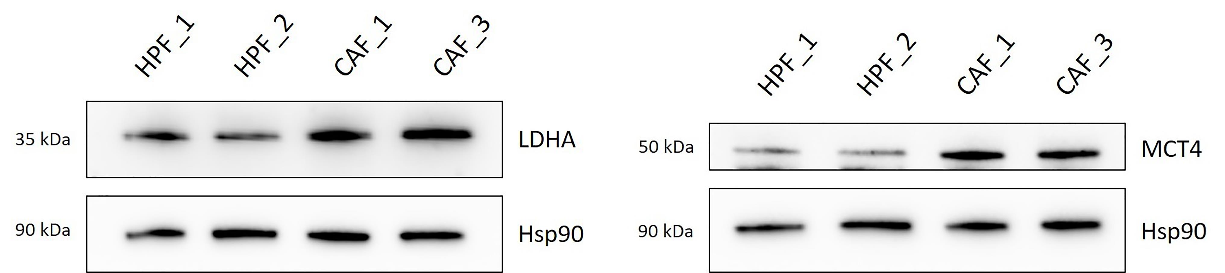
Figure 5. Validation of key lactate metabolism markers in cancer-associated fibroblasts (CAFs). Representative western blot images of MCT4 and LDHA expression on selected HPFs (healthy prostate fibroblasts) and CAFs (n = 2 explants). Hsp90 was used as the loading control. CAFs display higher levels of LDH-A and MCT4 compared to the healthy counterparts.
General notes and troubleshooting
General notes
1. Work in aseptic conditions at all times, use only sterile materials, and prepare complete medium with an appropriate antibiotic concentration (see Recipes) to prevent contamination.
2. Cutting tissue samples into small fragments with uniform borders promotes CAF migration.
3. This protocol is optimized for the isolation of CAFs from patients with prostate cancer.
4. Cells between passages 2 and 8 are recommended for experimental use. Also, consider the low CAF replicative timing (36–72 h) before designing an experimental plan.
Troubleshooting
Problem 1: Absence of CAFs outgrowth.
Possible causes: Small-sized or fibrotic biopsy, tissue degradation, unknown patient-specific factors.
Solution: Collect additional tissue samples of appropriate size to improve the sample size and process within 1–2 h to prevent degradation.
Problem 2: Isolation of low-activated CAFs (see Figure 2, CAF_2).
Possible causes: Inter-patient biological variability and heterogeneity of the CAF population.
Solutions: Collect samples preferably from high Gleason Group patient cohorts; increase the sample size of tumor explants.
Problem 3: Low viability after thawing.
Possible cause: Temperature scaling.
Solution: Backup of several samples.
Acknowledgments
This study was funded by the Italian Ministry of University and Research within the PRIN2022 PNRR and PRIN2022 programs (Progetti di Ricerca di Rilevante Interesse Nazionale 2022) in the framework of the National Recovery and Resilience Plan (PNRR), Mission 4—Component 2—Investment 1.1 “Research Projects of Relevant National Interest (PRIN),” funded by the European Union – NextGenerationEU – Project Code P2022CE7SP – CUP B53D23033040001 and Project Code 2022F5JLSE – CUP B53D23021510006 (to EG).
Data presented in this study were partially generated using grants from Associazione Italiana Ricerca sul Cancro (AIRC, grant IG 24731 to PC); European Union, National Recovery and Resilience Plan (PNRR), Mission 4 Component 2 – Investment 1.4 – National Center (NC) for Gene Therapy and Drugs based on RNA technology – NextGenerationEU – Project Code CN00000041 – CUP B93D21010860004 (to EG) and creation and strengthening of “Innovative ecosystems,” construction of “territorial R&D leaders”—TUSCANY HEALTH ECOSYSTEM (THE) NextGenerationEU—Project code ECS_00000017—CUP B83C22003920001 (to PC).
The following figures were created using BioRender: Graphical overview, BioRender.com/xhttps://biorender.com/e475idr.
Competing interests
The authors declare no conflict of interest.
Ethical considerations
Human prostate fibroblasts (HPFs and CAFs) were isolated from surgical explants after patient informed consent, according to the Ethics Committee (Comitato Etico Regionale per la Sperimentazione Clinica della Regione Toscana, Area Vasta Centro, CEAVC-2018-256).
References
- Rebello, R. J., Oing, C., Knudsen, K. E., Loeb, S., Johnson, D. C., Reiter, R. E., Gillessen, S., Van der Kwast, T. and Bristow, R.G. (2021). Prostate cancer. Nat Rev Dis Primers. 7(1): 9. https://doi.org/10.1038/s41572-020-00243-0
- Pederzoli, F., Raffo, M., Pakula, H., Ravera, F., Nuzzo, P. V. and Loda, M. (2022). “Stromal cells in prostate cancer pathobiology: friends or foes?”. Br J Cancer. 128(6): 930–939. https://doi.org/10.1038/s41416-022-02085-x
- Tuxhorn, J. A., Ayala, G. E. and Rowley, D. R. (2001). Reactive stroma in human prostate cancer: induction of myofibroblast phenotype and extracellular matrix remodeling. J Urol. 2472–2483. https://doi.org/10.1097/00005392-200112000-00126
- Sahai, E., Astsaturov, I., Cukierman, E., DeNardo, D. G., Egeblad, M., Evans, R. M., Fearon, D., Greten, F. R., Hingorani, S. R., Hunter, T., et al. (2020). A framework for advancing our understanding of cancer-associated fibroblasts. Nat Rev Cancer. 20(3): 174–186. https://doi.org/10.1038/s41568-019-0238-1
- Rantanen, F., Murumägi, A., Arjama, M., Välimäki, K., Multamäki, E., Mirtti, T., Rannikko, A., Pellinen, T., Ungureanu, D., Kallioniemi, O., et al. (2025). Molecular profiling of ex vivo prostate cancer CAF models captures stromal heterogeneity and drug vulnerabilities. Cell Death Discovery. 11(1): e1038/s41420–025–02792–3. https://doi.org/10.1038/s41420-025-02792-3
- Lavie, D., Ben-Shmuel, A., Erez, N. and Scherz-Shouval, R. (2022). Cancer-associated fibroblasts in the single-cell era. Nat Cancer. 3(7): 793–807. https://doi.org/10.1038/s43018-022-00411-z
- Yang, D., Liu, J., Qian, H. and Zhuang, Q. (2023). Cancer-associated fibroblasts: from basic science to anticancer therapy. Exp Mol Med. 55(7): 1322–1332. https://doi.org/10.1038/s12276-023-01013-0
- Zhang, D., Wang, Y., Shi, Z., Liu, J., Sun, P., Hou, X., Zhang, J., Zhao, S., Zhou, B. P., Mi, J., et al. (2015). Metabolic Reprogramming of Cancer-Associated Fibroblasts by IDH3α Downregulation. Cell Rep. 10(8): 1335–1348. https://doi.org/10.1016/j.celrep.2015.02.006
- Giannoni, E., Bianchini, F., Masieri, L., Serni, S., Torre, E., Calorini, L. and Chiarugi, P. (2010). Reciprocal Activation of Prostate Cancer Cells and Cancer-Associated Fibroblasts Stimulates Epithelial-Mesenchymal Transition and Cancer Stemness. Cancer Res. 70(17): 6945–6956. https://doi.org/10.1158/0008-5472.can-10-0785
- Pardella, E., Comito, G., Ippolito, L., Pranzini, E., Iozzo, M., Gangarossa, G., Virgilio, F., Bua, S., Nocentini, A., et al. (2025). Targeting Carbonic Anhydrase IX/XII Prevents the Anti-ferroptotic Effect of Stromal Lactic Acid in Prostate Carcinoma. Mol Oncol. 19 (9): 2515–2536. https://doi.org/10.1002/1878-0261.70083
- Comito, G., Iscaro, A., Bacci, M., Morandi, A., Ippolito, L., Parri, M., Montagnani, I., Raspollini, M. R., Serni, S., Simeoni, L., et al. (2019). Lactate modulates CD4+ T-cell polarization and induces an immunosuppressive environment, which sustains prostate carcinoma progression via TLR8/miR21 axis. Oncogene. 38(19): 3681–3695. https://doi.org/10.1038/s41388-019-0688-7
- Sharon, Y., Alon, L., Glanz, S., Servais, C. and Erez, N. (2013). Isolation of Normal and Cancer-associated Fibroblasts from Fresh Tissues by Fluorescence Activated Cell Sorting (FACS). J Visualized Exp. e3791/4425–v. https://doi.org/10.3791/4425-v
- Mun, K., Han, J., Roh, P., Park, J., Kim, G., Hur, W., Jang, J., Choi, J., Yoon, S., You, Y., et al. (2023). Isolation and characterization of cancer-associated fibroblasts in the tumor microenvironment of hepatocellular carcinoma. J Liver Cancer. 23(2): 341–349. https://doi.org/10.17998/jlc.2023.04.30
- Ippolito, L., Comito, G., Parri, M., Iozzo, M., Duatti, A., Virgilio, F., Lorito, N., Bacci, M., Pardella, E., Sandrini, G., et al. (2022). Lactate Rewires Lipid Metabolism and Sustains a Metabolic–Epigenetic Axis in Prostate Cancer. Cancer Res. 82(7): 1267–1282. https://doi.org/10.1158/0008-5472.can-21-0914
- Fiaschi, T., Marini, A., Giannoni, E., Taddei, M. L., Gandellini, P., De Donatis, A., Lanciotti, M., Serni, S., Cirri, P., Chiarugi, P., et al. (2012). Reciprocal Metabolic Reprogramming through Lactate Shuttle Coordinately Influences Tumor-Stroma Interplay. Cancer Res. 72(19): 5130–5140. https://doi.org/10.1158/0008-5472.can-12-1949
Article Information
Publication history
Received: Dec 1, 2025
Accepted: Jan 22, 2026
Available online: Feb 6, 2026
Published: Mar 5, 2026
Copyright
© 2026 The Author(s); This is an open access article under the CC BY-NC license (https://creativecommons.org/licenses/by-nc/4.0/).
How to cite
Gangarossa, G., Grillo, C., Roccabianca, S., Pranzini, E., Iozzo, M., Venditti, G., Bertoli, G., Ippolito, L., Giannoni, E., Comito, G. and Chiarugi, P. (2026). Non-Enzymatic Isolation of Cancer-Associated Fibroblasts From Human Prostate Tumor Explants. Bio-protocol 16(5): e5614. DOI: 10.21769/BioProtoc.5614.
Category
Cancer Biology > General technique > Tumor microenvironment
Cell Biology > Cell isolation and culture > Cell isolation
Do you have any questions about this protocol?
Post your question to gather feedback from the community. We will also invite the authors of this article to respond.