- Protocols

- Articles and Issues
- For Authors
- About
- Become a Reviewer

Novel Experimental Approach to Investigate Immune Control of Vascular Function: Co-culture of Murine Aortas With T Lymphocytes or Macrophages
Published: Vol 15, Iss 17, Sep 5, 2025 DOI: 10.21769/BioProtoc.5440 Views: 3547
Reviewed by: Andrea GramaticaAnonymous reviewer(s)
Original research article
The authors used this protocol in:
Feb 2025

Protocol Collections
Comprehensive collections of detailed, peer-reviewed protocols focusing on specific topics


Related protocols
Abstract
Cardiovascular disease, the current leading cause of death worldwide, is a multifactorial disorder that involves a strong contribution of both the innate and adaptive immune systems. Overactivation of the immune system and inappropriate secretion of pro-inflammatory cytokines lead to vascular impairments and the development of cardiovascular disorders, including hypertension, atherosclerosis, and peripheral artery disease. Lymphocytes, macrophages, and dendritic cells can all secrete pro-inflammatory cytokines. This makes it challenging to isolate a specific subset of immune cells, particularly cytokines, and their contribution to vascular dysfunction remains difficult to elucidate. To solve this problem, our laboratory has developed the novel “immune cell-aorta” co-culture system described herein. This experimental protocol enables investigators to isolate an immune cell of interest and identify the cytokine(s) at the origin of vascular alterations.
Key features
• Novel ex vivo approach combining the culture of one population of immune cells with blood vessels.
• No direct contact between the cells and the blood vessels; the model enables studying the role of immune cell–derived factors or cytokines on vascular function.
• Blood vessels can subsequently be used for functional (wire/pressure myography), molecular (western blot, quantitative real-time RT-PCR), and histological studies.
Keywords: Co-cultureGraphical overview
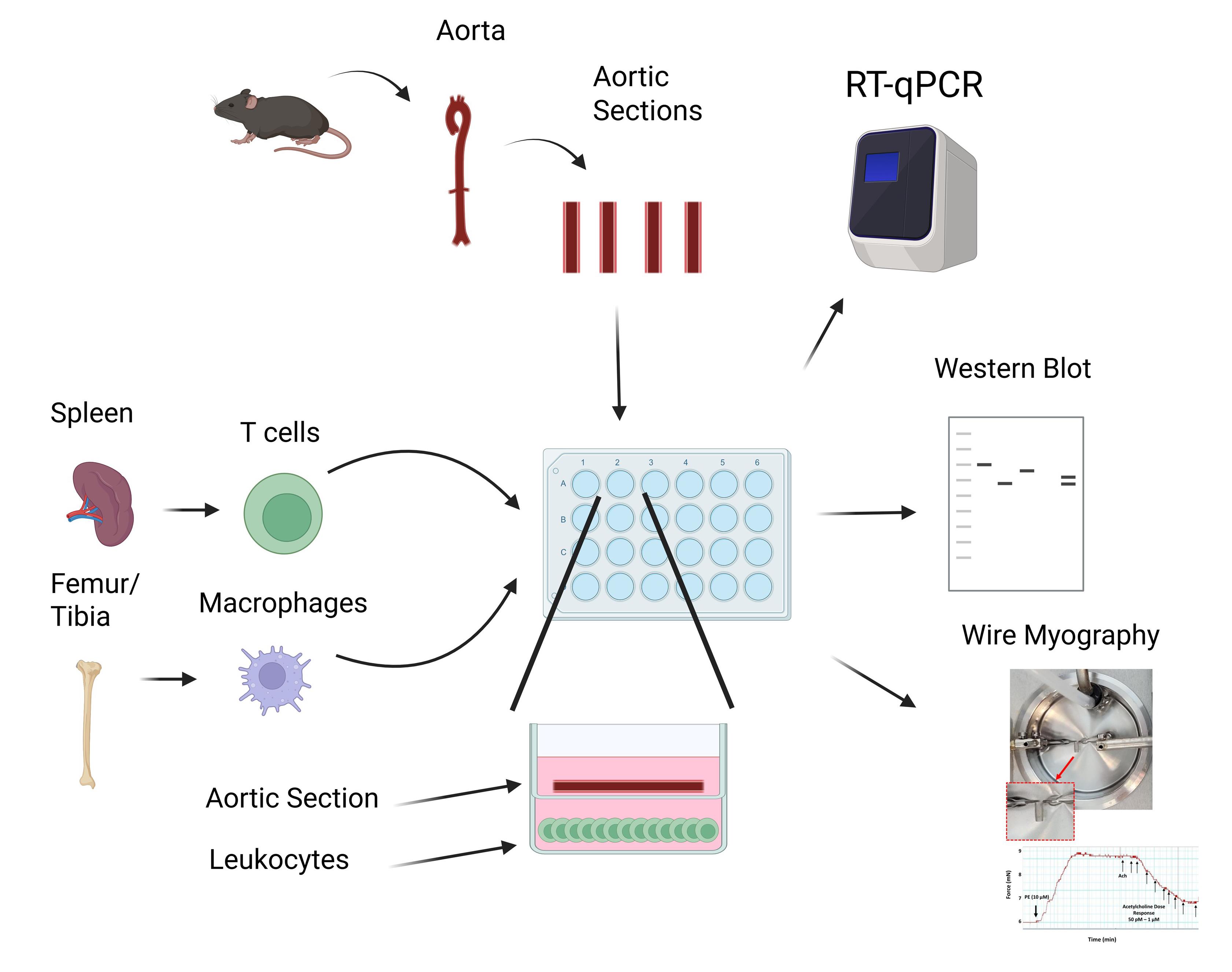 T cells and macrophages isolated from murine spleens and bone marrow, respectively, are cultured and plated in a 24-well plate. A murine aorta is excised, cleaned, sectioned, and placed into a transwell assay with 200 µL of fresh media. After overnight incubation, vessels may be extracted and cells and vessels analyzed via RT-qPCR, western blot, or wire myography.
Background
Cardiovascular disease (CVD), including heart failure, atherosclerotic diseases, and stroke [1], is the primary cause of death worldwide and is currently on the rise [2]. Its etiopathology is complex and multifactorial, and involves, among other mechanisms, an overactivation of the immune system [3]. Hypertension, the leading modifiable cardiovascular disease risk factor, as well as atherosclerosis and other cardiovascular disorders, have all been shown to have a strong immune component and involve the contribution of both the innate [4] and adaptive immune systems [3]. Compelling evidence from both human and rodent studies highlights roles for monocytes [5], macrophages [6], dendritic cells [7], and T lymphocytes (T cells) [8]. Notably, excessive production of pro-inflammatory cytokines by the latter immune cells has been shown to cause vascular inflammation and dysfunction [9–11], both precursors of hypertension and CVD. Pro-inflammatory cytokines can act directly on blood vessels to increase their contractile phenotype [12] and vascular oxidative stress [13], and to promote endothelial nitric oxide synthase (eNOS) uncoupling [14]. However, identifying the type of immune cells producing the pro-inflammatory cytokines and the cytokine involved in the vascular alterations remains challenging; hence, the development of the protocol described here.
Macrophages can generally be split into two groups, M1 and M2 macrophages [15]. M1 macrophages, which have more of a pro-inflammatory phenotype, produce TNFα, IL-1β, IL-6, and IL-8, which contribute to endothelial dysfunction [16]. Conversely, M2 macrophages are seen as cardioprotective [17]. In our protocol, bone marrow–derived macrophages were grown and differentiated from hematopoietic stem cells using macrophage colony-stimulating factor. Macrophages can further be differentiated into the M1 subtype via the addition of lipopolysaccharide (LPS) and IFN-γ, whereas the addition of IL-4 will differentiate macrophages into the M2 subtype [18].
With respect to T cells, the two main subtypes are cytotoxic CD8+ T cells and helper CD4+ T cells [19]. The cytotoxic CD8+ T cells directly interact with and destroy infected cells via the production of pro-inflammatory cytokines such as IFN-γ and TNFα [20,21], which contribute to endothelial dysfunction via eNOS uncoupling [22] and increasing oxidative stress [23,24]. Meanwhile, CD4+ T helper cells, as opposed to CD8+ T cells, work to modulate the immune response as opposed to directly killing infected cells [25]. Naïve CD4+ T cells undergo differentiation into effector CD4+ T cells, including those with pro-inflammatory phenotypes such as Th1 [26] and Th17 [27,28], along with the anti-inflammatory T regulatory cells (Tregs) [28]. The pro-inflammatory effects of Th1 and Th17 are modulated via IFNγ, TNFα, and IL-17A [29,30]. Like IFNγ and TNFα, IL17A has also been linked to endothelial dysfunction, vascular inflammation, and hypertension [31]. Conversely, IL-10 produced by Tregs reduces inflammation and has endothelial-protective activity [32]. In addition, Tregs are increased in a female rat model of hypertension and decrease blood pressure, which demonstrates their effect on blood pressure as well as sex-specific effects [33].
Taken together, T cells and macrophages can secrete pro- and anti-inflammatory cytokines, which induce endothelial dysfunction through inflammation, downregulation, or uncoupling of eNOS, and increased oxidative stress. Our novel co-culture system provides a novel approach to investigate how cytokines produced by individual macrophage or T-cell subsets affect blood vessels, which was not previously possible. In addition to looking at vessel function, the cytokine-containing media can be analyzed to identify and quantify the cytokines present via Multiplex and ELISA. Additionally, analysis of the immune cells themselves, via flow cytometry, RT-qPCR, and western blotting, can provide more evidence of altered cytokine production.
Materials and reagents
Biological materials
1. Murine spleen collected from male and female 8–12-week-old mice for isolation of CD3+, CD4+, or CD8+ T cells
2. Murine bone marrow isolated from male and female 8–12-week-old mice for differentiation of bone marrow–derived hematopoietic stem cells into macrophages
3. Male and female murine thoracic and abdominal aortas isolated from 8–12-week-old mice for co-culture (aortas are isolated 72 h after T-cell isolation or macrophage terminal differentiation)
Reagents
1. Phosphate-buffered saline (PBS) (Fisher, catalog number: SH30256LS)
2. Bovine serum albumin (BSA) (Millipore Sigma, catalog number: 3117332001)
3. 2 mM EDTA (Millipore Sigma, catalog number: E9884-500G)
4. MACS® BSA stock solution (Miltenyi, catalog number: 130-091-376)
5. MACS® rinsing solution (Miltenyi, catalog number: 130-091-222)
6. RPMI 1640 (Fisher, catalog number: 72-400-047)
7. FBS (R&D Systems, catalog number: S11550)
8. Pen/Strep (Fisher, catalog number: SV30010)
9. TexMACS medium, 500 mL (Miltenyi, catalog number: 130-097-196)
10. Mouse IL-2 IS premium-grade TexMACS (Miltenyi, catalog number: 130-120-332)
11. Mouse M-CSF (Miltenyi, catalog number: 130-094-129)
12. 2-Mercaptoethanol (BME) (Fisher, catalog number: BP176-100)
13. Penicillin-Streptomycin (Pen/Strep) (Fisher, catalog number: SV30010)
14. Amphotericin (Fisher, catalog number: MT30003CF)
Solutions
1. T-cell isolation buffer (see Recipes)
2. T-cell culture media (see Recipes)
3. Naïve CD3+ T-cell separation media (see Recipes)
4. Macrophage differentiation/culture media (see Recipes)
Recipes
1. T-cell isolation buffer
| Reagent | Final concentration | Quantity or volume |
|---|---|---|
| Sterile PBS | n/a | 20 mL |
| BSA | 0.5% | 10 mg |
| EDTA | 2 mM | 11.7 mg |
| Total | n/a | 20 mL |
Prepare a solution containing PBS, pH 7.2, 0.5% BSA, and 2 mM EDTA by diluting MACS® BSA stock solution 1:20 with autoMACS® rinsing solution.
2. T-cell culture media
| Reagent | Final concentration | Quantity or volume |
|---|---|---|
| RPMI 1640 media | n/a | 17 mL |
| FBS | 10% | 2 mL |
| Pen/Strep | 5% | 1 mL |
| Total | n/a | 20 mL |
3. Naïve CD3+ T-cell separation media
Prepare T-cell separation media by mixing 500 mL of TexMACS medium with mouse IL-2 IS premium-grade TexMACS, plus 10% FBS, 0.01 mM BME, 1:100 Pen/Strep, and 1:100 Amphotericin (add 50 U/mL of IL-2 to the media used for culture only right before use).
BME is prepared by diluting to 1:1,000. 70 μL of BME is added to each 100 mL of media.
IL-2 is prepared by dissolving it in 500 μL of PBS (stock 500 U/μL); 1 μL of diluted solution is added per 10 mL of media.
4. Macrophage differentiation/culture media
| Reagent | Final concentration | Quantity or volume |
|---|---|---|
| RPMI 1640 media | N/A | 17 mL |
| FBS | 10% | 2 mL |
| Pen/Strep | 5% | 1 mL |
| M-CSF | 20 ng/mL | 20 μL |
| Total | n/a | 20 mL |
Laboratory supplies
A. T-cell isolation
1. 1.5 mL Eppendorf tube (one per mouse) (Fisher, catalog number: 21-402-903)
2. 50 mL conical tubes (one per mouse) (Fisher, catalog number: 14-955-239)
3. 40 μM cell strainers (one per mouse) (Fisher, catalog number: 223-635-47)
4. 10 mL serological pipette (Fisher, catalog number: 12-567-603)
5. Plunger of a 1 mL syringe (one per mouse) (Fisher, catalog number: 14-817-179)
6. Pan T-Cell Isolation kit II (CD3+ T cells) (Miltenyi Biotech, catalog number: 130-095-130), CD4+ T-Cell Isolation kit (Miltenyi Biotech, catalog number: 130-104-454), CD8a+ T-Cell Isolation kit (Miltenyi Biotech, catalog number: 130-104-075)
7. LS columns (one per mouse) (Miltenyi Biotech, catalog number: 130-042-401)
8. Magnetic separator (Miltenyi, catalog number: 130-042-302; MidiMACS™ Separator on MACS® MultiStand, catalog number: 130-042-303)
9. 24-well plate with 0.4 μm polyester transwell inserts (Costar, catalog number: 3470)
B. Macrophage isolation and differentiation
1. 5 mL Eppendorf tube (one per mouse) (Fisher, catalog number: 21-402-903)
2. 100 μM cell strainers (one per mouse) (Fisher, catalog number: 22-363-549)
3. 50 mL conical tubes (one per mouse) (Fisher, catalog number: 14-955-239)
4. Plunger of a 1 mL syringe (one per mouse) (Fisher, catalog number: 14-817-179)
5. 10 mL syringe (Fisher, catalog number: 14-955-459)
6. 27-gauge ½” hypodermic needles (one per mouse) (Fisher, catalog number: 14-840-82)
7. 150 mm untreated culture plate (Thermofisher, catalog number: 150468)
Equipment
1. Dissection tools (micro scissors, scissors, forceps) (Fine Science Tools, catalog numbers: 15023-10, 14061-10, 14001-14, 11252-20, and 11274-20)
2. PipetBoy (BrandTech BRAND accu-jet pro Fisher Scientific, catalog number: 13-688-567)
3. Pasteur pipette
4. Refrigerated centrifuge for splenocytes isolation (Thermo Sorvall, model: ST16R with rotor, model: 73003181)
5. Refrigerated centrifuge for T-cell centrifugation (Labnet Prism R Refrigerated Microcentrifuge, model: C2500-R)
5. Water bath (Fisherbrand, model: Isotemp SWB15)
6. Cell culture incubator (37 °C, 5% CO2) (Heracell 150i)
7. Cell culture hood (Thermo Scientific, model: 1300 Series A2)
8. Dissection microscope (Olympus, model: SZ61)
9. Cell counter (Life Technologies Countess II)
Software and datasets
1. Prism v10 (GraphPad)
2. Microsoft Excel v16.90.2
3. BioRender (https://www.biorender.com)
Procedure
A. Mouse euthanasia
1. Take 8–12-week-old mice of the desired strain and anesthetize them.
B. CD3+, CD4+, or CD8+ T-cell isolation
1. Once euthanized, bring the mouse under a biosafety cabinet, spray the whole animal with 70% ethanol, and, using sterile tools, excise the murine spleen as quickly as possible, place it in a 1.5 mL Eppendorf tube filled with cold sterile PBS, and place it on ice.
2. Place the 40 μm cell strainer over the 50 mL conical tube under sterile conditions in a cell culture hood.
3. Remove the spleen from cold, sterile PBS and transfer it to a cell strainer. Use the back of the plunger of a 1 mL syringe to break up the spleen against the filter. Wash the filter with cold, sterile PBS and transfer the splenocytes into the conical tube (Figure 1).
4. Spin down the splenocytes at 1,500 rpm for 10 min (300× g) in 50 mL conical tubes, discard the supernatant, and resuspend the splenocytes in isolation buffer.
5. Follow Miltenyi’s manufacturer instructions to isolate T cells. The Miltenyi isolation protocol is done via negative selection to prevent erroneous T-cell activation. If using a different kit, be sure they also isolate via negative separation (Figure 2).
6. Spin down T cells at 1,500 rpm for 10 min (300× g) and resuspend in 10 mL of cold, sterile PBS. Count cells.
7. Spin down T cells at 1,500 rpm for 10 min (300× g) and resuspend in T-cell culture media at a concentration of 12,000,000 cells/mL.
8. Pipette 1 mL of cell suspension (12,000,000 cells) into each well of the 24-well plate.
Note: Do not leave the transwell insert in.
9. Incubate T cells for 72 h in a cell culture incubator (at 37 °C and 5% CO2).
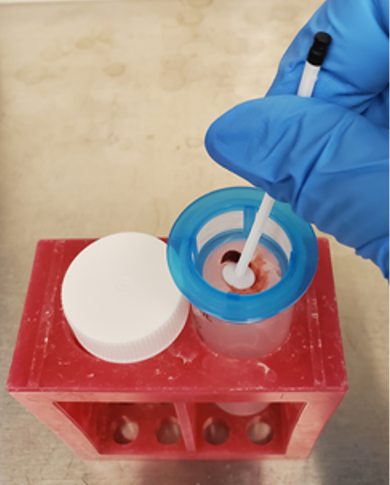
Figure 1. Visualization of spleen pulverization. Place a 40 μM strainer over a 50 mL conical tube in a general tube holder under sterile conditions. Using the back of a 1 mL pipette, “squish” the spleen through the filter and continually wash with cold, sterile PBS to facilitate the transfer of all cells. When finished, the liquid in the conical tube will be a cloudy red, and a translucent “shell” of the spleen will be left on the filter. From this step, continue to centrifugation.
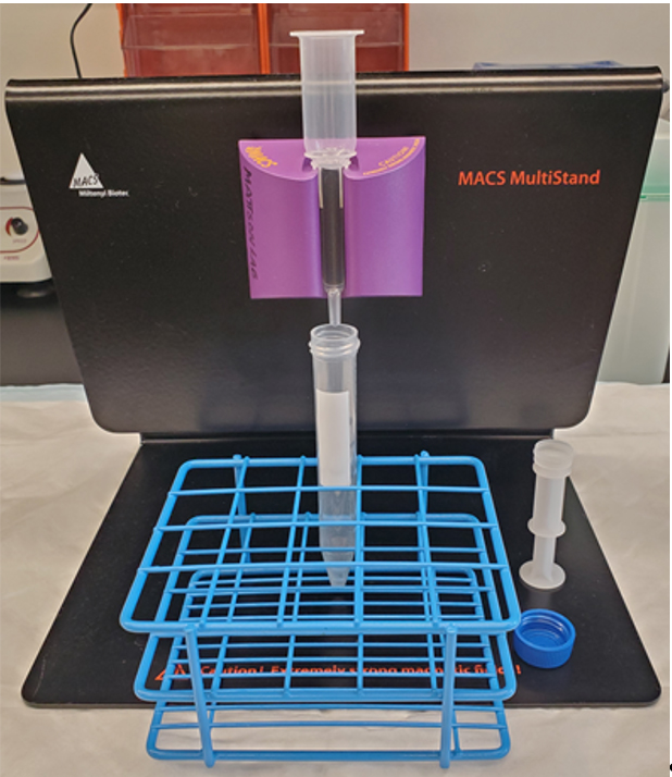
Figure 2. Setup of a Miltenyi magnetic separator. The setup of the magnetic separator involves a magnetic stand (black), a strong magnet tailored to the size of your magnetic column (purple), the LS magnetic column, and a collection tube as shown. It is possible to use the smaller MS column; however, the flow rate through the column will take longer and this will risk increased cell death. This should be performed under sterile conditions, and if desired, you may keep the collection tube on ice to increase the chances of cell survival.
C. Bone marrow–derived macrophages isolation and differentiation
1. After euthanasia, isolate the left and right tibia and femur and place them in cold sterile PBS. It is essential to ensure all tissue is removed from the bone. Letting them sit in PBS for 1 h will loosen the remaining adhering tissue (Figure 3).
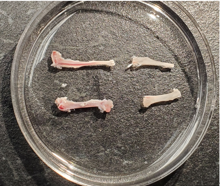
Figure 3. Visualization of bones before and after flushing of the bone marrow. After cleaning the bones of all tissue, the bone marrow will be visible within the bone as a crimson line, shown on the left. Once flushed, the crimson coloring will be cleared from the bone and will be a consistent, opaque color throughout, as seen on the right.
2. Set up 50 mL conical tubes with a 100 μM filter on top.
3. In the cell culture hood, under sterile conditions, carefully cut the ends of the tibia and femur and flush them using 10 mL of RPMI complete media with a 27G × 1/2” hypodermic needle into the 100 μm filter. The bone marrow will be visible within the bone as a maroon color, and once flushed, it will be opaque, like the rest of the bone.
4. Similar to the spleen, use the back of the plunger of a 1 mL syringe to push the bone marrow through the filter, and wash the filter with 10 mL of RPMI complete media.
5. Centrifuge cells at 1,500 rpm for 10 min (300× g), resuspend in 10 mL of RPMI complete media, and plate in a 150 mm untreated culture dish.
6. Once plated, induce differentiation by adding 20 ng/mL of M-CSF every other day for 6 days.
7. Once differentiation is complete, remove macrophages under sterile conditions using a Pasteur pipette and spin down at 1500 rpm for 10 min (300× g).
8. Count cells and resuspend to a concentration of 12,000,000 cells/mL in new, complete media. Plate 1 mL (12,000,000 cells) per well in a 24-well plate.
Note: Do not leave the transwell insert in.
9. Incubate in a cell culture incubator at 37 °C and 5% CO2 and treat every other day for 6 days. Proceed to use for co-culture.
D. Thoracic and abdominal aorta isolation
1. After euthanasia and decapitation, cut from the neck, through the ribs to the gonads. During this process, it is imperative to ensure you do not cut the heart, liver, or intestines, as doing so may damage the integrity of the vessel.
2. Cut the diaphragm carefully to avoid cutting the aorta. Cutting along the bottom rib should ensure this does not occur. From the top of the ribcage, carefully cut outward toward the left and right sides of the mouse to open up the ribcage and remove it to expose the thoracic cavity.
3. At this point, it is helpful to pin down the mouse. One dissection pin (or hypodermic needle) through each paw and four on either corner to pull the skin back is ideal (Figure 4A).
4. Carefully remove the lungs by grabbing each lobe, lifting, and removing as much as possible without stretching or harming the thoracic aorta. If this portion is difficult to do without cutting the aorta, you may skip this step and remove the lungs with the aorta and remove them under the dissection microscope.
5. Remove the liver, stomach, and intestines by gently lifting up the liver using forceps and carefully cutting down the midline of the animal. Looking down at the thoracic aorta while performing this step will help visualize where the aorta is and prevent damaging it. By the end of this step, you should have a clear view of the heart, thoracic aorta, and abdominal aorta (Figure 4B).
Note: It is essential to leave the kidneys, as removing them will damage the abdominal aorta.
6. Taking care not to stretch the thoracic or abdominal aorta, pull up on the heart and excise the aorta along with the perivascular adipose tissue (PVAT) by cutting between the aorta and spine down to the aortic bifurcation. If you find it difficult to remove without damaging the aorta, you can remove a portion of the spine as well as the aorta. This will ensure the entire aorta is excised undamaged (Figure 4C).
7. Once the aorta is removed with the heart, transfer to a dissection dish containing cold PBS. Using dissecting pins or hypodermic needles, elongate and pin down the aorta by fixing one in the heart and one either in the aorta bifurcation or the PVAT at the end of the abdominal aorta. During this step, take care not to stretch the aorta, as doing so will damage the vessel’s integrity.
8. Using a dissecting scope, scissors, and forceps, carefully remove the PVAT, kidneys, abdominal muscles, and spine that may be attached to the thoracic and abdominal aorta. Again, be careful not to stretch or cut the aorta during this process. (Figure 4D).
9. Once cleaned, cut the aorta above the renal bifurcation, separating the thoracic and abdominal aortas, and further cut the abdominal and thoracic aortas in half again, creating four equal-length aortic sections.
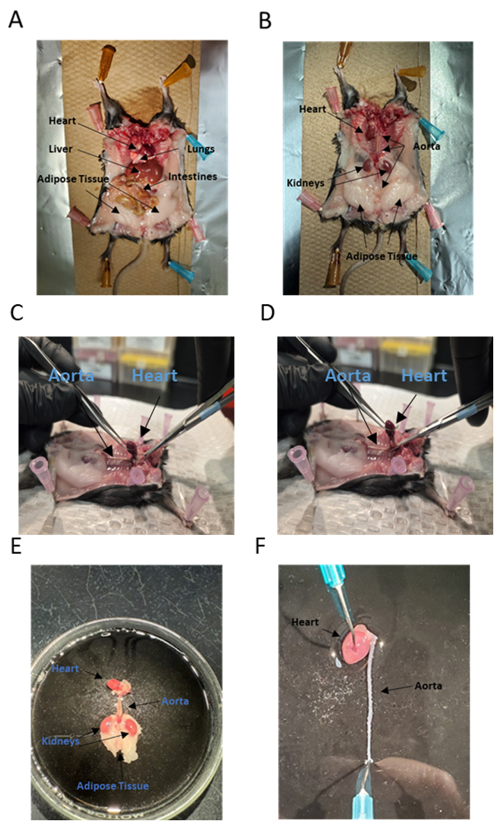
Figure 4. Isolation and cleaning of the thoracic and abdominal aorta. (A) Once the head is removed, cut from the neck, opening down to the gonads of the mouse, and open to a fully flayed position. (B) From this point, the internal organs (lungs, liver, spleen, intestines) may be removed. Do not remove the kidneys. (C, D) Grab the heart using forceps and carefully cut along the spine down to the aorta bifurcation, taking care not to cut the aorta in the process. (E) Once removed, you will have an isolated, unclean aorta. (F) Use fine-tipped forceps and micro scissors to carefully clean off the perivascular adipose tissue (PVAT), kidneys, and any abdominal muscle that is attached to the thoracic and abdominal aortas. Ideally, the aorta will naturally clear itself of blood during this step. If blood does coagulate within the aorta, cut it into fourths as described in step D9; then, you will be able to carefully remove it using fine-tipped forceps.
E. Aorta co-culture
1. Once the T cells/macrophages have been in culture for 72 h, place the filter insert and add 200 μL of fresh TexMACS media with 10% FBS, 0.01 mM 2-Mercaptoethanol, 1:100 Pen/Strep, 1:100 Amphotericin, and 50 U/mL Interleukin 2 into the top of the insert.
2. Place one half of the abdominal and one half of the thoracic aorta into individual wells corresponding to one condition, and the other halves into the individual control wells. If pressed for resources, you may combine the thoracic and abdominal aortas into one transwell of the same condition (Figure 5).
3. Allow the vessels to incubate overnight (16 h) and gently remove the vessels from the media with forceps.
4. Samples can be flash frozen for RT-qPCR or western blot.
5. We have used these vessels for myography and saw a slight decrease in the constrictive capacity of the vessel. When mounting on the myograph, remove the vessels from the transwell using fine-tip forceps, taking care to grab the very end of the vessel to prevent damage, and place them in warm physiological salt solution to transfer for mounting on the wire myograph. Take care not to use cold physiological salt solution, as it will shock the vessels and cause damage. A more in-depth explanation may be found in [34].
6. The media may be extracted via pipette and analyzed for cytokine concentration via ELISA.
7. The T cells or macrophages lying at the bottom of the plate may be removed by a cell scraper and analyzed via RT-qPCR or western blot.
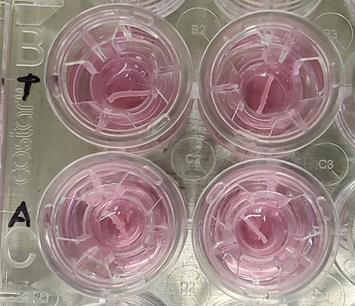
Figure 5. Co-culture of the thoracic and abdominal aorta. Once divided into four sections, place the transwell inserts into the 24-well plate. Add 200 µL of fresh media into the insert and place one-half of each aorta in each transwell. If pressed for resources, you may combine the thoracic and abdominal aortas from the same condition into one transwell.
Data analysis
One of the strengths of this technique is the ability to generate a lot of data from one experiment. In our hands, we have used this to:
1. RT-qPCR or western blot of the vessel and the immune cells themselves to look for upregulation of oxidative stress markers, endothelial dysfunction, and cytokine production, respectively.
2. Testing the vascular reactivity of the vessels via wire myography.
3. Analysis of cytokine concentration in the media via ELISA and multiplex assay.
Validation of protocol
The protocol has been used and validated in the following research article:
Kress et al. [34]. CD4+ T-cells Expressing Viral Proteins Induce HIV-Associated Endothelial Dysfunction and Hypertension Through Interleukin 1α-Mediated Increases in Endothelial NADPH Oxidase 1. Circulation.
General notes and troubleshooting
General notes
1. When isolating the spleen for T-cell isolation, place it immediately on ice. Furthermore, during the steps of T-cell isolation, keep the different steps on ice for as long as possible while completing the steps as fast as possible. Doing so will ensure the highest probability of T-cell survival. A refrigerated centrifuge is necessary.
2. Similar to T cells, when isolating macrophages, bones must be placed on ice for transport, and effort should be made to keep bone marrow on ice during straining. A refrigerated centrifuge is ideal.
3. While difficult, isolating the vessels should be done carefully so as not to cut them. Like T cells and macrophages, this must be done on ice if it takes longer than 15 min.
Troubleshooting
Problem 1: Low viability of T cells.
Possible causes: The isolation time was too long, or samples were not kept on ice.
Solution: In addition to keeping the spleen on ice, keeping the conical tubes on ice during pulverization and magnetic separation will result in the highest survival likelihood.
Problem 2: Difficulty with keeping the aorta intact.
Possible cause: Not being careful enough when removing the aorta and/or cleaning.
Solution: Try different forceps and microdissection scissors to find what you prefer. If you have not done this before, practice more until you are proficient.
Problem 3: Contamination of the culture dish.
Possible cause: Not performing the cell culture steps under sterile conditions.
Solution: Spray down the cell culture hood, equipment, and hands with ethanol when performing all steps in the cell culture hood.
Problem 4: Potential interferences with aorta resident immune cells.
Possible causes: Incomplete removal of aorta perivascular adipose tissue where immune cells reside, or use of aorta from mice with inflammatory disease.
Solution: Clean the aorta from surrounding perivascular adipose tissue and use the aorta from young healthy mice.
Acknowledgments
Conceptualization: TC Kress, EJ Belin de Chantemèle; Investigation: TC Kress, CT Barris, S Kennard, Writing—Original Draft: TC Kress, CT Barris, S Kennard, EJ Belin de Chantemèle; Writing—Review & Editing: TC Kress, CT Barris, S Kennard, EJ Belin de Chantemèle; Funding acquisition: EJ Belin de Chantemèle; Supervision: EJ Belin de Chantemèle.
This work was supported by grants NIH R01s (R01HL155265, R01AR082307, P01HL1605571, R01HL176323, and R01HL175471) to EJ Belin de Chantemele, AHA 21PRE830396 to TC Kress, and F31HL168963 to CT Barris.
The protocol was originally described and validated in: Circulation, DOI: 10.1161/CIRCULATIONAHA.124.070538. Graphical overview was created in BioRender. Belin de chantemele, E. (2025) https://BioRender.com/11kfw0l.
Competing interests
The authors have no competing interests.
Ethical considerations
All animal investigations were conducted in an American Association for the Accreditation of Laboratory Animal Care-accredited facility with studies approved by the Institutional Animal Care and Use Committee (Protocol #2011-0108). For euthanasia procedures, mice were first anesthetized in a closed chamber using 5% isoflurane (1 L/min O2) before decapitation.
References
- Bailin, S. S., Gabriel, C. L., Wanjalla, C. N. and Koethe, J. R. (2020). Obesity and Weight Gain in Persons with HIV. Curr HIV/AIDS Rep. 17(2): 138–150. https://doi.org/10.1007/s11904-020-00483-5
- Zheng, S., Hedl, M. and Abraham, C. (2015). Twist1 and Twist2 Contribute to Cytokine Downregulation following Chronic NOD2 Stimulation of Human Macrophages through the Coordinated Regulation of Transcriptional Repressors and Activators. J Immunol. 195(1): 217–226. https://doi.org/10.4049/jimmunol.1402808
- Martín, P. and Sánchez-Madrid, F. (2025). T-cells in cardiac health and disease. J Clin Invest. 135(2): e1172/jci185218. https://doi.org/10.1172/jci185218
- Chen, R., Zhang, H., Tang, B., Luo, Y., Yang, Y., Zhong, X., Chen, S., Xu, X., Huang, S., Liu, C., et al. (2024). Macrophages in cardiovascular diseases: molecular mechanisms and therapeutic targets. Signal Transduction Targeted Ther. 9(1): e1038/s41392–024–01840–1. https://doi.org/10.1038/s41392-024-01840-1
- Wenzel, P. (2018). Monocytes as immune targets in arterial hypertension. Br J Pharmacol. 176(12): 1966–1977. https://doi.org/10.1111/bph.14389
- Harwani, S. C. (2018). Macrophages under pressure: the role of macrophage polarization in hypertension. Translational Research 191: 45–63. https://doi.org/10.1016/j.trsl.2017.10.011
- Lu, X. and Crowley, S. D. (2022). Actions of Dendritic Cells in the Kidney during Hypertension. Compr Physiol. 12(4): 4087–4101. https://doi.org/10.1002/j.2040-4603.2022.tb00240.x
- Zhang, J. and Crowley, S. D. (2015). Role of T lymphocytes in hypertension. Curr Opin Pharmacol. 21: 14–19. https://doi.org/10.1016/j.coph.2014.12.003
- Mikolajczyk, T. P., Szczepaniak, P., Vidler, F., Maffia, P., Graham, G. J. and Guzik, T. J. (2021). Role of inflammatory chemokines in hypertension. Pharmacol Ther. 223: 107799. https://doi.org/10.1016/j.pharmthera.2020.107799
- Schwartz, D. M., Burma, A. M., Kitakule, M. M., Luo, Y. and Mehta, N. N. (2020). T-cells in Autoimmunity-Associated Cardiovascular Diseases. Front Immunol. 11: e588776. https://doi.org/10.3389/fimmu.2020.588776
- Chen, M., Li, X., Wang, S., Yu, L., Tang, J. and Zhou, S. (2020). The Role of Cardiac Macrophage and Cytokines on Ventricular Arrhythmias. Front Physiol. 11: e01113. https://doi.org/10.3389/fphys.2020.01113
- Vila, E. and Salaices, M. (2005). Cytokines and vascular reactivity in resistance arteries. Am J Physiol Heart Circ Physiol. 288(3): H1016–H1021. https://doi.org/10.1152/ajpheart.00779.2004
- Kofler S, Nickel T and Weis M. (2005). Role of cytokines in cardiovascular diseases: a focus on endothelial responses to inflammation. Clin Sci. 108(3): 205–213. https://doi.org/10.1042/cs20040174
- Theofilis, P., Sagris, M., Oikonomou, E., Antonopoulos, A. S., Siasos, G., Tsioufis, C. and Tousoulis, D. (2021). Inflammatory Mechanisms Contributing to Endothelial Dysfunction. Biomedicines. 9(7): 781. https://doi.org/10.3390/biomedicines9070781
- Yunna, C., Mengru, H., Lei, W. and Weidong, C. (2020). Macrophage M1/M2 polarization. Eur J Pharmacol. 877: 173090. https://doi.org/10.1016/j.ejphar.2020.173090
- Gui, Z., Zhang, X., Han, Q., Hang, Z., Tan, R., Gu, M. and Wang, Z. (2023). Macrophage polarization induces endothelium-to-myofibroblast transition in chronic allograft dysfunction. Ren Fail. 45(1): e2220418. https://doi.org/10.1080/0886022x.2023.2220418
- Fujiu, K., Wang, J. and Nagai, R. (2014). Cardioprotective function of cardiac macrophages. Cardiovasc Res. 102(2): 232–239. https://doi.org/10.1093/cvr/cvu059
- Orecchioni, M., Ghosheh, Y., Pramod, A. B. and Ley, K. (2020). Corrigendum: Macrophage Polarization: Different Gene Signatures in M1(LPS+) vs. Classically and M2(LPS–) vs. Alternatively Activated Macrophages. Front Immunol. 11: e00234. https://doi.org/10.3389/fimmu.2020.00234
- Steier, Z., Kim, E. J. Y., Aylard, D. A. and Robey, E. A. (2024). The CD4 Versus CD8 T-cell Fate Decision: A Multiomics-Informed Perspective. Annu Rev Immunol. 42(1): 235–258. https://doi.org/10.1146/annurev-immunol-083122-040929
- Uhl, L. F. K., Cai, H., Oram, S. L., Mahale, J. N., MacLean, A. J., Mazet, J. M., Piccirilli, T., He, A. J., Lau, D., Elliott, T., et al. (2023). Interferon-γ couples CD8+ T-cell avidity and differentiation during infection. Nat Commun. 14(1): e1038/s41467–023–42455–4. https://doi.org/10.1038/s41467-023-42455-4
- Sun, L., Su, Y., Jiao, A., Wang, X. and Zhang, B. (2023). T-cells in health and disease. Signal Transduction Targeted Ther. 8(1): 235. https://doi.org/10.1038/s41392-023-01471-y
- Łuczak, A., Madej, M., Kasprzyk, A. and Doroszko, A. (2020). Role of the eNOS Uncoupling and the Nitric Oxide Metabolic Pathway in the Pathogenesis of Autoimmune Rheumatic Diseases. Oxid Med Cell Longevity. 2020: 1–15. https://doi.org/10.1155/2020/1417981
- Chen, X., Andresen, B., Hill, M., Zhang, J., Booth, F. and Zhang, C. (2008). Role of Reactive Oxygen Species in Tumor Necrosis Factor-alpha Induced Endothelial Dysfunction. Curr Hypertens Rev. 4(4): 245–255. https://doi.org/10.2174/157340208786241336
- Hubackova, S., Kucerova, A., Michlits, G., Kyjacova, L., Reinis, M., Korolov, O., Bartek, J. and Hodny, Z. (2015). IFNγ induces oxidative stress, DNA damage and tumor cell senescence via TGFβ/SMAD signaling-dependent induction of Nox4 and suppression of ANT2. Oncogene. 35(10): 1236–1249. https://doi.org/10.1038/onc.2015.162
- Luckheeram, R. V., Zhou, R., Verma, A. D. and Xia, B. (2012). CD4+T-cells: Differentiation and Functions. Clinical and Developmental Immunology 2012: 1–12. https://doi.org/10.1155/2012/925135
- Romagnani, S. (2000). T-cell subsets (Th1 versus Th2). Ann Allergy Asthma Immunol. 85(1): 9–21. https://doi.org/10.1016/s1081-1206(10)62426-x
- Mitsdoerffer, M., Lee, Y., Jäger, A., Kim, H. J., Korn, T., Kolls, J. K., Cantor, H., Bettelli, E. and Kuchroo, V. K. (2010). Proinflammatory T helper type 17 cells are effective B-cell helpers. Proc Natl Acad Sci USA. 107(32): 14292–14297. https://doi.org/10.1073/pnas.1009234107
- Lei, H., Schmidt-Bleek, K., Dienelt, A., Reinke, P. and Volk, H. D. (2015). Regulatory T-cell-mediated anti-inflammatory effects promote successful tissue repair in both indirect and direct manners. Front Pharmacol. 6: e00184. https://doi.org/10.3389/fphar.2015.00184
- Gallego, A., Vargas, J. A., Castejón, R., Citores, M. J., Romero, Y., Millán, I. and Durántez, A. (2003). Production of intracellular IL‐2, TNF‐α, and IFN‐γ by T-cells in B‐CLL. Cytometry Part B: Clinical Cytometry. 56: 23–29. https://doi.org/10.1002/cyto.b.10052
- Korn, T., Bettelli, E., Oukka, M. and Kuchroo, V. K. (2009). IL-17 and Th17 Cells. Annu Rev Immunol. 27(1): 485–517. https://doi.org/10.1146/annurev.immunol.021908.132710
- Karbach, S., Croxford, A. L., Oelze, M., Schüler, R., Minwegen, D., Wegner, J., Koukes, L., Yogev, N., Nikolaev, A., Reißig, S., et al. (2014). Interleukin 17 Drives Vascular Inflammation, Endothelial Dysfunction, and Arterial Hypertension in Psoriasis-Like Skin Disease. Arterioscler, Thromb, Vasc Biol. 34(12): 2658–2668. https://doi.org/10.1161/atvbaha.114.304108
- Yi, L., Weng, T., Nie, P., Zhu, L., Gao, M., Jia, H., Yang, S., Li, X., Zhang, L., Xu, Y., et al. (2022). Overexpression of interleukin‐10 in engineered macrophages protects endothelial cells against LPS‐induced injury in vitro. FEBS Open Bio. 12(3): 605–615. https://doi.org/10.1002/2211-5463.13365
- Belanger, K. M., Crislip, G. R., Gillis, E. E., Abdelbary, M., Musall, J. B., Mohamed, R., Baban, B., Elmarakby, A., Brands, M. W., Sullivan, J. C., et al. (2020). Greater T Regulatory Cells in Females Attenuate DOCA-Salt–Induced Increases in Blood Pressure Versus Males. Hypertension. 75(6): 1615–1623. https://doi.org/10.1161/hypertensionaha.119.14089
- Kress, T. C., Barris, C. T., Kovacs, L., Khakina, B. N., Jordan, C. R., Bruder-Nascimento, T., Stepp, D. W., MacArthur, R., Patel, V. S., Chen, J., et al. (2025). CD4 + T-cells Expressing Viral Proteins Induce HIV-Associated Endothelial Dysfunction and Hypertension Through Interleukin 1α–Mediated Increases in Endothelial NADPH Oxidase 1. Circulation. 151(16): 1187–1203. https://doi.org/10.1161/circulationaha.124.070538
Article Information
Publication history
Received: May 26, 2025
Accepted: Jul 30, 2025
Available online: Aug 19, 2025
Published: Sep 5, 2025
Copyright
© 2025 The Author(s); This is an open access article under the CC BY-NC license (https://creativecommons.org/licenses/by-nc/4.0/).
How to cite
Kress, T. C., Barris, C. T., Kennard, S. and Belin de Chantemèle, E. J. (2025). Novel Experimental Approach to Investigate Immune Control of Vascular Function: Co-culture of Murine Aortas With T Lymphocytes or Macrophages. Bio-protocol 15(17): e5440. DOI: 10.21769/BioProtoc.5440.
Category
Immunology > Immune cell function > Lymphocyte
Immunology > Immune cell function > Macrophage
Medicine > Cardiovascular system
Do you have any questions about this protocol?
Post your question to gather feedback from the community. We will also invite the authors of this article to respond.