- Protocols

- Articles and Issues
- For Authors
- About
- Become a Reviewer

Assessing Human Treg Suppression at Single-Cell Resolution Using Mass Cytometry
Published: Vol 15, Iss 16, Aug 20, 2025 DOI: 10.21769/BioProtoc.5424 Views: 2949
Reviewed by: Alka MehraAnonymous reviewer(s)
Original research article
The authors used this protocol in:

Feb 2025

Protocol Collections
Comprehensive collections of detailed, peer-reviewed protocols focusing on specific topics


Abstract
Regulatory T cells (Tregs) are essential for maintaining immune balance by controlling the activation and expansion of other immune cells. Conventional suppression assays often rely on co-culturing purified cell populations, which limits multiplexed phenotyping and physiological relevance. This protocol describes a high-dimensional, single-cell assay for profiling Treg-mediated suppression within a peripheral blood mononuclear cell (PBMC) system. Tregs are first isolated by cell sorting and then reintroduced into autologous PBMCs at defined ratios. A 52-marker mass cytometry (CyTOF) panel is used to quantify cell division and phenotypic responses across multiple immune subsets. This approach allows for integrated analysis of Treg function with broad compatibility for patient profiling and drug evaluation.
Key features
• Quantifies Treg-mediated suppression in autologous PBMCs at single-cell resolution.
• Enables high-dimensional phenotyping and proliferation tracking across multiple immune subsets using a 52-marker CyTOF panel.
• Maintains physiological relevance by assessing suppression in a complex PBMC environment.
• Compatible with patient-derived samples and drug perturbation experiments for translational immunology applications
Keywords: Regulatory T cellsGraphical overview

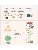
Single-cell suppression profiling of human Tregs. On the first day of the procedure, peripheral blood mononuclear cells (PBMCs) are thawed and CFSE-stained. Subsequently, Tregs are isolated by magnetic- and flow-assisted cell sorting. The Treg-negative PBMCs and the Tregs are mixed together in desired ratios and stimulated for 5 days. On day 5, cells are harvested and stained for mass cytometry (CyTOF). On day 6, data is acquired, normalized, and exported. On day 7 to approximately day 14, data is analyzed mainly in R.
Background
Regulatory T cells (Tregs) are a specialized subset of CD4+ T cells that play a central role in immune tolerance by suppressing activation and expansion of conventional T cells and other immune populations. Treg dysfunction has been implicated in a wide range of diseases, including autoimmunity, chronic inflammation, cancer, and infectious diseases [1,2]. To study Treg activity in vitro, suppression assays have traditionally relied on co-cultures of sorted Tregs and responder T cells, typically CD4+CD25- or CD8+ T cells, in the presence of polyclonal stimulation [3–5]. Suppressive function is commonly read out by measuring proliferation, cytokine production, or activation marker expression in the responder cells [3–5]. While these assays have been foundational for dissecting Treg biology, they are limited by their reductionist nature and the need for purified cell populations [5–9].
Standard suppression assays are often conducted with only one or two responder cell types, thereby excluding interactions with other immune populations that are part of the physiological context in vivo. Furthermore, they offer limited resolution for multiplexed phenotyping and functional tracking of individual cells. More recently, high-dimensional single-cell technologies such as mass cytometry, also known as cytometry by time of flight (CyTOF), have enabled simultaneous measurement of dozens of parameters across diverse immune subsets. CyTOF is a single-cell analysis technology that combines flow cytometry with mass spectrometry. In CyTOF, antibodies are tagged with metal isotopes rather than fluorophores, enabling the simultaneous quantification of over 50 parameters without spectral overlap. Cells are ionized, and the attached metal tags are detected by time-of-flight mass spectrometry, allowing precise quantification of marker expression [10–12]. This technology has opened new avenues for functional immune profiling [13–18]. However, protocols leveraging this technology to assess Treg function in a more holistic, system-level context have remained underdeveloped until recently [19].
The protocol presented here, referred to as single-cell suppression profiling of human Tregs (scSPOT), integrates reconstitution of sorted Tregs into autologous peripheral blood mononuclear cells (PBMCs) at controlled ratios, followed by in vitro stimulation and mass cytometry–based analysis [19]. Using the recommended 52-marker CyTOF panel, this approach allows simultaneous assessment of Treg suppressive effects on multiple immune cell types, including CD4+ and CD8+ T cells, B cells, NK cells, and myeloid subsets. Importantly, it captures both proliferative responses and phenotypic shifts within a complex immune environment, without the need for prior responder cell purification. The protocol is well-suited for evaluating inter-individual variation, analyzing patient samples, or screening drug effects on immune regulation.
Compared to conventional suppression assays, scSPOT offers improved physiological relevance by preserving the cellular diversity of PBMCs and provides high-resolution, multidimensional data at the single-cell level. While the approach requires access to mass cytometry infrastructure, it enables a deeper and broader view of Treg function and its impact on the immune landscape. As such, it represents a valuable tool for both basic research and translational immunology applications, especially in contexts where immune modulation is of therapeutic interest. Although we provide a detailed protocol for mass cytometry staining, if mass cytometry is not available, measurement of cells at the end of this protocol can be done with an alternative single-cell profiling method with sufficient depth to capture the relevant immune populations, such as spectral flow cytometry.
Materials and reagents
Biological materials
1. Human PBMCs from healthy donors (collected under local ethical approval or purchased from Stem Cell Technologies, catalog number: 70025.2)
Reagents
1. RPMI 1640 (Nacalai Tesque, catalog number: 30263-95)
2. Fetal bovine serum (FBS), heat-inactivated (Gibco, catalog number: 10437028)
3. Penicillin-Streptomycin-Glutamine (P/S-L-glu) (Thermo Fisher Scientific, catalog number: 10378016)
4. Ultra-LEAFTM purified anti-human CD3 antibody (BioLegend, clone UCHT1, catalog number: 300438)
5. Recombinant human IL-2 (Shionogi, catalog number: Imunace35)
6. Ipilimumab biosimilar (BioXCell, catalog number: SIM0004)
7. Human IgG1 isotype control (BioXCell, catalog number: BE0297)
8. Tazemetostat (Selleck Chemicals, catalog number: S7128)
9. DMSO (Sigma, catalog number: D6250)
10. Pierce Universal nuclease (Thermo Fisher Scientific, catalog number: 88702)
11. CellTrace CFSE Cell Proliferation kit (Invitrogen, catalog number: C34554)
12. EasySepTM Release Human CD4 Positive Selection kit (Stem Cell Technologies, catalog number: 17752)
13. PBS (Nacalai Tesque, catalog number: 14249-24)
14. EDTA (Nacalai Tesque, catalog number: 06894-14)
15. eBioscienceTM Foxp3/Transcription Factor Staining Buffer set (eBioscience, catalog number: 00-5523-00)
16. PierceTM 16% formaldehyde (w/v), methanol-free (Thermo Scientific, catalog number: 28906)
17. Cell acquisition solution (Standard BioTools, catalog number: 201240)
18. EQ four element calibration beads (Standard BioTools, catalog number: 201078)
19. BSA (Sigma, catalog number: A7906)
20. 10× PBS (Rockland, catalog number: MB-008)
21. FACS sorting antibodies:
a. Anti-CD45RA, BV711 (BioLegend, clone HI100, catalog number: 304138, titration: 1:20)
b. Anti-CD4, APC-Cy7 (BD, clone RPA-T4, catalog number: 557871, titration: 1:20)
c. Anti-CD127, AF647 (BioLegend, clone A019D5, catalog number: 351318, titration: 1:20)
d. Anti-CXCR5, BV421 (BD, clone RF8B2, catalog number: 562747, titration: 1:20)
e. Anti-CD45RO, PerCP-Cy5.5 (BD, clone UCHL1, catalog number: 560607, titration: 1:20)
f. Anti-CD25, PE (BD, clone M-A251, catalog number: 555432, titration: 1:5)
g. Anti-CD19, V500 (BD, clone HIB19, catalog number: 561121, titration: 1:20)
22. Mass cytometry antibodies: all self-conjugated antibodies (all aside from Standard BioTools) were conjugated as previously described [19,20] and stored at a stock concentration of 0.5 mg/mL unless otherwise stated
Barcoding and viability
a. Human Fc block (BioLegend, catalog number: 422302, titration: 1:50)
b. Anti-CD45, 89Y (Standard BioTools, clone HI30, catalog number: 3089003B, titration: 1:1:50)
Note: Labels such as 89Y refer to the specific metal isotopes used for mass-tagging antibodies, where the number (89) indicates the mass number of the isotope and the letter (Y) represents the chemical element (yttrium in this case). These isotopes act as unique tags detected by the mass cytometer, allowing simultaneous quantification of many markers. This naming convention is consistently applied throughout the manuscript.
c. Anti-CD45, 113In/115In/194Pt/195Pt/196Pt/198Pt (BioLegend, clone HI30, catalog number: 304002, titration: 1:100)
d. DCED Palladium (Sigma, catalog number: 574902, stock concentration: 500 μM, titration: 1:500, final concentration: 1 μM)
e. Alternative viability dye: Cell-IDTM Cisplatin-198Pt (Standard BioTools, catalog number: 201198, titration: 1:2,000)
Pre-stain
Pre-stain contains antibodies that stain better at 37 °C as well as de novo biosynthesis tagging reagents
a. Anti-CD45RA, 153Eu (BioLegend, clone HI100, catalog number: 304102, titration: 1:100)
b. Anti-CXCR3, 154Sm (BioLegend, clone G025H7, catalog number: 353733, titration: 1:100)
c. Anti-CCR7, 155Gd (BioLegend, clone G043H7, catalog number: 353202, titration: 1:100)
d. Anti-CCR4, 160Gd (BioLegend, clone L291H4, catalog number: 359402, titration: 1:100)
e. Anti-CCR6, 163Dy (BioLegend, clone G034E3, catalog number: 353427, titration: 1:50)
f. Anti-CXCR5 (BD, clone RF8B2, catalog number: 552118, titration: 1:25)
g. Puromycin (Sigma, catalog number: P8833, concentration: 1 mg/mL, titration: 1:300)
h. BrU (Sigma, catalog number: 850187, concentration: 5 mM, titration: 1:20)
i. IdU (Standard BioTools, catalog number: 201192A, concentration: 125 μM, titration: 1:600)
Surface stain
a. Anti-CD21, 110Cd (BioLegend, clone Bu32, catalog number: 354902, titration: 1:100)
b. Anti-CD4, 111Cd (BioLegend, clone RPA-T4, catalog number: 300502, titration: 1:100)
c. Anti-CD8, 112Cd (BioLegend, clone RPA-T8, catalog number: 301002, titration: 1:50)
d. Anti-CD39, 114Cd (BioLegend, clone A1, catalog number: 328221, titration: 1:100)
e. Anti-CD19, 116Cd (BioLegend, clone HIB19, catalog number: 302202, titration: 1:100)
f. Anti-CD86, 147Sm (BioLegend, clone IT2.2, catalog number: 305402, titration: 1:50)
g. Anti-IgD, 151Eu (BioLegend, clone IA6-2, catalog number: 348202, titration: 1:100)
h. Anti-PD-L1 (CD274), 156Gd (Standard BioTools, clone 29E.2A3, catalog number: 3156026B, titration: 1:100)
i. Anti-CD27, 158Gd (Standard BioTools, clone L128, catalog number: 3158010B, titration: 1:200)
j. Anti-TIM3, 159Tb (Standard BioTools, clone F38-2E2, catalog number: 3159037B, titration: 1:100)
k. Anti-Biotin (for CXCR5), 165Ho (Standard BioTools, clone 1D4C5, catalog number: 3165012B, titration: 1:50)
l. Anti-PD1, 166Er (BioLegend, clone EH12.2H7, catalog number: 329941, titration: 1:100)
m. Anti-CD25, 169Tm (Standard BioTools, clone 2A3, catalog number: 3169003B, titration: 1:200)
n. Anti-CD98, 171Yb (BD, clone UM7F8, catalog number: 556074, titration: 1:200)
o. Anti-CD38, 172Yb (Standard BioTools, clone HIT2, catalog number: 3172007B, titration: 1:100)
p. Anti-CD11c, 173Yb (BioLegend, clone S-HCL-3, catalog number: 371502, titration: 1:100)
q. Anti-HLA-DR, 174Yb (BioLegend, clone L243, catalog number: 307602, titration: 1:400)
r. Anti-TIGIT, 209Bi (Standard BioTools, clone MBSA43, catalog number: 3209013B, titration: 1:100)
Intracellular stain
a. Anti-H3K27me3, 140Ce (Active Motif, clone 323, catalog number: 61017, titration: 1:200)
b. Anti-Puromycin, 141Pr (Merck, clone 12D10, catalog number: MABE343, titration: 1:200)
c. Anti-Cleaved Caspase-3, 142Nd (Standard BioTools, clone D3E9, catalog number: 3142004A, titration: 1:100)
d. Anti-TCF1, 143Nd (BioLegend, clone 7F11A10, catalog number: 655202, titration: 1:50)
e. Anti-FITC (for CFSE), 144Nd (Standard BioTools, clone FIT-22, catalog number: 3144006B, titration: 1:100)
f. Anti-Helios, 145Nd (BioLegend, clone 22F6, catalog number: 137202, titration: 1:100)
g. Anti-OGDH, 146Nd (Thermo Fisher Scientific, polyclonal, catalog number: PA528195, titration: 1:100)
i. Anti-XBP1, 148Nd (Thermo Fisher Scientific, polyclonal, catalog number: PA5-27650, titration: 1:100)
h. Anti-Cytochrome C, 149Sm (BioLegend, clone 6H2.B4, catalog number: 612302, titration: 1:200)
i. Anti-Granzyme B, 150Nd (Novus, clone CLB-GB11, catalog number: NBP1-50071, titration: 1:100)
j. Anti-CPT1A, 152Sm (Abcam, clone 8F6AE9, catalog number: ab128568, titration: 1:100)
k. Anti-T-bet, 157Gd (BioLegend, clone 4B10, catalog number: 644802, titration: 1:100)
l. Anti-VDAC1/Porin, 161Dy (Abcam, clone 20B12AF2, catalog number: ab14734, titration: 1:100)
m. Anti-Foxp3, 162Dy (eBioscience, clone 236A/E7, catalog number: 14-4777-82, titration: 1:200)
n. Anti-Cyclin B1, 164Dy (Standard BioTools, clone GNS-1, catalog number: 3164010A, titration: 1:200)
o. Anti-GLUT1, 167Er (Abcam, clone EPR3915, catalog number: ab196357, titration: 1:200)
p. Anti-Ki67, 168Er (Standard BioTools, clone Ki67, catalog number: 3168001B, titration: 1:200)
q. Anti-CTLA4, 170Er (Standard BioTools, clone 14D3, catalog number: 3170005B, titration: 1:50)
r. Anti-BrdU (for BrU), 175Lu (BD, clone 3D4, catalog number: 555627, titration: 1:50)
s. Anti-OPA1, 176Yb [Abcam, clone EPR11057(B), catalog number: ab240143, titration: 1:100]
DNA stain
a. Cell-IDTM Intercalator-Rh (103Rh) (Standard BioTools, catalog number: 201103A, titration: 1:500)
Solutions
1. PBSE (see Recipes)
2. Complete RPMI (cRPMI) (see Recipes)
3. cRPMI + DNAse (see Recipes)
4. PBS with 0.1% FBS (see Recipes)
5. CyFACS (see Recipes)
Recipes
Note: PBSE, cRPMI, and PBS with 0.1% FBS solutions are made with sterile ingredients and handled sterile. Especially, myeloid cells depend on the quality of the FBS, and thus, we recommend making new solutions every week.
1. PBSE, 50 mL
49 mL of PBS (should be free of Ca++ and Mg++)
1 mL of FBS (2% final)
100 μL of EDTA (0.5 M stock, 1 mM EDTA final)
2. Complete RPMI (cRPMI), 50 mL
45 mL of RPMI
5 mL of FBS (10% final)
0.5 mL of P/S-L-glu (1% final)
3. cRPMI + DNAse, 50 mL
45 mL of RPMI
5 mL of FBS (10% final)
0.5 mL of P/S-L-glu (1% final)
4 μL of Pierce Universal nuclease stock (stock is 25 kU/100 μL → 250 U/μL; final: 500 U/25 mL)
4. PBS with 0.1% FBS, 10 mL
10 mL of PBS (should be free of Ca++ and Mg++)
10 μL of FBS (1,000× dilution)
5. CyFACS, 500 mL
50 mL of 10× PBS
2 mL of EDTA (0.5 M stock, 2 mM EDTA final)
0.5 g of BSA (0.1% final)
Top up to 500 mL with Milli-Q water. Filter-sterilize with a 0.2 μm filter. Store at 4 °C for up to 1 year.
Laboratory supplies
1. 15 mL and 5 mL round-bottom polystyrene tubes (for magnetic separation and FACS sorting)
2. 96-well round-bottom plates (Nunc, catalog number: 163320)
3. Neubauer chamber (for manual cell counting)
4. Inverted light microscope (for visual well checks)
5. 35 μm cell strainer (BD, catalog number: 352235)
6. Nalgene filters with PES membrane, 0.2 μm pore size, 1,000 mL capacity (Thermo Scientific, catalog number: 567-0020)
Equipment
1. Aria III cell sorter (BD Biosciences)
2. Centrifuge (suitable for 5 and 15 mL tubes with 4 °C option)
3. CO2 incubator (humidified, 37 °C, 5% CO2)
4. Water bath (37 °C)
5. CyTOF Helios (Standard BioTools) or equivalent mass cytometer
6. Standard biological safety cabinet (class II)
Software and datasets
1. CyTOF acquisition software (Standard BioTools, v. 7.0.8493)
2. Cytobank (Beckman Coulter, v. 10.5) - license required
3. R software, v. 4.3.0 - open source
a. Premessa v. 0.3.4
b. CATALYST v. 1.26.0
c. ggplot2 v. 3.4.4
d. diffcyt v. 1.22.0
e. scran v. 1.30.0
f. rstatix v. 0.7.2
4. GitHub repositories:
a. https://github.com/jonasns/scSPOT
b. https://github.com/jonasns/scDivisionPro
5. Zenodo archives:
a. scSPOT dataset, https://doi.org/10.5281/zenodo.10828517
b. scSPOT code, https://doi.org/10.5281/zenodo.14056251
c. scDivisionPro code, https://doi.org/10.5281/zenodo.14064605
Procedure
A. PBMC thawing
1. Prepare a 37 °C water bath.
2. Prewarm cRPMI (complete RPMI) with DNase at 37 °C.
3. Take cells from the liquid nitrogen tank. Normal ice is ok for 1–2 samples if processed quickly. Otherwise, transport them on dry ice.
4. Critical step for cell viability: Thaw cells in a 37 °C water bath for exactly 2 min.
5. Critical step for cell viability: Work fast but carefully in steps A5–7. Inside the hood, slowly pipette cells from the cryovial into a 15 mL tube.
6. Wash the vial with 1 mL of prewarmed 10% FBS RPMI (with 20 U/mL of DNase) and slowly add to the tube.
7. Add 3 × 1 mL of warm RPMI slowly to the tube, swirling each time.
8. Centrifuge at 400× g for 5 min at room temperature (RT).
9. Decant and add another 5 mL of RPMI at RT.
10. Centrifuge at 400× g for 5 min at RT.
11. Resuspend in 2 mL of RT RPMI, resuspend with p1000 (not tilting the tube), and count.
12. Either use immediately or place on ice if many samples are being processed.
B. CFSE staining
Note: The following steps use the CellTrace CFSE Cell Proliferation kit.
1. Defrost PBMC as per the normal method (see steps A1–12).
2. Prewarm PBS with 0.1% FBS to 37 °C.
3. Prepare CFSE solution according to the manufacturer’s instructions:
a. Add 18 μL of DMSO to a new vial of CFSE stock to a 5 mM solution.
b. Thaw reagents at RT before opening them.
c. Aliquots can be stored at -20 °C.
4. Centrifuge cells to remove culture media (at 400× g for 5 min at RT).
If there are only a few cells, wash in PBS with 0.1% FBS before staining (to reduce the FBS present).
5. Prepare CFSE staining buffer immediately before adding to cells: add 1 μL of CFSE (5 mM stock, final concentration 1 μM) to 2,500 μL of RT PBS with 0.1% FBS (this will result in 2 μM, and we will use 30 μL/106 cells).
6. Remove as much culture media as possible by pipetting and gently resuspend the cells with RT PBS with 0.1% FBS at a concentration of 30 μL/106 cells.
7. Gently mix the cells with the CFSE staining buffer at a concentration of 30 μL/106 cells (i.e., 2× dilution to give a final 1 μM).
8. Incubate at RT for 10 min in the dark.
9. Add a minimum of 10 volumes of ~37 °C cRPMI (i.e., containing FBS, and no DNase!).
10. Critical step for cell viability: Mix gently (e.g., by turning the tube) and incubate for 5 min at RT (to remove free CFSE).
11. Centrifuge at 400× g for 5 min at RT.
12. Wash twice in 5 mL of RT cRPMI (400× g for 5 min at RT). Flick the tube carefully to resuspend.
C. EasySepTM release buffer prep
1. Dilute the EasySepTM release buffer (concentrate) 40× with PBSE on the day of use. You need a minimum of 2.5 mL per isolation.
2. Per isolation: 65 μL of 40× release buffer + 2,535 μL of PBSE.
D. CD4+ T-cell isolation
Note: The following steps use the EasySep Release Human CD4 Positive Selection kit.
1. Resuspend cells from step B12 in 1 mL of PBSE and transfer to a polystyrene (PS) FACS tube. Wash the 15 mL tube twice with 1 mL of PBSE by pipetting and transferring to the FACS tube (do not filter unless clumpy). If filtering:
a. Resuspend in 2 mL of PBSE. Filter through a filter-lid FACS tube.
b. Use 1 mL extra to wash the tube and the filter.
2. Centrifuge at 400× g for 5 min at RT.
Note: Use a polystyrene round-bottom tube for all steps.
3. Resuspend cells at 1 × 10⁸ cells/mL in PBSE (minimum 250 μL, maximum 2 mL). Use 250 μL if you have less than 25 million cells.
4. Add selection cocktail (from the EasySep Release Human CD4 Positive Selection Kit) to the sample at 100 μL/mL of sample and 25 μL if less than 25 million cells.
5. Mix and incubate at RT for 3 min.
6. Vortex releasable RapidSpheresTM (30 s).
Note: Particles should appear evenly dispersed.
7. Add releasable RapidSpheresTM to the sample: 100 μL/mL of sample or 25 μL if less than 25 million cells.
8. Mix and incubate at RT for 3 min.
9. Top up the sample to 2.5 mL with PBSE. Mix by gently pipetting up and down 2–3 times.
10. Place the tube (without the lid) into the magnet and incubate at RT for 10 min.
11. Put the centrifuge at 4 °C.
12. Pipette the supernatant (SN) to a new PS FACS tube. It contains CD4- cells.
Note: Be careful not to disturb the bead-bound cells.
13. Repeat steps D9–12 one more time for the CD4- fraction.
14. Repeat steps D9–12 two extra times for the CD4+ fraction, but only incubate for 5 min in the magnet; also, do not keep the SN.
15. Put the CD4- fraction on ice until CD4+ purification is done.
16. For the CD4+ fraction: Add release buffer (1×) to top up the sample to 2.5 mL. Mix by gently pipetting up and down 2–3 times.
17. Incubate at RT for 3 min (during these incubations, you can count the CD4- fraction). Optional: Take a sample for the unstained control on Aria III: 300 μL on a filter-tube + 300 μL extra wash).
18. Place the tube (without the lid) into the magnet and incubate at RT for 5 min.
19. Pipette the SN into a new tube. It contains the CD4+ fraction.
20. Isolated cells (in the new tube) are ready for use.
E. Staining for sorting
1. Spin down cells (400× g for 5 min at 4 °C).
2. Resuspend in 2–3 mL of PBSE (so that they are not in release buffer).
3. Spin down cells (400× g for 5 min at 4 °C).
4. Resuspend in 2–3 mL of PBSE.
5. Spin down cells (400× g for 5 min at 4 °C).
6. Remove SN from cells (as much as possible; it is expected to have 20 μL of residual volume).
7. Add the antibody (Ab) cocktail as described in Table 1.
Table 1. FACS panel recipe
| Antibody | Master mix for 1 donor (μL) | Target |
|---|---|---|
| CD45RA BV711 | 5 | Naïve cells |
| CD4 APC-Cy7 | 5 | CD4+ T cells |
| CD127 AF647 | 5 | Low on Tregs |
| CXCR5 Bv421 | 5 | T follicular regulatory cells (Tfr) |
| CD45RO PerCP-Cy5.5 | 5 | Effector cells |
| CD25 PE | 20 | Tregs |
| CD19 v500 | 5 | B cells |
| PBSE total 100 μL/sample | 50 |
8. Incubate for 30 min at 4 °C in the dark.
9. Resuspend in 2–3 mL of PBSE.
10. Spin down cells (400× g for 5 min at 4 °C).
11. Resuspend in 2–3 mL of PBSE.
12. Spin down cells (400× g for 5 min at 4 °C).
13. Resuspend in 1 mL of PBSE for sorting.
14. Filter through a filter-lid FACS tube.
Note: Filter each tube immediately before sorting to avoid re-clumping of cells.
15. Also, prepare and bring with you to the sorter 5 × 5 mL PS FACS tubes with 0.7–1 mL of PBSE.
Critical step for cell recovery: Coat the inner surface of the tube with FBS by gently tilting the tube with the PBSE. Do not shake it, as it will create bubbles. As cells are electrostatically charged after passing through the sorter, the coating of the tube with FBS minimizes plastic adherence.
F. Aria III sorting
1. Sort at 5,000–8,000 events/s, preferably at a flow rate below 7.
G. Sorting gating strategy
1. See an example gating strategy in Figure 1 [reproduced from Søndergaard et al. [19], Figure S10, licensed under CC BY-NC-ND 4.0 (https://creativecommons.org/licenses/by-nc-nd/4.0/)]
2. All cells: lymphocyte gate, singlets, CFSE+, CD4+.
3. Make a gate for Tregs (CD25hi, CD127lo) -> continue gating with these.
4. Also, make a “NOT Treg gate.”
5. Make a gate for Tfr cells (CXCR5+).
6. Also, make a “NOT Tfr gate” -> continue gating with these.
7. Make a gate for naïve cells (CD45RA+, CD45RO-) -> “nTreg.”
8. Make a gate for effector cells (CD45RA-, CD45RO+) -> “eTreg.”
• Sorting tube 1:
“Tfr”
• Sorting tube 2:
“nTreg”
• Sorting tube 3:
“eTreg”
• Sorting tube 4:
“NOT Treg gate”
Note: This tube may fill up. Change the tube during the run. If sufficient cells are collected, sorting for this tube can be stopped as needed or by pre-selecting a predefined stopping event threshold.
9. Sort all donors and keep on ice until finished. Remember to purity check! Then, plate all donors. Purity check is done by passing the sorted sample through the sorter again and recording the events. It is recommended to wash the sample line thoroughly before each purity check (backflush 10–20 s).

Figure 1. Gating strategy and purity control of flow-associated cell sorting. (A) Cells were gated for lymphocytes, forward scatter (FSC) and side-scatter (SSC) singlets, CD4 T cells (excluding potential B cell duplicates), and CFSE+ cells, before identifying Tregs by CD127-low, CD25-high expression. Tregs were gated on CXCR5 to identify Tfr. Non-Tfr was subsequently divided into eTregs (CD45RO+CD45RA-) and nTregs (CD45RO-CD45RA+). (B) purity control of the four different sorted populations: non-Tregs, nTregs, eTregs, and Tfr. The figure is representative of 15 donors done in 8 independent experiments. Reproduced from Søndergaard et al., Nature Communications Figure S10 [19], licensed under CC BY-NC-ND 4.0 (https://creativecommons.org/licenses/by-nc-nd/4.0/).
H. Cell plating
1. Use a 96-well U-bottom plate.
2. Critical step for keeping cells viable during culture: Dilute PBSE from sorting by topping up the tube with 3 mL of cRPMI before centrifugation.
3. Spin down cells (400× g for 5 min at 4 °C) in the FACS tubes.
4. Discard SN. The leftover volume is usually ~40 μL.
5. For low resuspension volume, always measure it precisely (using a pipette).
6. Resuspend Tregs at 0.4 × 10⁶ cells/mL in cRPMI (to give 20,000 in 50 μL).
7. Resuspend NOT Tregs at 2 × 10⁶/mL in cRPMI (to give 100,000 in 50 μL).
8. Plate 100,000/well in a round-bottom 96-well plate.
a. Culture medium: cRPMI.
b. Final cell concentration: 106/mL.
c. Avoid outer wells; add liquid (sterile PBS, PBSE, or MQ water) to outer wells to reduce dehydration.
9. 0× Treg condition: 100,000 “NOT Treg”: 50 μL. 50 μL of medium.
10. 20,000 Treg condition: 100,000 “NOT Treg”: 50 μL. 20,000 Treg subset: 50 μL.
11. Add further Treg ratios as desired.
12. Optimally, we would recommend plating five wells per condition and pooling these for the CyTOF staining. However, if there is a lack of material, a lower number of wells can be plated. It is possible to only plate one well per condition and get reliable results if the user has extensive prior experience with the protocol.
I. Stimulation conditions and final concentrations
1. To calculate the checkpoint/inhibitor/CD3-IL2 stimulation mix concentrations, a 4× dilution in the well must be kept in mind.
2. Final concentration: 1 μg/mL ultra-LEAFTM Purified anti-human CD3 Antibody.
3. Final concentration: 2 U/mL recombinant human interleukin-2 (Imunace35).
4. Example checkpoint/inhibitor final concentrations:
• 10 μg/mL anti-CTLA4 (Ipilimumab biosimilar) and human IgG1.
• 500 nM Tazemetostat reconstituted in DMSO at 20 mM.
5. Plate 100 μL of cells, 50 μL of checkpoint/inhibitor solution, and 50 μL of Anti-CD3+ IL2 stimuli (in that order).
6. Incubate in a humidified 5% CO2 incubator for 5 days.
Example calculation for 100 wells anti-CD3+IL2-stimuli:
1. 100 wells → 5,000 μL final volume. Concentrations are 4× the final concentration.
2. 4991 μL of cRPMI
3. 7.12 μL of CD3 (702.5× dilution of 2.81 mg/mL stock. Note: Concentration is batch dependent).
4. 2 μL of IL2 (2,500× dilution of 5× pre-diluted solution of 100 U/μL stock).
J. Harvesting of incubated PBMCs
Notes on the harvesting of incubated PBMCs:
1. Prepare RT CyFACS and 1 mL of CyPBS buffer (for viability staining) in advance.
2. Ab mixes can be made on the day, up to 6 hours before. However, for the pre-stain, add CyFACS and puromycin/IdU/BrU right before. For others, just add CyFACS right before.
3. Use filter tips.
4. Follow local regulations for waste disposal.
5. Below, we present a protocol for barcoding samples in a 96-well plate. However, if there are a few conditions, replicate wells can simply be harvested by pipetting into a 15 mL tube and barcoded in the tube. Barcoding in CyTOF refers to the use of metal isotope-labeled reagents to uniquely label cells from different samples, enabling them to be pooled and stained together, thereby minimizing batch effects [21,22]. Following staining, the barcoded cells are deconvoluted by gating to assign each cell back to its original sample.
1. After 5 days in culture, cells have changed morphology depending on the stimulation conditions. It is, therefore, recommended to check the cells in the microscope and take notes on their appearance (this can be done the evening before).
2. Critical steps for getting all cells off the plate: After 5 days in culture, some myeloid cells will be stuck to the plastic, but can be released by incubating in PBS on ice, followed by pipetting.
a. Spin down the plate (400× g for 5 min at 4 °C).
b. Pipette off ~190 μL and discard.
c. Empty non-used wells (otherwise, they will spill over during vortex).
d. Resuspend by “gentle vortex” at a setting that does not cause fluid to spill out. This can either be done with a soft wrist or with a plate vortexer.
Note: Instead of vortexing, cells may be resuspended by pipetting the volume in step J2e if desired.
e. Use a multichannel pipette to add 75 μL of 4 °C CyPBS to each well.
f. Place on ice for 30–60 min.
3. Prepare barcode Ab mixes in a 96-well U-bottom plate (not cell culture–treated) during incubation following the dilutions given in the Reagents section.
Note: Ab mixes may also be prepared in individual Eppendorf tubes if desired.
a. The barcodes are anti-CD45 Abs tagged with a unique metal.
b. We used 7 different Abs, and the Ab cocktail must be unique per sample.
c. If ≤ 7 samples, one Ab is used per sample. If 8–21 samples, two Abs are combined per sample (7-choose-2). If 22–35 samples, three Abs are combined per sample (7-choose-3).
Note: All individual Ab mixes must contain the FC-block.
4. Continue harvesting the cells after incubation is done.
a. Use a multichannel pipette to resuspend PBMCs. Set the pipette to 50 μL when pipetting (to avoid creating bubbles). Harvest each condition into one well (max volume is ~300 μL).
Note: Each condition means multiple wells of the same sample. We can only plate 100,000 cells per well, but preferably stain 500,000 for CyTOF; hence, at this step, they are pooled.
b. Add 75 μL of 4 °C CyPBS to empty wells (which may still contain some cells) and spin down (400× g for 5 min at 4 °C).
c. Pipette off ~200 μL from “collection well” and discard.
d. Use the 75 μL in the other wells to resuspend and transfer (i.e., just use what is already there for the transfer).
e. Repeat steps J4b–d as many times as needed to get all cells in one well.
f. Use an inverted light microscope to check the empty wells to make sure that all cells have been harvested.
g. If ok, spin down (400× g for 5 min at 4 °C to RT), aspirate SN as much as possible (pipette down to 50 μL), and add barcode Abs as described in step K1.
K. CyTOF staining
1. Add barcodes and Fc block in CyFACS buffer (50 μL of Ab-mix added to 50 μL of cells). Remove from ice and incubate at RT for 30 min.
2. Set the centrifuge to RT.
3. When incubation is done, add 150 μL of RT CyFACS on top and spin down (400× g for 5 min at RT).
4. Pipette off 200 μL and discard.
5. Gently vortex the plate as described above.
6. Add 200 μL of RT CyFACS on top and spin down (400× g for 5 min at RT).
7. Pipette off 200 μL and discard.
8. Repeat steps K6–7 one more time.
9. Resuspend each sample in 200 μL of CyFACS buffer. Mix/pool samples into a 15 mL tube.
10. If desired, rinse wells one more time and put in the same 15 mL tube.
11. Centrifuge at 400× g for 5 min at RT.
12. Wash in 2 mL of cRPMI (centrifuge at 400× g for 5 min at RT). Critical step for accurate staining volume: Remove the supernatant by decantation, then carefully aspirate the residual hanging drop using a P1000 pipette, leaving approximately 50 μL of final volume.
Note: This means to hold the tube upside down and use a P1000 pipette to pipette off any fluid from the rim of the tube.
13. Add pre-stains, mix, and incubate for 30 min at 37 °C.
14. Wash 3× 2 mL of CyFACS (centrifuge at 400× g for 5 min at RT).
a. Tap-resuspend when washing.
b. First, add 2 mL on top of the staining solution.
c. Critical step for accurate staining volume: Remove the supernatant by decantation, then carefully aspirate the residual hanging drop using a P1000 pipette, leaving approximately 50 μL of final volume.
15. Add surface stains, mix, and incubate for 45 min at RT (50 μL of Ab-mix to 50 μL of cells).
16. Wash 3× 2 mL of CyFACS (centrifuge at 400× g for 5 min at RT) and pipette the final supernatant to 50 μL.
17. During the final spin, prepare DCED palladium in 1 mL of CyPBS at 1:500.
Note: Alternative viability dyes may also be used.
a. Store DCED at -20 °C (500 μM stock, 2 μL aliquots).
b. Dilute it in CyPBS. Take 50 mL sterile from stock.
c. 2 μL of DCED stock + 1 mL of CyPBS. Pipette to resuspend.
18. Pipette mix and incubate for 5 min at RT.
19. Wash 2× in 5 mL of CyFACS (centrifuge at 400× g for 5 min at 4 °C) (all 4 °C from now on). Pipette the final supernatant down to 50 μL.
20. Add 1 mL of fix/perm buffer and incubate for 45 min at 4 °C.
a. The solution is a ratio of 1:3 of eBio fix/perm concentrate to eBio fix/perm diluent (250 μL + 750 μL).
b. Pipette mix the 1 mL buffer solution with the cells.
c. After this step, cells are fixed.
21. Prepare Invitrogen 10× perm buffer:
a. Dilute with fresh MQ (should be 18.2 mV). Keep on ice.
b. Make 25 mL: 2.5 mL of stock + 22.5 mL of MQ.
22. Wash 2× in 5 mL of perm buffer (centrifuge at 800× g for 5 min at 4 °C). Critical step for accurate staining volume: Remove the supernatant by decantation, then carefully aspirate the residual hanging drop using a P1000 pipette, leaving approximately 50 μL of final volume.
23. Add the intracellular Ab cocktail, mix, and incubate for 45 min at 4 °C (50 μL of Ab-mix to 50 μL of cells).
24. Wash twice with 5 mL of perm buffer and then once with 2 mL of CyPBS (centrifuge at 800× g for 5 min at 4 °C).
25. Prepare 1 mL of 2% formaldehyde (FA) with 1:500 Rhodium: 125 μL of 16% FA, 875 μL of CyPBS, and 2 μL of Rhodium.
26. Add 2% FA Rhodium and mix by pipetting.
27. Wrap in tinfoil and put it in the fridge overnight or up to 48 h. FA with no Rhodium can be used for up to one week.
Note: Storing cells in FA may be done up to 14 days, but it is preferable to acquire within 48 h.
L. CyTOF data acquisition
Acquire data on a mass cytometer according to the manufacturer’s instructions.
Data analysis
A detailed code for performing data analysis is available on GitHub (https://github.com/jonasns/scSPOT, https://github.com/jonasns/scDivisionPro) and Zenodo archive:
• scSPOT dataset: https://doi.org/10.5281/zenodo.10828517
• scSPOT code: https://doi.org/10.5281/zenodo.14056251
• scDivisionPro code: https://doi.org/10.5281/zenodo.14064605
1. Normalization and gating
a. Normalize and concatenate .fcs files using CyTOF software.
Note: FCS (Flow Cytometry Standard) is a file format developed by the International Society for Advancement of Cytometry (ISAC) to store data generated by flow and mass cytometers.
b. Perform gating of single, viable, and debarcoded cells in Cytobank (v. 10.5).
c. Export gated fcs files for further analysis in R.
2. Data import and preprocessing in R
https://github.com/jonasns/scSPOT/blob/main/240321_scSPOT_code_PART1.Rmd
a. Import FCS files into R.
b. Use the Premessa package to remove unused channels:
• DNA channels
• Barcode channels
• Gaussian parameters
• Viability markers
3. Initial clustering and cell type annotation
https://github.com/jonasns/scSPOT/blob/main/240321_scSPOT_code_PART1.Rmd
a. Perform top-level FlowSOM clustering using CATALYST [23].
b. Manually merge clusters into major immune populations based on canonical markers:
• CD4 T cells
• CD8 T cells
• B cells
• NK cells
• Myeloid cells
• Metabolome-low cells
• Markers used for clustering: CD4, CD8, CD19, CyclinB1, IgD, PD-L1, Tbet, CXCR5, Puro, CD86, CytC, CD11c, HLA-DR
4. Subclustering by lineage
a. For each main population (CD4 T cells, CD8 T cells, B cells, myeloid cells), perform a new round of FlowSOM clustering and manual annotation in separate SingleCellExperiment (SCE) objects.
b. Merge SCEs after subclustering is completed.
5. CD4 T-cell subtypes
https://github.com/jonasns/scSPOT/blob/main/240321_scSPOT_code_PART1.Rmd
a. Identify Tregs using FlowSOM based on Foxp3, Helios, CD25, CD4, CD39, PD1, Ki67, CTLA4, CD45RA, CCR7, cCasp3, TCF1, CFSE, OGDH, XBP1, GzmB, Puro, and CD98.
b. Subset Tfr from Tregs by manual gating on CXCR5.
c. Further cluster Tregs into eTreg and nTreg based on CD45RA, CD25, Foxp3, Ki67, CFSE, CD39, CCR6, and TCF1.
d. Classify remaining CD4 T cells into Naïve, Th1, Th2, Th17, Tfh, and metabolome-low (markers: CXCR5, CCR7, CD45RA, CXCR3, CD25, Tbet, CCR4, CCR6, CD98, CD38, TCF1, CytC, CFSE, Ki67, CyclinB1, HLA-DR, and CD39).
6. CD8 T cells
https://github.com/jonasns/scSPOT/blob/main/240321_scSPOT_code_PART1.Rmd
a. Cluster into naïve, central memory (CM), effector memory (EM), and TEMRA subsets using CD45RA, CCR7, Ki67, CD27, CD38, and CFSE.
7. B cells
https://github.com/jonasns/scSPOT/blob/main/240321_scSPOT_code_PART2.Rmd
a. Cluster into naïve, memory, activated, and plasmablasts using IgD, CD27, CD38, CD11c, Tbet, CD98, CD39, CytC, CXCR5, CCR6, and CCR7.
8. Myeloid cells
https://github.com/jonasns/scSPOT/blob/main/240321_scSPOT_code_PART2.Rmd
a. Classify into HLA-DR low, intermediate, and high groups using HLA-DR, Puro, CD11c, CD86, and CCR7.
Note: Further classification is not possible due to marker loss after culture.
9. Downstream analysis: differential abundance and expression
(https://github.com/jonasns/scSPOT/blob/main/240321_scSPOT_code_PART3.Rmd)
a. Use diffcyt for comparisons between experimental conditions [24].
b. Visualize results using volcano plots created with ggplot2.
c. Use scran::findMarkers() instead of diffcyt when comparing cell types or division states.
• Use test.type = "t" for fold change.
• Use test.type = "wilcox" for FDR adjustment.
d. Generate density UMAPs using stat_density_2d() from ggplot2 with:
• geom = "polygon", contour = TRUE, aes(fill = after_stat(level)), bins = 10
e. Use scDP to assign division stages (div0, div1, etc.)
https://github.com/jonasns/scSPOT/blob/main/240409_scSPOT_code_PART5.Rmd
https://github.com/jonasns/scDivisionPro
f. Calculate:
• % in each division = (# in divX/total cells) × 100
• % proliferated = % of cells not in div0
• Division index = (total divisions)/(initial number of cells)
Validation of protocol
This protocol has been used and validated in the following research article:
Søndergaard et al. [19]. Single cell suppression profiling of human regulatory T cells. Nat Commun. 16(1): 1325. https://doi.org/10.1038/s41467-024-55746-1
General notes and troubleshooting
Troubleshooting
1. Low Treg recovery after sorting.
• Make sure the FACS tube is coated with FBS as described.
• Optimize side streams of the sorter.
• Titrate the antibody amount used for staining.
2. Low CFSE signal.
• Do an extra wash with PBS + 0.1% FBS before staining (to lower FBS content).
• Make a fresh aliquot of CFSE.
• Prepare a CFSE diluted stock solution as close to starting the staining as possible.
Note: CFSE signal can be checked on the cell sorter or any flow cytometer right after staining. Include an unstained control to find the negative signal.
3. Poor cell viability post-stimulation.
• Treat cells gently during the entire procedure (avoid harsh pipetting).
• Make fresh culture medium every week.
• Do a cRPMI wash to dilute the sorting buffer before plating.
• Use magnetic enrichment prior to sorting so that only CD4 T cells are sorted. If directly sorting, other cell populations like myeloid cells may suffer from low viability.
Acknowledgments
This work was supported by the Japan Society for the Promotion of Science 23K11304 (J.N.S.), IFReC advanced postdoc program (J.N.S.), IFReC grant program for next generation principal investigators (J.B.W.), Nippon Foundation (J.B.W.), Takeda Foundation (J.B.W.), Japan Society for the Promotion of Science 16H06295 (S.S.), Japan Agency for Medical Research and Development JP19gm0010005 (S.S.), and Leading Advanced Projects for Medical Innovation (S.S.).
This protocol was used in [19].
Competing interests
The authors declare no competing interests.
Ethical considerations
Human PBMCs from healthy volunteer donor blood were either collected under ethical approval numbers 895 and 897 (Osaka University Research Ethics Committee) after informed consent or purchased from Stemcell Technologies (cat. 70025.2).
References
- Sakaguchi, S., Mikami, N., Wing, J. B., Tanaka, A., Ichiyama, K. and Ohkura, N. (2020). Regulatory T Cells and Human Disease. Annu Rev Immunol. 38(1): 541–566. https://doi.org/10.1146/annurev-immunol-042718-041717
- Bittner, S., Hehlgans, T. and Feuerer, M. (2023). Engineered Treg cells as putative therapeutics against inflammatory diseases and beyond. Trends Immunol. 44(6): 468–483. https://doi.org/10.1016/j.it.2023.04.005
- Takahashi, T., Kuniyasu, Y., Toda, M., Sakaguchi, N., Itoh, M., Iwata, M., Shimizu, J. and Sakaguchi, S. (1998). Immunologic self-tolerance maintained by CD25+CD4+ naturally anergic and suppressive T cells: induction of autoimmune disease by breaking their anergic/suppressive state. Int Immunol. 10(12): 1969–1980. https://doi.org/10.1093/intimm/10.12.1969
- Thornton, A. M. and Shevach, E. M. (1998). CD4+CD25+ Immunoregulatory T Cells Suppress Polyclonal T Cell Activation In Vitro by Inhibiting Interleukin 2 Production. J Exp Med. 188(2): 287–296. https://doi.org/10.1084/jem.188.2.287
- Venken, K., Thewissen, M., Hellings, N., Somers, V., Hensen, K., Rummens, J. L. and Stinissen, P. (2007). A CFSE based assay for measuring CD4+CD25+ regulatory T cell mediated suppression of auto-antigen specific and polyclonal T cell responses. J Immunol Methods. 322: 1–11. https://doi.org/10.1016/j.jim.2007.01.025
- McMurchy, A. N. and Levings, M. K. (2011). Suppression assays with human T regulatory cells: A technical guide. Eur J Immunol. 42(1): 27–34. https://doi.org/10.1002/eji.201141651
- Canavan, J. B., Afzali, B., Scottà, C., Fazekasova, H., Edozie, F. C., Macdonald, T. T., Hernandez-Fuentes, M. P., Lombardi, G. and Lord, G. M. (2012). A rapid diagnostic test for human regulatory T-cell function to enable regulatory T-cell therapy. Blood. 119(8): e57–e66. https://doi.org/10.1182/blood-2011-09-380048
- Long, A. E., Tatum, M., Mikacenic, C. and Buckner, J. H. (2017). A novel and rapid method to quantify Treg mediated suppression of CD4 T cells. J Immunol Methods. 449: 15–22. https://doi.org/10.1016/j.jim.2017.06.009
- Brusko, T. M., Hulme, M. A., Myhr, C. B., Haller, M. J. and Atkinson, M. A. (2007). Assessing the In Vitro Suppressive Capacity of Regulatory T Cells. Immunol Invest. 36: 607–628. https://doi.org/10.1080/08820130701790368
- Spitzer, M. H. and Nolan, G. P. (2016). Mass Cytometry: Single Cells, Many Features. Cell. 165(4): 780–791. https://doi.org/10.1016/j.cell.2016.04.019
- Hartmann, F. J. and Bendall, S. C. (2019). Immune monitoring using mass cytometry and related high-dimensional imaging approaches. Nat Rev Rheumatol. 16(2): 87–99. https://doi.org/10.1038/s41584-019-0338-z
- Iyer, A., Hamers, A. A. J. and Pillai, A. B. (2022). CyTOF® for the Masses. Front Immunol. 13: e815828. https://doi.org/10.3389/fimmu.2022.815828
- Hartmann, F. J., Mrdjen, D., McCaffrey, E., Glass, D. R., Greenwald, N. F., Bharadwaj, A., Khair, Z., Verberk, S. G. S., Baranski, A., Baskar, R., et al. (2020). Single-cell metabolic profiling of human cytotoxic T cells. Nat Biotechnol. 39(2): 186–197. https://doi.org/10.1038/s41587-020-0651-8
- Levine, L. S., Hiam-Galvez, K. J., Marquez, D. M., Tenvooren, I., Madden, M. Z., Contreras, D. C., Dahunsi, D. O., Irish, J. M., Oluwole, O. O., Rathmell, J. C., et al. (2021). Single-cell analysis by mass cytometry reveals metabolic states of early-activated CD8+ T cells during the primary immune response. Immunity. 54(4): 829–844.e5. https://doi.org/10.1016/j.immuni.2021.02.018
- Cheung, P., Vallania, F., Warsinske, H. C., Donato, M., Schaffert, S., Chang, S. E., Dvorak, M., Dekker, C. L., Davis, M. M., Utz, P. J., et al. (2018). Single-Cell Chromatin Modification Profiling Reveals Increased Epigenetic Variations with Aging. Cell. 173(6): 1385–1397.e14. https://doi.org/10.1016/j.cell.2018.03.079
- Behbehani, G. K. (2018). Cell Cycle Analysis by Mass Cytometry. Methods Mol Biol. 1686: 105–124. https://doi.org/10.1007/978-1-4939-7371-2_8
- Good, Z., Borges, L., Vivanco Gonzalez, N., Sahaf, B., Samusik, N., Tibshirani, R., Nolan, G. P. and Bendall, S. C. (2019). Proliferation tracing with single-cell mass cytometry optimizes generation of stem cell memory-like T cells. Nat Biotechnol. 37(3): 259–266. https://doi.org/10.1038/s41587-019-0033-2
- Kimmey, S. C., Borges, L., Baskar, R. and Bendall, S. C. (2019). Parallel analysis of tri-molecular biosynthesis with cell identity and function in single cells. Nat Commun. 10(1): 1–11. https://doi.org/10.1038/s41467-019-09128-7
- Søndergaard, J. N., Tulyeu, J., Priest, D., Sakaguchi, S. and Wing, J. B. (2025). Single cell suppression profiling of human regulatory T cells. Nat Commun. 16(1): 1325. https://doi.org/10.1038/s41467-024-55746-1
- Priest, D. G., Ebihara, T., Tulyeu, J., Søndergaard, J. N., Sakakibara, S., Sugihara, F., Nakao, S., Togami, Y., Yoshimura, J., Ito, H., et al. (2024). Atypical and non-classical CD45RBlo memory B cells are the majority of circulating SARS-CoV-2 specific B cells following mRNA vaccination or COVID-19. Nat Commun. 15(1): 6811. https://doi.org/10.1038/s41467-024-50997-4
- Lai, L., Ong, R., Li, J. and Albani, S. (2015). A CD45-based barcoding approach to multiplex mass-cytometry (CyTOF). Cytometry A. 87(4): 369–374. https://doi.org/10.1002/cyto.a.22640
- Mei, H. E., Leipold, M. D. and Maecker, H. T. (2016). Platinum-conjugated antibodies for application in mass cytometry. Cytometry A. 89(3): 292–300. https://doi.org/10.1002/cyto.a.22778
- Crowell, H. L., Chevrier, S., Jacobs, A., Sivapatham, S., Tumor Profiler Consortium, Bodenmiller, B. and Robinson, M. D. (2020). An R-based reproducible and user-friendly preprocessing pipeline for CyTOF data. F1000Res. 9: 1263. https://doi.org/10.12688/f1000research.26073.2
- Weber, L. M., Nowicka, M., Soneson, C. and Robinson, M. D. (2019). diffcyt: Differential discovery in high-dimensional cytometry via high-resolution clustering. Commun Biol. 2(1): 1–11. https://doi.org/10.1038/s42003-019-0415-5
Article Information
Publication history
Received: May 21, 2025
Accepted: Jul 22, 2025
Available online: Aug 1, 2025
Published: Aug 20, 2025
Copyright
© 2025 The Author(s); This is an open access article under the CC BY-NC license (https://creativecommons.org/licenses/by-nc/4.0/).
How to cite
Søndergaard, J. N., Tulyeu, J., Priest, D., Sakaguchi, S. and Wing, J. B. (2025). Assessing Human Treg Suppression at Single-Cell Resolution Using Mass Cytometry. Bio-protocol 15(16): e5424. DOI: 10.21769/BioProtoc.5424.
Category
Immunology > Immune mechanisms > In vitro model
Cell Biology > Cell isolation and culture > Co-culture
Immunology > Immune cell staining > Mass cytometry
Do you have any questions about this protocol?
Post your question to gather feedback from the community. We will also invite the authors of this article to respond.