- Protocols

- Articles and Issues
- For Authors
- About
- Become a Reviewer

Measurement of Intracellular ROS in Caenorhabditis elegans Using 2’,7’-Dichlorodihydrofluorescein Diacetate
.jpg)
Published: Vol 8, Iss 6, Mar 20, 2018 DOI: 10.21769/BioProtoc.2774 Views: 17059
Reviewed by: Neelanjan BoseMichael EnosAnonymous reviewer(s)
Original research article
The authors used this protocol in:

Oct 2017
Advertisement


Protocol Collections
Comprehensive collections of detailed, peer-reviewed protocols focusing on specific topics


Related protocols
Abstract
Reactive oxygen species (ROS) are generated during normal metabolic processes under aerobic conditions. Since ROS production initiates harmful radical chain reactions on cellular macromolecules, including lipid peroxidation, DNA mutation, and protein denaturation, it has been implicated in a wide spectrum of diseases such as cancer, cardiovascular disease, ischemia-reperfusion and aging. Over the past several decades, antioxidants have received explosive attention regarding their protective potential against these deleterious reactions. Accordingly, many analytical methodologies have been developed for the evaluation of the antioxidant capacity of compounds or complex biological samples. Herein, we introduce a simple and convenient method to detect in vivo intracellular ROS levels photometrically in Caenorhabditis elegans using 2’,7’-dichlorofluorescein diacetate (H2DCFDA), a cell permeant tracer.
Keywords: Reactive oxygen speciesBackground
In situ detection of intracellular reactive oxygen species (ROS) levels in the living organism using fluorescent probe 2’,7’-dichlorofluorescein diacetate (H2DCFDA) has been broadly performed by those researchers who work in the field of oxidative stress and related diseases. The non-polar and non-ionic probe, H2DCFDA, can easily penetrate the cellular membrane and is enzymatically deacetylated by esterases. This biochemical reaction turns H2DCFDA into the non-fluorescent compound H2DCF which is then rapidly oxidized to highly fluorescent 2’,7’-dichlorofluorescein (DCF) in the presence of ROS (Figure 1A). Therefore, fluorescence signals from H2DCFDA probe demonstrate important information for the quantification of ROS at single cell level (Labuschagne and Brenkman, 2013). The Caenorhabditis elegans model system provides an excellent in vivo experimental environment for evaluating molecular mechanisms of ROS pathophysiology due to their short lifespan, simplicity, and ease of genetic manipulation (Labuschagne and Brenkman, 2013; Miranda-Vizuete and Veal, 2017; Yoon et al., 2017) (Figure 1B). Here, we describe a simple protocol to measure the levels of time-course ROS generation in C. elegans using H2DCFDA under normal and heat- or chemically-induced oxidative stress conditions. Using this protocol, we determined the effects of H2DCFDA concentration and number of tested worms on DCF fluorescence signal (Figure 2).
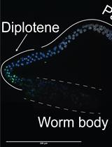
Figure 1. ROS detection. A. Production of florescent DCF by intracellular ROS. B. Measurement of intracellular ROS using molecular probe (H2DCFDA) in C. elegans.
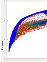
Figure 2. Change in DCF fluorescence signals depends on experimental conditions. A. The expression of gcs-1(promoter)::GFP (oxidative stress marker) transgene by oxidative stress. Heat (30 °C for 2 h) induced the expression of gcs-1(promoter)::GFP transgene in the intestines of live nematodes. B. Concentration-response curves were plotted in terms of the mean value of DCF fluorescence signals induced by 12.5, 25, 50, and 100 μM of H2DCFDA using 50 nematodes for each experiment. C. Changes in DCF fluorescence signals depending on the number of tested nematodes (10, 20, 50, and 100) were assessed using 25 μM of H2DCFDA.
Materials and Reagents
- 1.5 ml microcentrifuge tube (Corning, Axygen®, catalog number: MCT-150-C )
- Conical tube, 15 ml (Corning, Falcon®, catalog number: 352096 )
- Slide glass (VWR, catalog number: 48300-025 )
- 96-well microplate, black (SPL Life Sciences, catalog number: 30496 )
- 60 mm Petri dish (SPL Life Sciences, catalog number: 10060 )
- Platinum wire, 0.2 mm (Alfa Aesar, catalog number: 45093 )
- Caenorhabditis elegans strain, wild-type [N2] (Caenorhabditis Genetics Center, University of Minnesota: https://cgc.umn.edu)
- Escherichia coli OP50 strain (Caenorhabditis Genetic Center)
- Distilled water
- 2’,7’-Dichlorofluorescein diacetate (Sigma-Aldrich, catalog number: D6883 )
- Dimethyl sulfoxide (DMSO) (Sigma-Aldrich, catalog number: D2650 )
- Methyl viologen dichloride hydrate (Merck, catalog number: 856177 )
- Cholesterol (Sigma-Aldrich, catalog number: C8667 )
- Ethanol (Sigma-Aldrich, catalog number: E7023 )
- Sodium chloride (NaCl) (Sigma-Aldrich, catalog number: S5886 )
- Bacto-peptone (BD, BactoTM, catalog number: 211677 )
- Agar (Sigma-Aldrich, catalog number: A1296 )
- Calcium chloride (CaCl2) (Sigma-Aldrich, catalog number: C1016 )
- Magnesium sulfate (MgSO4) (Sigma-Aldrich, catalog number: M7506 )
- Potassium phosphate, dibasic (K2HPO4) (Sigma-Aldrich, catalog number: P3786 )
- Potassium phosphate, monobasic (KH2PO4) (Sigma-Aldrich, catalog number: 795488 )
- Sodium phosphate dibasic (Na2HPO4) (Sigma-Aldrich, catalog number: S3264 )
- Sodium hydroxide (NaOH) (Sigma-Aldrich, catalog number: S8045 )
- Sodium hypochlorite (NaClO) (Sigma-Aldrich, catalog number: 425044 )
- 5 mg/ml cholesterol (see Recipes)
- Nematode growth medium (NGM) agar plates (see Recipes)
- M9 buffer (see Recipes)
- Bleaching solution (see Recipes)
Equipment
- Shaking incubator (Benchtop Shaking Incubator, VWR, model: Model 1570 )
- Micropipette (VWR International)
- Incubators for stable temperature (VWR SIGNATURE 20-cuft Model 2020 B.O.D. -10 °C to 45 °C Low Temp Incubator) (VWR, Advanced Instruments, model: Model 2020 )
- Tabletop centrifuge (VWR, model: VWR® Mini Centrifuge C1413V )
- Dissecting stereomicroscope (Stereo Microscope with Apochromatic Optics Leica S8 APO) (Leica Microsystems, model: Leica S8 APO )
- Fluorophotometer (Promega, model: GloMax®-Multi Microplate Multimode Reader )
- Vortex mixer (Standard Heavy-Duty Vortex Mixer, VWR, catalog number: 97043-562 )
- Autoclave (Hanshin Medical, model: HS-2321SD )
Software
- Microsoft Office 2017 Excel (Microsoft Corporation, Redmond, USA)
- IBM SPSS statistics 24 (IBM Corporation, New York, USA)
Procedure
- Measuring intracellular ROS of nematodes under normal culture conditions
- Preparation of a synchronized nematode population
- Wash a plate containing full adults and eggs with 1 ml of M9 buffer.
- Transfer nematode suspension into a microcentrifuge tube.
- Spin down at 3,884 x g (rcf; 8,500 rpm) for 1 min and remove the supernatant.
- Add 200 μl of Distilled Water (D.W.) and 800 μl of bleaching solution.
- Wait until over 90% of nematodes have lysed and several embryos are released [please see Section 3.2 in Bianchi and Driscoll (2006)].
Note: Check for lysis every 30 sec after 3 min. Vortexing at maximum speed for about 2-3 sec helps with bleach lysis. - Spin down at 3,884 x g (rcf; 8,500 rpm) for 1 min and remove the supernatant.
Note: Be careful not to disturb the pellet. - Wash three times with M9 buffer (1 ml each): Spin down at 137 x g (rcf; 1,000 rpm) for 1 min and remove the supernatant.
- Transfer embryos with 1 ml of M9 buffer to a conical tube containing 1 ml of M9 (total 2 ml).
- Incubate overnight at 20 °C with shaking at 30 rpm to allow L1 larvae to hatch.
- Wash a plate containing full adults and eggs with 1 ml of M9 buffer.
- Counting animals and preparation of nematode solution
- Resuspend the larva pellet and spread 10 μl of the suspension onto a glass slide. Do this in triplicate.
- Count the number of nematodes using a dissecting stereomicroscope and calculate the average number of L1 larvae in each drop.
- Add an appropriate volume of M9 buffer to dilute the larva suspension.
- Shake gently and count again.
- Repeat Steps A2b-A2d until the number of nematodes arrives to around 50 per 10 μl.
- Resuspend the larva pellet and spread 10 μl of the suspension onto a glass slide. Do this in triplicate.
- Preparation of H2DCFDA reaction solution
- Prepare a concentrated stock (50 mM) by dissolving 24.365 mg of H2DCFDA in 1 ml of cell culture grade DMSO.
- Split into 100 μl aliquots in black microcentrifuge tubes and store at -20 °C (stable for at least 3 months).
- Dilute stock solution to 50 μM with M9 buffer as required (volume required = number of test wells x 50 μl).
Note: Working solution should be prepared shortly before use.
- Prepare a concentrated stock (50 mM) by dissolving 24.365 mg of H2DCFDA in 1 ml of cell culture grade DMSO.
- Determination of intracellular ROS levels using a fluorophotometer
- Turn on the fluorophotometer and set plate settings.
- Add 40 μl of M9 buffer into the wells of a black 96-well plate.
- Add 10 μl of nematode solution (prepared in Step A2) into each well.
- Add 50 μl of H2DCFDA reaction solution to a final concentration of 25 μM.
- Prepare blank wells by adding 50 μl of M9 and 50 μl of H2DCFDA reaction solution.
- Immediately, insert the plate into the fluorophotometer and let gently shake for 30 sec.
- Quantify the fluorescence intensity at an excitation wavelength of 490 nm and an emission wavelength of 510-570 nm.
- Measure fluorescence signal every hour (maximum 6 h) and export raw data through USB port (see Figure 2A).
- Turn on the fluorophotometer and set plate settings.
Note: All steps should be done at RT. - Preparation of a synchronized nematode population
- Measuring intracellular ROS of nematodes under oxidative stress conditions
- Paraquat-induced oxidative stress
- Make 50 mM paraquat solution by dissolving methyl viologen dichloride hydrate (paraquat) powder in D.W.
- Prepare arrested L1 larvae as described in Step A1.
- Add 1 part 50 mM paraquat to 9 parts L1-containing M9 buffer (1:10 dilution) and incubate at 20 °C for 2 h with shaking at 30 rpm in the shaking incubator.
- Wash three times with M9 buffer (1 ml each): Spin down at 137 x g (rcf; 1,000 rpm) for 1 min and remove the supernatant.
- Prepare nematode solution as described in Step A2.
- Quantify intracellular ROS levels as described in Steps A3 and A4.
- Make 50 mM paraquat solution by dissolving methyl viologen dichloride hydrate (paraquat) powder in D.W.
- Heat-induced oxidative stress
- Prepare arrested L1 larvae and nematode solution as described in Steps A1 and A2.
- Incubate nematodes at 30 °C for 2 h with shaking at 30 rpm in the shaking incubator.
- Quantify intracellular ROS levels as described in Steps A3 and A4.
Note: Maintain the fluorophotometer temperature at 30 °C throughout the measurement.
- Prepare arrested L1 larvae and nematode solution as described in Steps A1 and A2.
- Paraquat-induced oxidative stress
Data analysis
- For each condition, use at least 20 animals to obtain accurate results.
- Each experiment should be performed at least three independent times.
- Blank-subtracted raw data (DCF fluorescence level) are normalized to those in wild-type (or untreated) nematodes and are presented as mean ± standard deviation using Microsoft Office 2017 Excel software.
- The one-way ANOVA with Tukey’s post-hoc test is carried out using IBM SPSS statistics 24 to analyze the statistical significance of differences between groups.
Notes
- As is the case with many other fluorescent probes, H2DCFDA is photoactive, and thus, minimal exposure to light when handling is vital.
- Whole nematodes should be used instead of lysed nematodes when measuring intracellular ROS using H2DCFDA in C. elegans. Lysis causes disruption of outer cuticle and internal membranes followed by the release of intracellular metal ions such as iron. Since free iron can produce ROS by itself through the Fenton reaction, ROS levels may be overestimated in lysed nematodes compared to measurements in the whole nematode.
- Since H2DCFDA readily diffuses into cells until it reaches equilibrium, a time course of increased basal fluorescence signal can be detected. The gradual increasing tendency is shifted to steep upcurve at the time point of animal death. Therefore, taking measurements within 4 or 6 h after starting experiment is appropriate.
- Adult nematodes (age-synchronized) can also be used to test antioxidant capacity of compounds under oxidative stress conditions with minor modifications. Using a small number of nematodes is recommended (10-20 worms/well). Also, increasing paraquat concentration is required to induce pronounced oxidative stress in adult nematodes (over 20 mM, final concentration). However, a relatively low concentration of paraquat should be given to aged nematodes.
Recipes
- 5 mg/ml cholesterol
5 mg cholesterol powder is dissolved in 1 ml of 100% ethanol and stored at 4 °C - Nematode growth medium (NGM) agar plates (per L)
3 g of NaCl
2.5 g of peptone
17 g of agar
1 ml of cholesterol solution (5 mg/ml)
1 ml of CaCl2 (1 M)
1 ml of MgSO4 (1 M)
25 ml of potassium phosphate (1 M, pH 6.0)
975 ml of distilled water
Autoclave and cool to 50 °C
Pour 10 ml of NGM agar into 60 mm Petri dish and let harden for 1 h - M9 buffer
3 g of KH2PO4
6 g of Na2HPO4
5 g of NaCl
1 ml of MgSO4 (1 M)
Make up to 1 L with distilled water
Autoclave - Bleaching solution
6.25 ml of 4 M NaOH
2.5 ml of NaOCl
50 ml of M9 buffer
Prepare freshly before use
Acknowledgments
This work was supported in part by the National Research Foundation of South Korea (NRF-2017R1C1B5015695) to DSC, Brody Brothers Endowment Grant (21602-664261), NIH (1R15GM112174-01A1), NSF (MCB1714264) to M-H.L. The Caenorhabditis Genetics Center (CGC) is supported by the National Institutes of Health – Office of Research Infrastructure Programs (P40 OD010440). This protocol has been adapted from Yoon et al., 2017. The authors have declared that no competing interests exist.
References
- Bianchi, L. and Driscoll, M. (2006). Culture of embryonic C. elegans cells for electrophysiological and pharmacological analyses. WormBook 1-15.
- Labuschagne, C. F. and Brenkman, A. B. (2013). Current methods in quantifying ROS and oxidative damage in Caenorhabditis elegans and other model organism of aging. Ageing Res Rev 12(4): 918-930.
- Miranda-Vizuete, A. and Veal, E. A. (2017). Caenorhabditis elegans as a model for understanding ROS function in physiology and disease. Redox Biol 11: 708-714.
- Yoon, D. S., Choi, Y., Cha, D. S., Zhang, P., Choi, S. M., Alfhili, M. A., Polli, J. R., Pendergrass, D., Taki, F. A., Kapalavavi, B., Pan, X., Zhang, B., Blackwell, T. K., Lee, J. W. and Lee, M. H. (2017). Triclosan disrupts SKN-1/Nrf2-mediated oxidative stress response in C. elegans and human mesenchymal stem cells. Sci Rep 7(1): 12592.
Article Information
Copyright
© 2018 The Authors; exclusive licensee Bio-protocol LLC.
How to cite
Yoon, D. S., Lee, M. and Cha, D. S. (2018). Measurement of Intracellular ROS in Caenorhabditis elegans Using 2’,7’-Dichlorodihydrofluorescein Diacetate. Bio-protocol 8(6): e2774. DOI: 10.21769/BioProtoc.2774.
Category
Developmental Biology > Cell signaling > Stress response
Biochemistry > Other compound > Oxygen
Cell Biology > Cell signaling > Intracellular Signaling
Do you have any questions about this protocol?
Post your question to gather feedback from the community. We will also invite the authors of this article to respond.