- Submit a Protocol
- Receive Our Alerts
- Log in
- /
- Sign up
- My Bio Page
- Edit My Profile
- Change Password
- Log Out
- EN
- EN - English
- CN - 中文
- Protocols
- Articles and Issues
- For Authors
- About
- Become a Reviewer
- EN - English
- CN - 中文
- Home
- Protocols
- Articles and Issues
- For Authors
- About
- Become a Reviewer
Transplantation of Mesenchymal Cells Including the Blastema in Regenerating Zebrafish Fin
Published: Vol 7, Iss 2, Jan 20, 2017 DOI: 10.21769/BioProtoc.2109 Views: 9554
Reviewed by: Michelle GoodyAnonymous reviewer(s)

Protocol Collections
Comprehensive collections of detailed, peer-reviewed protocols focusing on specific topics
Related protocols

Wounding Zebrafish Larval Epidermis by Laceration
Andrew S. Kennard [...] Julie A. Theriot
Dec 20, 2021 3433 Views

Long-term in toto Imaging of Cellular Behavior during Nerve Injury and Regeneration
Weili Tian [...] Hernán López-Schier
May 5, 2023 2401 Views

Quantifying Cell Proliferation Through Immunofluorescence on Whole-Mount and Cryosectioned Regenerating Caudal Fins in African Killifish
Augusto Ortega Granillo [...] Alejandro Sánchez Alvarado
Dec 20, 2023 2644 Views
Abstract
Regeneration of fish fins and urodele limbs occurs via formation of the blastema, which is a mass of mesenchymal cells formed at the amputated site and is essential for regeneration. The blastema transplantation, a novel technique developed in our previous studies (Shibata et al., 2016; Yoshinari et al., 2012) is a useful approach for tracking and manipulating the blastema cells during fish fin regeneration.
Keywords: ZebrafishBackground
Cell transplantation studies are routinely performed during the early embryonic stage in animal models such as fish, amphibians and mammals, but targeting the transplanted cells to specific tissues has been difficult. Blastema transplantation developed in our studies is an efficient method for introducing mesenchymal donor cells into host fin ray. It enables us to track cell fate and/or manipulate cell signaling such as fibroblast growth factor (Fgf) during fish fin regeneration. Actually, in our recently published work, we transplanted blastema cells, which carried the hsp70l:dominant-negative fgf receptor and the β-actin:dsRed2 transgenes, into a wild-type blastema region and mosaically inhibited Fgf signaling in a subset of fin ray mesenchymal cells (Shibata et al., 2016). This method is applicable for analyzing other cell signals and for tracking cell fate by live cell imaging.
Materials and Reagents
- 50 ml disposable syringe (Terumo, catalog number: SS-50ESZ )
- Stainless steel surgical blade, No.10 (FEATHER Safety Razor, catalog number: No. 10 )
- Plastic dish (9 cm diameter) (As One, catalog number: GD90-15 )
- Glass capillary (1 x 90 mm, without filament) (Narishige, catalog number: G-1 )
- Dissection needle: This is made by attaching 30 G needle (BD, catalog number: 305106 ) to a P1000 pipette chip (BM Equipment, catalog number: BIO1000RF ) whose tip is truncated (Figure 1)
Note: A handmade tool for removing the wound epidermis from the regenerate and for dissecting the blastema. This needle is different from that for transplantation (see Procedure B).
Figure 1. Dissection needle - Syringe filter unit (0.22 µm) (EMD Millipore, catalog number: SLGV033RB )
- Zebrafish transplantation donor strain: Tg(Olactb:loxP-dsred2-loxP-egfp), which constitutively expresses the DsRed2 ubiquitously (Yoshinari et al., 2012). For simplicity, we refer the line as Tg(β-actin:dsred2) in this manuscript
- Zebrafish transplantation host: a wild-type strain, which has been maintained in our facility by inbred breeding
- Agarose (Nacalai Tesque, catalog number: 01028-85 )
- Tricaine (3-aminobenzoic acid ethyl ester) (Sigma-Aldrich, catalog number: A5040 )
- Fetal bovine serum (FBS) (any brand can be used)
- Leibowitz’s L-15 medium (powder) (Thermo Fisher Scientific, GibcoTM, catalog number: 41300070 )
- 1 M Tris-HCl (pH 9)
- L-15 medium (see Recipes)
- 20x tricaine stock solution (see Recipes)
Equipment
- Microwave oven
- Micropipette puller (Sutter Instrument, model: P-97 )
- Forceps
- Glass dish (5 cm diameter)
- Surgical blade handle (FEATHER Safety Razor, catalog number: No. 3 )
- Microforge (Narishige, model: MF-900 )
- Three-dimensional coarse manual manipulator (Narishhige, model: M-152 ) installed on a magnet stand
- P20 micropipette
- Fluorescence stereomicroscope
Procedure
- Preparation of agar gel stage (Figure 2)
- Mix agarose and water to make a 2% agarose solution and melt in a microwave oven.
- Pour the melted agarose into a plastic dish up to a half of dish depth and leave at room temperature for 20-30 min.
- Further fill the dish with the melted 2% agarose solution to approximately full depth and leave at room temperature for 20-30 min.
- Make a rectangular well (2 x 4 cm square, 0.5 cm depth) at the center of gel stage by cutting and removing upper layer of the gel.

Figure 2. Agarose gel stage
- Preparation of transplantation needle
- Pull the glass capillary with the micropipette puller to make a needle with a thin and long shaft. We routinely pull the glass capillary using the same conditions as for microinjection needles (our settings: heat 570, pull 150, velocity 120, time 60).
- Set the pulled needle on the microforge, break the needle with forceps at a site where the inside diameter is approximately 20 μm. Make the end as blunt as possible with forceps. Using the microforge heater, make the needle end smooth and further make a harpoon.

Figure 3. Transplantation needle. Scale bar = 50 μm.
- Preparation of donor mesenchymal cells of whole regenerate including blastema (Video 1)Video 1. Transplantation of mesenchymal cells including the blastema in regenerating zebrafish fin
- Two days before transplantation, anaesthetize the donor zebrafish with 1x tricaine solution (~30 ml or enough volume to cover the fish body) in a plastic dish and cut their caudal fins with a scalpel. Note that the fins of host fish need to be amputated simultaneously. The amputated fish were returned to aquarium and normally fed until transplantation.
- On the day of transplantation (2 days post amputation [dpa]), anaesthetize the donor fish with tricaine and cut the fin regenerate with a scalpel.
- Transfer the fin regenerate with forceps to a glass dish containing 5-10 ml L-15 medium supplemented with 0.01% FBS.
- Remove the wound epidermis from the regenerate (mesenchymal part) using the dissection needles as forceps. Peel the epidermis from the proximal side and remove the whole epidermis by gently pulling the epidermis from the edge as if taking off a T-shirt.
- Divide the mesenchymal part of the regenerate to respective fin ray units. If necessary, the blastema can be further cut into smaller sizes.
- Transplantation (Video 1)
- Set the transplantation needle into the needle holder of manipulator.
- Anaesthetize the host zebrafish with tricaine and place it on the agarose stage (Figure 4 and Figure 5).

Figure 4. Zebrafish fin and the transplantation needle
Figure 5. Microscope and manipulator set up - Make an opening on the surface of wound epidermis using the transplantation needle. Push the needle onto the regenerate surface and tap the head of manipulator knob to break the epidermis.
- Transfer a piece of donor mesenchymal tissue including the blastema to site of the epidermal opening using a P20 micropipette.
- Push the donor mesenchymal tissue with the transplantation needle to insert into the host regenerate. Hook the donor mesenchymal tissue with harpoon of the needle and force to insert into the host regenerate. Because proximal side of the donor mesenchyme is rigid, it is easier to hook the proximal side. Note that it is difficult to push the tissue with thinner needles. Transplantation can be performed for several fin rays at a time, but either of dorsal or ventral side of the fin should be blank as a regeneration control.
- Put the operated fish back to aquarium (Figure 6).
Notes: - Only regenerating fins are amenable to transplantation. Removing the epidermis from the regenerate is easily done with 1.5-2.5 dpa regenerating fin. The host tissue should also be 1.5-2.5 dpa. Only the thick region in the regenerating fin can be used as a host tissue. The transplanted cells in the blastema proliferate to produce lots of progeny cells within fin ray, and this is a hallmark that the transplanted cells incorporated as a part of fin mesenchyme.
- Though we have not traced the transplanted cells beyond 14 dpa, in some cases we observed a significant decrease of donor cells, which could be due to the transplant rejection by host immune system.

Figure 6. A representative example of blastema transplantation. The fin of the Tg(β-actin:dsred2) was amputated 2 days before transplantation, and the blastema tissue was transplanted into the blastema region of wild-type host. Operated fish were anaesthetized and placed on the agarose gel stage, and the pictures were taken under a fluorescence stereomicroscope. Arrowheads, amputation sites; dpt, days post transplantation. Scale bars = 200 μm.
Recipes
- L-15 medium
- Dissolve L-15 powder according to the manufacturer’s instruction
- Sterilize through a 0.22-μm filter
- 20x tricaine stock solution
- Dissolve 2 g of tricaine in 489.5 ml of distilled water
- Add 10.5 ml 1 M Tris-HCl (pH 9) to bring the pH to 7
- Store the solution in aliquots at -30 °C
- Dilute the tricaine stock to 20 times with fish water to make 1x tricaine solution, which is used for anesthesia
Acknowledgments
This protocol was adapted from our previous works (Yoshinari et al., 2012; Shibata et al., 2016). The works were supported by the Grant-in-Aid for Scientific Research from Japan Society for the Promotion of Science (JSPS).
References
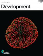
- Shibata, E., Yokota, Y., Horita, N., Kudo, A., Abe, G., Kawakami, K. and Kawakami, A. (2016). Fgf signalling controls diverse aspects of fin regeneration. Development 143(16): 2920-2929.
- Yoshinari, N., Ando, K., Kudo, A., Kinoshita, M. and Kawakami, A. (2012). Colored medaka and zebrafish: transgenics with ubiquitous and strong transgene expression driven by the medaka beta-actin promoter. Dev Growth Differ 54(9): 818-828.
Article Information
Copyright
© 2017 The Authors; exclusive licensee Bio-protocol LLC.
How to cite
Shibata, E., Ando, K. and Kawakami, A. (2017). Transplantation of Mesenchymal Cells Including the Blastema in Regenerating Zebrafish Fin. Bio-protocol 7(2): e2109. DOI: 10.21769/BioProtoc.2109.
Category
Developmental Biology > Cell growth and fate > Regeneration
Stem Cell > Pluripotent stem cell > Blastema cell
Do you have any questions about this protocol?
Post your question to gather feedback from the community. We will also invite the authors of this article to respond.
Share
Bluesky
X
Copy link


