- Protocols

- Articles and Issues
- For Authors
- About
- Become a Reviewer

Bacterial Intracellular Sodium Ion Measurement using CoroNa Green
Published: Vol 7, Iss 1, Jan 5, 2017 DOI: 10.21769/BioProtoc.2092 Views: 10475
Reviewed by: Arsalan DaudiKanika GeraAnonymous reviewer(s)
Original research article
The authors used this protocol in:

Mar 2016
Advertisement


Protocol Collections
Comprehensive collections of detailed, peer-reviewed protocols focusing on specific topics


Related protocols
Abstract
The bacterial flagellar type III export apparatus consists of a cytoplasmic ATPase complex and a transmembrane export gate complex, which are powered by ATP and proton motive force (PMF) across the cytoplasmic membrane, respectively, and transports flagellar component proteins from the cytoplasm to the distal end of the growing flagellar structure where their assembly occurs (Minamino, 2014). The export gate complex can utilize sodium motive force in addition to PMF when the cytoplasmic ATPase complex does not work properly. A transmembrane export gate protein FlhA acts as a dual ion channel to conduct both H+ and Na+ (Minamino et al., 2016). Here, we describe how to measure the intracellular Na+ concentrations in living Escherichia coli cells using a sodium-sensitive fluorescent dye, CoroNa Green (Minamino et al., 2016). Fluorescence intensity measurements of CoroNa Green by epi-fluorescence microscopy allows us to measure the intracellular Na+ concentration quantitatively.
Keywords: BacteriaBackground
Measurements of intracellular Na+ concentrations by fluorescence imaging techniques are able to be more accurately and quantitatively performed at single cell levels, because background noise of each cell can be removed by image analysis procedures. Lo et al. have established a protocol for measurement of the cytoplasmic Na+ concentrations in living E. coli cells using a sodium-sensitive fluorescent dye, Sodium Green and have shown that the cytoplasmic Na+ concentration maintains around 10 mM in E. coli over a wide range of 0 to 100 mM of the external Na+ concentrations (Lo et al., 2006). Because CoroNa Green, which is a sodium-sensitive fluorescent dye too, shows much higher cell permeability than Sodium Green, we have developed a CoroNa Green-based protocol to measure the intracellular Na+ concentrations in E. coli. (Minamino et al., 2016). This protocol allows us to quite easily and reproducibly measure the intracellular Na+ concentration of E. coli cells overexpressing FlhA or PomAB complex, both of which have the Na+ channel activity.
Materials and Reagents
- 1.5 ml Eppendorf tubes
- Aluminum foil
- Slide
- 24 x 32 mm coverslip (thickness: 0.12-0.17 mm) (Matsunami Glass, catalog number: C024321 )
- 18 x 18 mm coverslip (thickness: 0.12-0.17 mm) (Matsunami Glass, catalog number: C018181 )
- Double-sided tape (NICHIBAN, catalog number: NW-5 )
- Pipette tips
- Filter paper
- E. coli BL21(DE3) cell (Novagen)
- pBAD24 expression vector (Guzman et al., 1995)
- pNH319 (pBAD24/ N-His-FLAG-FlhA) (Minamino et al., 2016)
- pBAD-PomΔplug (pBAD24/ PomA + PomB[∆41-120]) (Minamino et al., 2016)
- Ampicillin sodium (Wako Pure Chemical Industries, catalog number: 014-23302 )
- L-arabinose (Wako Pure Chemical Industries, catalog number: 010-04582 )
- CoroNa Green-AM (Thermo Fisher Scientific, Molecular ProbesTM, catalog number: C36676 )
- Ethylenediamine-N, N, N', N'-tetraacetic acid, dipotassium salt, dihydrate (EDTA·2K) (Dojindo, catalog number: 340-01511 )
- Sodium chloride (Wako Pure Chemical Industries, catalog number: 192-13925 )
- Gramicidin (Thermo Fisher Scientific, catalog number: G6888 )
- Carbonyl cyanide 3-chlorophenylhydrazone (CCCP) (Sigma-Aldrich, catalog number: C2759 )
- Bacto tryptone (BD, catalog number: 211705 )
- Potassium dihydrogenphosphate (Wako Pure Chemical Industries, catalog number: 164-22635 )
- Dipotassium hydrogenphosphate (Wako Pure Chemical Industries, catalog number: 164-04295 )
- T-broth (see Recipes)
Equipment
- haking incubator (30 °C, at 200 rpm) (TAITEC, model: BR-40LF )
- Centrifuge (able to hold 1.5 ml tube, spin at 6,000 x g) (TOMY SEIKO, model: MX-305 )
- Tube rotator (able to hold 1.5 ml tubes, rotate at 5 rpm) (WAKENBTECH, model: WKN-2210 )
- Single channel pipettes (1,000 µl, 100 µl) (Gilson, model: P-1000 , P-100 )
- Spectrophotometer (able to measure OD600) (Shimadzu, model: UV-1800 )
- Inverted fluorescence microscope (Olympus, model: IX-73 )
- 100x oil immersion objective lens (Olympus, model: UPLSAPO100XO , NA 1.4)
- sCMOS camera (Andor Technology, model: Zyla4.2 )
- Mercury light source system (Olympus, model: U-HGLGPS )
- Fluorescence mirror unit (Olympus, model: U-FGFP [Excitation BP 460-480; Emission BP 495-540])
Software
- Image J (National Institutes of Health, https://imagej.nih.gov/ij/)
- KaleidaGraph (Synergy Software)
- Microsoft Excel (Microsoft)
Procedure
Note: Carry out procedures at ca. 23 °C unless otherwise specified.
- Transform competent cells of the E. coli BL21(DE3) strain, which are treated with calcium chloride method, with a pBAD24-based plasmid encoding FlhA or the PomAB∆plug, which is a Na+ channel complex of the marine Vibrio flagellar motor (Morimoto and Minamino, 2014).
- Grow the resulting transformants overnight in duplicate in 5 ml T-broth containing 100 µg/ml ampicillin at 30 °C.
- Inoculate 50 µl of overnight culture of the E. coli BL21 (DE3) cells carrying the pBAD24-based plasmid into 5 ml of fresh T-broth containing 100 µg/ml ampicillin and incubate at 30 °C for 4 h with shaking at 150 rpm. (The cell density reaches an OD600 of ca. 1.0.)
- Add 0.2% arabinose (final concentration, w/v) to induce over-expression of FlhA or PomAB∆plug derived by a pBAD promotor on the pBAD24 vector and keep shaking for another 1 h.
- After 1 h: Transfer 200 µl of each culture to a 1.5 ml Eppendorf tube and then collect the cells by centrifugation (6,000 x g, 2 min).
- Suspend the cell pellet gently in 1.0 ml of fresh TB using a pipette (P-1000).
- Centrifuge at 6,000 x g for 2 min.
- Discard supernatant.
- Repeat steps 6-8 (2 x).
- Resuspend the cells in 100 µl of T-broth containing 40 µM CoroNa Green (Invitrogen) and 10 mM EDTA.
- Cover the tubes with aluminum foil to keep them in the dark.
- Rotate the tubes at 5 rpm for 60 min using a tube rotator.
- Centrifuge at 6,000 x g for 2 min.
- Discard supernatant.
- Suspend the cell pellet in 1.0 ml of fresh T-broth.
- Centrifuge at 6,000 x g for 2 min.
- Discard supernatant.
- Repeat steps 14-16 (3 x).
- Resuspend the cells in 500 µl of fresh T-broth.
- Make a tunnel slide by sandwiching double-sided tape between 24 x 32 mm coverslip (bottom side) and 18 x 18 mm coverslip (top side) (see Video 1).
 Video 1. Preparation for a tunnel slide
 - Add the cell suspension to the tunnel slide and leave for 5-10 min to attach the cells onto the coverslip surface.
- Wash out unbound cells by supplying 100 µl of fresh T-broth using a pipette (P-100). Absorb the excess amount of the medium with a piece of filter paper.
- Put the sample on an inverted fluorescence microscope.
- Select a 100x objective.
- Capture fluorescence images of CoroNa Green using a mirror unit for GFP (U-FGFP, Ex: 460–480, Em: 495–540, Olympus) by an sCMOS camera every 100 msec.
- Measure the fluorescence intensity of the cells stained with CoroNa Green in T-broth with 0 mM, 5 mM, 10 mM, 20 mM, 50 mM or 100 mM NaCl in the presence of 20 µM gramicidin and 5 µM CCCP.
Data analysis
Analyze fluorescent images by an image analysis software ImageJ (National Institutes of Health).
- Apply a rectangular mask for the fluorescent image of the bacterial cell body of 8 x 8 pixels to the ROI (region of interest).
- Subtract the total background intensity from each pixel value. The instrumental background intensity is defined as the mean pixel intensity within the ROI of a nearby cell-less region.
- Measure the intensity of CoroNa Green from more than 100 cells at each condition.
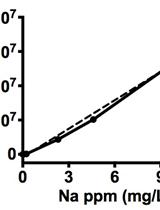
- Make the calibration curve from the data acquired at various external Na+ concentrations in the presence of 20 µM gramicidin and 5 µM CCCP, both of which equilibrate Na+ concentrations inside and outside E. coli cells (Figure 1).

Figure 1. Calibration curve for intracellular Na+ concentrations using CoroNa Green. The fluorescence intensity of the cells stained with CoroNa Green were measured in T-broth containing 20 µM gramicidin and 5 µM CCCP for 30 min at various Na+ concentrations (5 mM, 10 mM, 20 mM, 50 mM and 100 mM NaCl). The fluorescent intensities of intracellular CoroNa Green were plotted against external Na+ concentrations and then were fitted by the Hill equation (black line). Vertical bars indicate standard deviations. Because the staining efficiency with CoroNa Green is quite variable among E. coli cells, the standard deviations of fluorescence intensity become large at high Na+ concentrations. - Fit the calibration curve to the Hill equation by KaleidaGraph (Synergy Software).

- Estimate the intracellular Na+ concentration through the fluorescent intensity of CoroNa Green of each cell using the calibration curve.
- Calculate the average of the intracellular Na+ concentration and standard error of the mean from the data of more than 100 cells using Microsoft Excel (Microsoft).
Representative data

Figure 2. Measurement of intracellular Na+ concentrations using CoroNa Green. Effect of overexpression of FlhA or PomAB∆plug on intracellular Na+ concentrations in E. coli cells. Intracellular Na+ concentrations were measured with CoroNa Green in the presence and absence of 100 mM NaCl at an external pH of 7.0. The E. coli BL21(DE3) strain was transformed with pBAD24 (Vector, V), pNH319 (FlhA) or pBAD-Pom∆plug (PomAB∆plug). For each transformant, 200 cells were measured. Vertical bars indicate standard errors. (Modified from Minamino et al., 2016)
Notes
- To investigate the effect of extracellular sodium concentration, whole experiments are done using T-broth with or without 100 mM NaCl.
- 10 mM EDTA is required to allow CoroNa Green to be incorporated into the E. coli cells.
- To avoid contamination of sodium, EDTA·2K must be used (Do not use EDTA·2Na).
Recipes
- T-broth
1% Bacto tryptone, 10 mM potassium phosphate, pH 7.0 with or without 100 mM NaCl
Acknowledgments
This protocol was modified from a previous work (Lo et al., 2006). This research has been supported in part by JSPS KAKENHI Grant Numbers JP15K14498 and JP15H05593 to YVM, JP21227006 and JP25000013 to KN and JP26293097 to TM and MEXT KAKENHI Grant Numbers JP26115720 and JP15H01335 to YVM and JP23115008, JP24117004, JP25121718 and JP15H01640 to TM.
References
- Guzman, L. M., Belin, D., Carson, M. J., Beckwith. J. (1995). Tight regulation, modulation, and high-level expression by vectors containing the arabinose PBAD promoter. J Bacteriol 177(14): 4121-4130.
- Lo, C. J., Leake, M. C., Berry, R. M. (2006). Fluorescence measurement of intracellular sodium concentration in single Escherichia coli cells. Biophys J 90(1): 357-365.
- Minamino, T. (2014). Protein export through the bacterial flagellar type III export pathway. Biochim Biophys Acta 1843(8): 1642-1648.
- Minamino, T., Morimoto, Y. V., Hara, N., Aldridge, P. D. and Namba, K. (2016). The bacterial flagellar type III export gate complex is a dual fuel engine that can use both H+ and Na+ for flagellar protein export. PLoS Pathog 12(3): e1005495.
- Morimoto, Y. V. and Minamino, T. (2014). Structure and function of the bi-directional bacterial flagellar motor. Biomolecules 4(1): 217-234.
Article Information
Copyright
© 2017 The Authors; exclusive licensee Bio-protocol LLC.
How to cite
Readers should cite both the Bio-protocol article and the original research article where this protocol was used:
- Morimoto, Y. V., Namba, K. and Minamino, T. (2017). Bacterial Intracellular Sodium Ion Measurement using CoroNa Green. Bio-protocol 7(1): e2092. DOI: 10.21769/BioProtoc.2092.
- Minamino, T., Morimoto, Y. V., Hara, N., Aldridge, P. D. and Namba, K. (2016). The bacterial flagellar type III export gate complex is a dual fuel engine that can use both H+ and Na+ for flagellar protein export. PLoS Pathog 12(3): e1005495
Category
Microbiology > Microbial cell biology > Cell viability
Microbiology > Microbial cell biology > Cell-based analysis > Ion analysis
Biochemistry > Other compound > Ion > Sodium
Do you have any questions about this protocol?
Post your question to gather feedback from the community. We will also invite the authors of this article to respond.