- Protocols

- Articles and Issues
- For Authors
- About
- Become a Reviewer

Aniline Blue and Calcofluor White Staining of Callose and Cellulose in the Streptophyte Green Algae Zygnema and Klebsormidium
Published: Vol 6, Iss 20, Oct 20, 2016 DOI: 10.21769/BioProtoc.1969 Views: 14817
Reviewed by: Maria SinetovaRumen IvanovAnonymous reviewer(s)
Original research article
The authors used this protocol in:
Nov 2015
Advertisement


Protocol Collections
Comprehensive collections of detailed, peer-reviewed protocols focusing on specific topics


Abstract
Plant including green algal cells are surrounded by a cell wall, which is a diverse composite of complex polysaccharides and crucial for their function and survival. Here we describe two simple protocols to visualize callose (1→3-β-D-glucose) and cellulose (1→4-β-D-glucose) and related polysaccharides in the cell walls of streptophyte green algae. Untreated or algal cells heated in NaOH are incubated in Calcofluor white (binding to β-glucans including cellulose) or Aniline blue (binding to callose), respectively. Both dyes can be visualized by epifluorescence microscopy.
Background
Due to its easy and quick applicability, Aniline blue was used to visualize callose in various strains of Klebsormidium sp. and Zygnema sp., before more laborious fixation and immunolocalisation protocols were applied (Herburger and Holzinger, 2015). Applying Aniline blue staining and monoclonal antibodies against callose brought similar results (Herburger and Holzinger, 2015). Calcofluor white staining is the fastest way to visualize the 1→4-β-glucan fraction including cellulose of the cell wall, since no pre-treatments are required.
Materials and Reagents
- 2 ml Eppendorf tubes
- Epoxy coated microscopic slides with 8-12 wells (Thermo Fisher Scientific, Thermo ScientificTM, catalog number: 1014326410EPOXY ) and cover slips
- Algal cells or filaments (Figure 1A)
Note: In principle, this protocol is not restricted to green algae and can be applied to all plant cells surrounded by a cell wall, including cell cultures. - Sodium hydroxide pellets (NaOH) (EMD Millipore, catalog number: 106469 )
- A. bidest. (double distilled water)
- Sodium phosphate monobasic dihydrate (NaH2PO4·2H2O) (EMD Millipore, catalog number: 1063420250 )
- Sodium phosphate dibasic dihydrate (Na2HPO4·2H2O) (EMD Millipore, catalog number: 1065800500 )
- Culture medium (BBM [Bischoff and Bold, 1963], MBBM [Starr and Zeikus, 1993])
- Aniline blue diammonium salt (Sigma-Aldrich, catalog number: 415049 )
- Calcofluor white (Sigma-Aldrich, catalog number: 18909 )
Note: Adding Evans blue as a counterstain to diminish background fluorescence is not obligatory, since background fluorescence in the algae investigated is very low. - 0.2 M NaH2PO4·2H2O stock solution (see Recipes)
- 0.2 M Na2HPO4·2H2O stock solution (see Recipes)
- 50 ml of Sørensen’s phosphate buffer (see Recipes)
Equipment
- Glass Pasteur pipettes
- 25 ml beaker
- 1,000 ml and 500 ml volumetric flask
- 100 ml Schott flask
- Balance
- Pair of fine-pointed tweezers
- Metal rack (1.5 ml microfuge rack) (Thermo Fisher Scientific, Thermo ScientificTM, catalog number: 3166185 )
- Water bath or heat plate and beaker
- Tabletop centrifuge (Sigma Laborzentrifugen, model: Sigma 1-14 )
- Shaker (Thermo Fisher Scientific, Thermo ScientificTM, model: Compact Digital Microplate Shaker)
- Epifluorescence microscope capable of UV excitation and long pass detection, with high numerical aperture lens, connected to a camera for documentation (e.g., We used a ZEISS Axiovert 200M equipped with a 63 x 1.4 NA objective, a OSRAM HBO 50 Q/AC L1 CZ Mercury short ARC Photo optic lamp, Zeiss Filter Set 01 (excitation: band pass [BP] 365/12 nm; emission: long pass [LP] 397 nm and connected to a Axiocam MRc5 camera) (Carl Zeiss, model: Axiovert 200M )
Procedure
- Aniline blue staining
- Prepare 20 ml of 8 N NaOH from sodium hydroxide pellets in double distilled water (A. bidest.) in a 25 ml beaker.
- Prepare 50 ml of Sørensen’s phosphate buffer (0.1 M, pH = 8.0).
- Transfer algae to 2 ml Eppendorf tubes (Figure 1B) and wash them once with culture medium.
Note: Washing can be omitted if algae from lab cultures are investigated. Avoid mechanical stress or damage to the cells by using a pair of fine-pointed tweezers. This prevents additional short-term incorporation of callose in the cell walls. Single-celled organisms or cells surrounded by a sticky mucilage layer can be transferred easily by using glass pipettes. - Decant and discard culture medium and add 1.5 ml 8 N NaOH in the tube (Figure 1C).
Note: Centrifuge at ~1,000 x g to sediment biomass in order to remove liquid. - Incubate the tubes at 60 °C for 20 min.
Note: An easy way to do this is fixing the tubes in a metal rack in a pre-heated water bath or in a beaker on a heat plate. Maintain bath level at same level as the liquid in the tubes. - Decant and discard the 8 N NaOH and wash algae 3 x with A. bidest. for at least 5 min.
- Add 1% aniline blue (w/v) to freshly prepared Sørensen’s phosphate buffer.
- In the following, it is recommended to work under low light intensities (~5 μmol photons m-2 s-1) to prevent degradation of the dye.
- Stain algae in 1.5 ml of 1% aniline blue for 1 h (Figure 1D).
Note: Incubate in darkness under gentle shaking. - Wash algae 2 x with Sørensen’s phosphate buffer for 5 min and transfer to microscopic slides (mount in Sørensen’s phosphate buffer).
- Visualize aniline blue with an epifluorescence microscope (excitation: band pass [BP] 365/12 nm; emission: long pass [LP] 397 nm; Figure 1E).
Note: Aniline blue staining allows a quick and easy-to-use visualization of callose in plant’s cell walls. Furthermore, Aniline blue can be used to quantify the callose content in plant material spectrofluorimetrically (Köhle et al., 1985). For a more specific detection of callose at the confocal laser scanning and transmission electron microscopy level, commercially available monoclonal antibodies can be applied (Meikle et al., 1991; Herburger and Holzinger, 2015).
- Prepare 20 ml of 8 N NaOH from sodium hydroxide pellets in double distilled water (A. bidest.) in a 25 ml beaker.
- Calcofluor white staining
- Add 1% (v/v) Calcofluor white to 50 ml of A. dest.
- Transfer algae to 2 ml Eppendorf tubes and wash once with culture medium (Figure 1B).
Note: Washing can be omitted, if algal cultures are investigated. Single-celled organisms or cells surrounded by a sticky mucilage layer can be transferred easily by using glass pipettes. - Stain algae with 1.5 ml 1% Calcofluor white for 20 sec (Figure 1F).
Note: Krishnamurthy (1999) describes protocols using 0.01-0.7% (v/v) Calcofluor white in buffered or unbuffered solutions and incubation times up to 2 min. Although our plant material was rather small (algal filaments with a max. diameter of ~24 µm), a 1% aqueous solution of Calcofluor white produced the best results. Lower concentrations yielded very low fluorescence signals despite incubation times up to 10 min. - Wash algae twice in A. dest. for 30 sec and transfer immediately to microscopic slides (mount in A. dest.).
Note: To immobilize entire algal filaments in one optical plane, epoxy-coated microscopic slides can be used. This allows focusing the central area of algal filaments and reduces blur effects by filament’s top or bottom side (Figure 1G). - Visualize Calcofluor white with an epifluoresence microscope (excitation: band pass [BP] 365/12 nm; emission: long pass [LP] 397 nm; Figure 1G).
Note: Excitation at 400-410 is also fine. Calcofluor white may not specifically bind to cellulose and can also stain callose, chitin, mixed-linkage glycans or other cell wall polysaccharides (Krishnamurthy, 1999).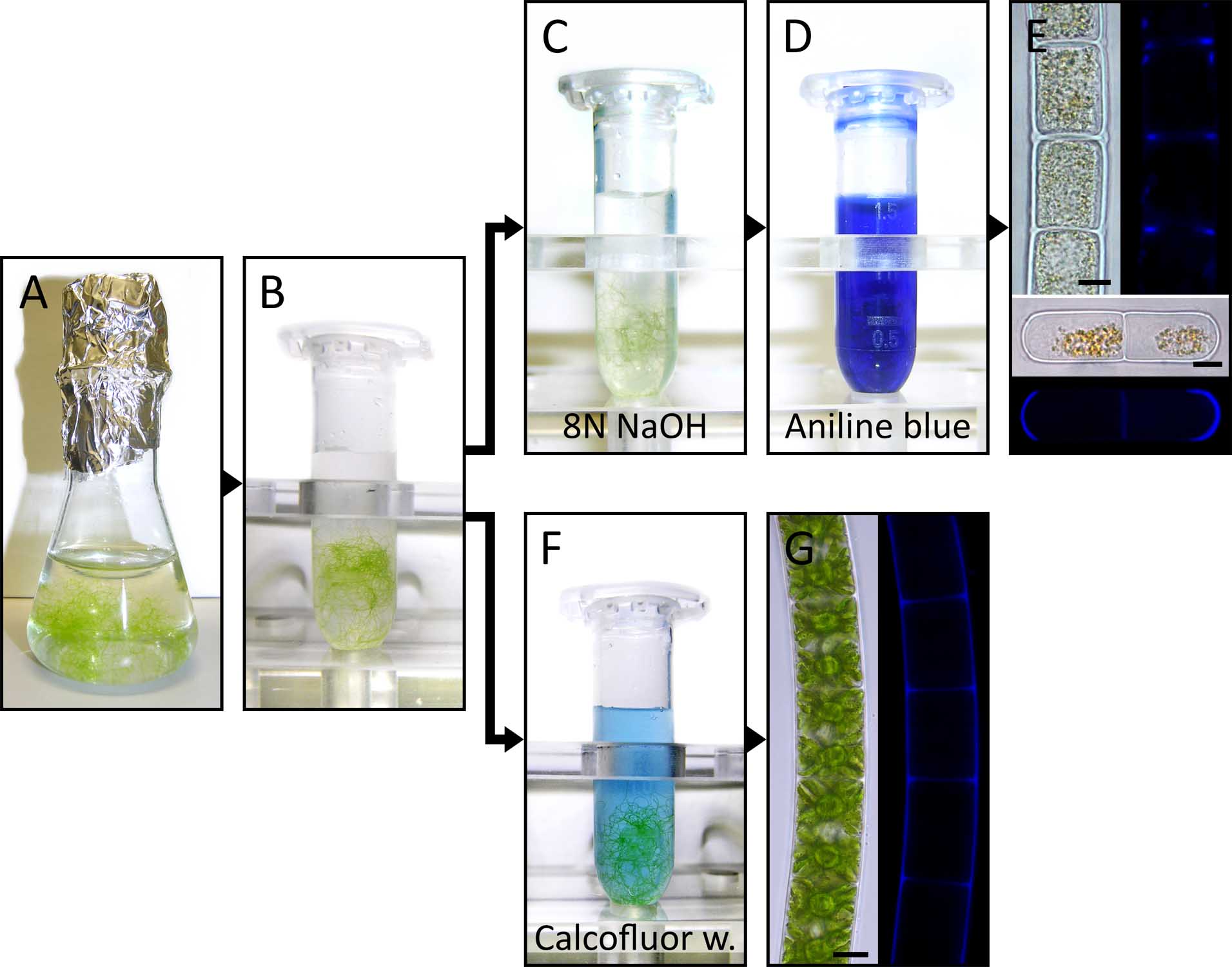
Figure 1. (A-G) Summary of the experimental procedure for Aniline blue (C-E) and Calcofluor white (F, G) staining. Aniline blue (E) and Calcofluor white (G) fluorescence is shown in blue. Bright field and corresponding fluorescence images in (E) and (G) are reprinted from Herburger and Holzinger (2015). Scale bars = 10 µm.
- Add 1% (v/v) Calcofluor white to 50 ml of A. dest.
Recipes
- 0.2 M NaH2PO4·2H2O stock solution
Add 15.60 g sodium phosphate monobasic dehydrate (NaH2PO4·2H2O) to 500 ml of A. bidest.
Note: Can be stored at 4 °C for several months. - 0.2 M Na2HPO4·2H2O stock solution
Add 35.61 g sodium phosphate dibasic dihydrate (Na2HPO4·2H2O) to 1,000 ml of A. bidest.
Note: Can be stored at 4 °C for several months. - 50 ml of Sørensen’s phosphate buffer (0.1 M, pH = 8.0)
Mix 2.7 ml of 0.2 M NaH2PO4·2H2O stock solution with 47.3 ml of 0.2 M Na2HPO4·2H2O stock solution (Sørensen, 1909) in a 100 ml Schott flask.
Acknowledgments
Aniline blue staining as applied by Herburger and Holzinger (2015) and described in this protocol is based on the fluorescence method of Currier et al. (1955), previously used for pollen tube staining (Linskens and Esser, 1957). Calcofluor white staining follows the protocol of Krishnamurthy (1999). The authors declare that there is no conflict of interests. The study was funded by Austrian Science Fund (FWF) projects P 24242-B16 and I 1951-B16 to A.H.
References
- Bischoff, H. W. and Bold, H. C. (1963). Phycological studies IV. Some soil algae from Enchanted Rock and related algal species. Univ Tex Publ 6318: 1-95.
- Currier, H. B., Esau, K. and Cheadle, V. I. (1955). Plasmolytic studies of phloem. Am J Bot 42: 68-81.
- Herburger, K. and Holzinger, A. (2015). Localization and quantification of callose in the streptophyte green algae Zygnema and Klebsormidium: Correlation with desiccation tolerance. Plant Cell Physiol 56(11): 2259-2270.
- Krishnamurthy, K. V. (1999). Methods in cell wall cytochemistry. CRC Press: 156-158.
- Köhle, H., Jeblick, W., Poten, F., Blaschek, W. and Kauss, H. (1985). Chitosan-elicited callose synthesis in soybean cells as a ca-dependent process. Plant Physiol 77(3): 544-551.
- Linskens, H. F. and Esser, K. L. (1957). Über eine spezifische Anfärbung der Pollenschläuche im Griffel und die Zahl der Kallosepfropfen nach Selbstung und Fremdung. Naturwissenschaften 44: 16-16.
- Meikle, P. J., Bonig, I., Hoogenraad, N. J., Clarke, A. E. and Stone, B. A. (1991). The location of (1-->3)-β-glucans in the walls of pollen tubes of Nicotiana alata using a (1-->3)-β-glucan-specific monoclonal antibody. Planta 185(1): 1-8.
- Sørenson, S. P. L. (1909). Enzymstudien. II, Über die Messung und die Bedeutung der Wasserstoffionenkonzentration bei enzymatischen Prozessen. Biochem Zeitschr 21: 131-304.
- Starr, R. C. and Zeikus, J. A. (1993). UTEX - the culture collection of algae atthe University of Texas at Austin 1993 list of cultures. J Phycol 29: 1-106.
Article Information
Copyright
© 2016 The Authors; exclusive licensee Bio-protocol LLC.
How to cite
Herburger, K. and Holzinger, A. (2016). Aniline Blue and Calcofluor White Staining of Callose and Cellulose in the Streptophyte Green Algae Zygnema and Klebsormidium. Bio-protocol 6(20): e1969. DOI: 10.21769/BioProtoc.1969.
Category
Plant Science > Phycology > Cell analysis
Plant Science > Plant cell biology > Cell imaging
Cell Biology > Cell imaging > Fluorescence
Do you have any questions about this protocol?
Post your question to gather feedback from the community. We will also invite the authors of this article to respond.