- Protocols

- Articles and Issues
- For Authors
- About
- Become a Reviewer

Liquid Luminescent DNA-precipitation Assay


Published: Vol 6, Iss 10, May 20, 2016 DOI: 10.21769/BioProtoc.1812 Views: 8779
Reviewed by: Jia LiGriselda Zuccarino-CataniaAnonymous reviewer(s)
Original research article
The authors used this protocol in:
Jul 2015
Advertisement


Protocol Collections
Comprehensive collections of detailed, peer-reviewed protocols focusing on specific topics


Abstract
Working on transcription factors requires studying interactions between protein and DNA. After identification of putative binding-sequences and motifs, Electrophoretic Mobility Shift Assay (EMSA) experiment is classically used to determine specific interactions of proteins and nucleic acids. This lengthy process is rather heavy-handed because of radioisotopically labeled DNA and autoradiographic visualization that are required for the experiments.
Liquid luminescent DNA precipitation assay provides rapid, reliable and quantitative results concerning protein-DNA interactions. This protein-DNA binding assay is based on solution hybridization between Digoxigenin-labeled (DIG) DNA and glutathione S-transferase (GST)-fused DNA binding protein bound to Glutathione Sepharose 4B beads (Figure 1), without electrophoresis (Toshiharu et al., 2008). Digoxigenin is a steroid found in plants. It is increasingly used as a label for nonradioactive detection of nucleic acids and proteins.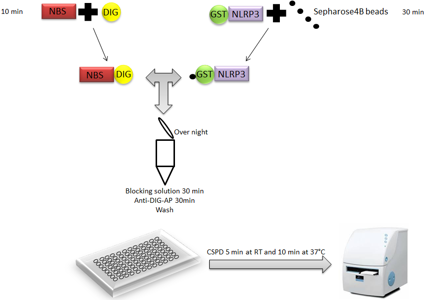
Figure 1. Representation of liquid chemiluminescent DNA pull-down assay. A Glutathione S-transferase (GST)-fused NLRP3 (GST-NLRP3) bound to Glutathione Sepharose 4B beads is incubated with a DIG-labeled double-stranded DNA fragment containing putative NLRP3 Binding Site (NBS) in protein-DNA binding buffer. After extensive washing, protein-DNA binding on beads is detected using anti-DIG antibody conjugated to alkaline phosphatase, which is measured by a chemiluminescent reaction using a luminometer Disodium 3-(4-methoxyspiro {1,2-dioxetane-3,2′-(5′-chloro) tricyclo [3.3.1.13, 7] decan}-4-yl) phenyl phosphate(CSPD).
Here, we described how we used this technique to demonstrate the interaction between NLRP3 protein and its DNA binding site (Bruchard et al., 2015).
Materials and Reagents
- BL21 bacteria (DE3) (Thermo Fisher Scientific, InvitrogenTM)
- pGEX-4T-1 vector (Addgene, catalog number: 27458001 )
- GST fusion NLRP3 protein
- Glutathione sepharose 4B (GE Healthcare, catalog number: 17-0756-01 )
- Potassium chloride (KCl) (Sigma-Aldrich, catalog number: P9541 )
- Sodium chloride (NaCl) (Sigma-Aldrich, catalog number: S7653 )
- Ethylenediaminetetraacetic acid (EDTA), pH 8 (Sigma-Aldrich, catalog number: E9884 )
- Dithiothreitol (DTT) (Sigma-Aldrich, catalog number: D0632 )
- Glycerol (Sigma-Aldrich, catalog number: G5516 )
- Triton X-100 (Sigma-Aldrich, catalog number: T9284 )
- Double-stranded oligonucleotides containing the putative binding sequence (Life Technologies)
- DIG gel shift kit (Roche Diagnostics, catalog number: 03353591910 )
- Disodium 3-(4-methoxyspiro {1, 2-dioxetane-3, 2′-(5′-chloro) tricyclo [3.3.1.13,7] decan}-4-yl) phenyl phosphate (CSPD) (chemiluminescent substrate)
Note: It is included in DIG gel shift kit. - DNAse/RNase free water (Thermo Fisher Scientific, catalog number: 11538646 )
- Tris-HCl (Sigma-Aldrich, catalog number: T5941 )
- Binding buffer (see Recipes)
- Washing buffer (see Recipes)
- Maleic acid buffer (see Recipes)
- 10x blocking solution (see Recipes)
- Detection buffer (see Recipes)
Equipment
- End-over-end rotator
- PerkinElmer Envision Plate Reader (PerkinElmer Inc., catalog number: 2104-0010A )
Procedure
GST fusion NLRP3 proteins were previously produced in BL21 bacteria. GST alone was used as a control. Briefly, classical cloning method was used to insert GST-NLRP3 sequence in pGEX-4T-1 vector. BL21 were transformed by a heat shock according to supplier’s instructions. 24 h later bacteria were lysate with 1% Triton X-100 solution.
- Day 1
Binding of GST-fusion protein and Glutathione Sepharose 4B beads- Glutathione Sepharose 4B was washed twice with 5 ml binding buffer and centrifuged at 500 x g for 5 min at room temperature.
- GST-fused proteins were purified with Glutathione Sepharose 4B. Three ml of bacteria lysate obtained after BL21 sonication were added to the prepared Glutathione Sepharose 4B in a final volume of 30 ml of binding buffer, and incubated for 30 min at room temperature with gentle agitation. Protein/Glutathione Sepharose 4B complexes were isolated by centrifugation at 500 x g for 5 min at room temperature and washed with 5 ml binding buffer. Centrifugation at 500 x g for 5 min allowed collecting Protein/Glutathione Sepharose 4B complexes.
DNA labeling reaction
The NLRP3-binding sequence (NBS) (5′-TCTGTTTTGGGAGGCAGAGCTTTGTTTCTATG-3′) was labeled with Digoxigenin through the use of a DIG Gel Shift Kit but Liquid chemiluminescent DNA pull-down assays can also be performed using biotinylated DNA. - Double-stranded oligonucleotides were diluted to 10 ng/µl with DNAse/RNase free water.
- Put the tubes containing 100 ng of double-strand DNA (10 µl) on ice and add 4 µl labeling buffer, 5 mM CoCl2-solution, 0.05 mM DIG-ddUTP solution and 200 U Terminal transferase. Mix by pipetting up and down.
- The tubes were centrifuged and incubated for 15 min at 37 °C in a dry block heater and put immediately on ice after this incubation.
- The reaction was stopped with 2 µl EDTA.
- DNA was then diluted to 4 ng/µl by adding 3 µl of water (DNAse/RNAse free).
Protein-DNA binding reaction - 2 µg of GST-proteins bound to Glutathione Sepharose 4B beads were incubated with 20 fmol DIG-labeled double-stranded DNA fragments in protein-DNA binding buffer from the DIG Gel Shift Kit in a final volume of 20 µl overnight at room temperature.
- Glutathione Sepharose 4B was washed twice with 5 ml binding buffer and centrifuged at 500 x g for 5 min at room temperature.
- Day 2
- GST-fused proteins bound to DNA were washed three times with 1 ml Washing Buffer from the kit and centrifuged each time at 500 x g for 5 min at room temperature.
- Then the beads were incubated in 500 µl 1x blocking solution (dilution of 10x blocking solution in maleic acid buffer) for 30 min at room temperature.
- GST-fused proteins bound to DNA were incubated with anti-DIG Fab fragments conjugated with 75 mU/ml alkaline phosphatase (from DIG gel shit kit) for 30 min at room temperature in 1x blocking solution.
- GST-fused proteins bound to DNA are washed twice with 5 ml Washing Buffer.
- After washes, the complexes were transferred in detection buffer to 96-well plates and were incubated for 5 min at room temperature with 1 μg/ml 3-(4-methoxyspiro {1,2-dioxetane-3,2′-(5′-chloro) tricyclo [3.3.1.13,7] decan}-4-yl) phenyl phosphate (CSPD) , followed by incubation for 10 min at 37 °C.
- Light emission was measured using a PerkinElmer Envision Plate Reader. Light detection was performed during 1 sec per well.
- GST-fused proteins bound to DNA were washed three times with 1 ml Washing Buffer from the kit and centrifuged each time at 500 x g for 5 min at room temperature.
Representative data
The emission signal from all the reagents mixed together without DNA-DIG was used for normalization and data are presented as arbitrary units. All experimental conditions were tested with GST-NLRP3 and GST alone. Double strand DNA labeled without DIG was tested alone and used as a competitor (Figure 2).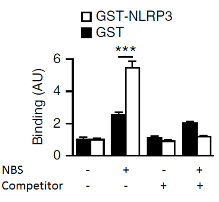
Figure 2. Representative data of liquid chemiluminescent DNA pull-down assay. Light emission was measured from the liquid chemiluminescent DNA pull-down assay to indicate binding. The histograms indicate relative light emission (arbritary units, AU). Glutathione S-transferase (GST)-NLRP3 was incubated with Digoxigenin (DIG)-labeled NBS with or without Competitor (non labelled NBS). Data are shown as means ± SEM.
Recipes
- Binding buffer
75 mM KCl
50 mM NaCl
1 mM EDTA
1 mM DTT
10% glycerol
0.1% Triton X-100 - Washing buffer
0.1 M maleic acid
0.15 M NaCl (pH 7.5)
0.3% (v/v) Tween 20 - Maleic acid buffer
0.1 M maleic acid
0.15 M NaCl
Adjust with NaOH to pH 7.5 - 10x blocking solution
10% (w/v) blocking reagent in maleic acid buffer - Detection buffer
0.1 M Tris-HCl
0.1 M NaCl (pH 9.5)
Acknowledgments
This protocol is adapted from Toshiharu et al. (2008). This project was supported by the French National Research Agency (“Investissements d’Avenir” program; ANR-11-LABX-0021), the Ligue nationale contre le cancer (F. G. and F. V.), the Institut National du Cancer (F. G.), Fondation pour la Recherche Médicale (F. G).
References
- Bruchard, M., Rebe, C., Derangere, V., Togbe, D., Ryffel, B., Boidot, R., Humblin, E., Hamman, A., Chalmin, F., Berger, H., Chevriaux, A., Limagne, E., Apetoh, L., Vegran, F. and Ghiringhelli, F. (2015). The receptor NLRP3 is a transcriptional regulator of TH2 differentiation. Nat Immunol 16(8): 859-870.
- Iwasaki, T., Miyazaki, W., Rokutanda, N. and Koibuchi, N. (2008). Liquid chemiluminescent DNA pull-down assay to measure nuclear receptor-DNA binding in solution. Biotechniques 45(4): 445-448.
Article Information
Copyright
© 2016 The Authors; exclusive licensee Bio-protocol LLC.
How to cite
Végran, F., Bruchard, M., Derangère, V. and Ghiringhelli, F. (2016). Liquid Luminescent DNA-precipitation Assay. Bio-protocol 6(10): e1812. DOI: 10.21769/BioProtoc.1812.
Category
Cell Biology > Cell-based analysis > Protein-DNA interactions
Do you have any questions about this protocol?
Post your question to gather feedback from the community. We will also invite the authors of this article to respond.