- Protocols

- Articles and Issues
- For Authors
- About
- Become a Reviewer

Isolation and Purification of Murine Microglial Cells for Flow Cytometry

Published: Vol 6, Iss 1, Jan 5, 2016 DOI: 10.21769/BioProtoc.1703 Views: 15510
Reviewed by: Sabine Le SauxJia Li
Original research article
The authors used this protocol in:
Apr 2015
Advertisement


Protocol Collections
Comprehensive collections of detailed, peer-reviewed protocols focusing on specific topics


Related protocols
Abstract
The detailed protocol is used to isolate different cell types from murine brain as glial cells, including microglia, using an enzymatic digestion that minimizes cellular mortality. A Percoll gradient (30% to 80%) separation allows a maximal recovery of isolated murine microglial cells prior to flow cytometry analysis.
Keywords: Flow CytometryMaterials and Reagents
- Syringe 10 ml (BD bioscience, catalog number: 309604 )
- Needle 22 G (BD bioscience, catalog number: 305155 )
- Syringe 1 ml 27 G (BD bioscience, catalog number: 309623 )
- 15 ml centrifugation tube (Thermo Fisher Scientific, catalog number: 339651 )
- 50 ml centrifugation tube (Thermo Fisher Scientific, catalog number: 339653 )
- 96 v-shaped wells plate (Thermo Fisher Scientific, catalog number: 249662 )
- 5 ml polystyrene FACS tube (BD Biosciences, Falcon®, catalog number: 352054 )
- 70 µm nylon filter (VWR International, catalog number: 352350 )
- Mice
- PBS without Ca2+/Mg2+ (BOLLÉ COMMUNICATIONS, Wisent, catalog number: 311-010-CL )
- Ice cold Dulbecco’s Phosphate Buffered Saline (DPBS) without Ca2+/Mg2+/ Dulbecco’s Phosphate Buffered Saline (Sigma-Aldrich, catalog number: D8537 )
- PBS 10x without Ca2+/Mg2+ (BOLLÉ COMMUNICATIONS, Wisent, catalog number: 311-012-CL )
- Nα-Tosyl-L-lysine chloromethyl ketone hydrochloride (TLCK) (Sigma-Aldrich, catalog number: T7254 )
- Liberase TL Research Grade (Roche Diagnostics, catalog number: 05401020001 )
- DNAse1 (Roche Diagnostics, catalog number: 11284932001 )
- HEPES (1 M) (Sigma-Aldrich, catalog number: H0887 )
- Fetal Bovine Serum (FBS) (Sigma-Aldrich, catalog number: F1051 )
- EDTA (Sigma-Aldrich, catalog number: E16144 )
- Percoll (GE Healthcare, Dharmacon, catalog number: 17-0891-01 )
- HBSS 10x without Ca2+/Mg2+ (BOLLÉ COMMUNICATIONS, Wisent, catalog number: 311-506-CL )
- HBSS without Ca2+/Mg2+ with red phenol (Life technologies, catalog number: 14170-112 )
Note: Currently, it is “Thermo Fisher Scientific, GibcoTM, catalog number: 14170-112”.
- Purified Rat anti-mouse CD16/CD32 (BD Bioscience, catalog number: 553141 )
- Anti-mouse CD45 PE/Cy5 (BD Bioscience, catalog number: 553082 )
- Anti-mouse CD11b Alexa 700 (eBioscience, catalog number: 56-0112-82 )
- Anti-mouse CD16/CD32 (1:100) (BD Biosciences, catalog number: 553142 )
- Live/Dead Fixable Blue Dead Cell (Life technologies, catalog number: L23105 )
Note: Currently, it is “Thermo Fisher Scientific, Molecular ProbesTM, catalog number: L23105”.
- Digestion medium (see Recipes)
- Ice-cold FACS buffer (see Recipes)
- Percoll dilution medium (see Recipes)
- PFA 4% (w/v) solution (see Recipes)
Equipment
- Tissue grinder Capacity 3 ml (VWR International, catalog number: 886000-0020 )
- Centrifuge Sorvall legend RT (Mandel, catalog number: 75004377 )
- Flow cytometer BD Canto II or LSR II (BD bioscience)
- Peristaltic pump, facultative (Cole-Parmer Instrument Company, catalog number: EW-78001-02 )
Software
- BD FACS Diva software (version 6.1.2)
Procedure
- Stage 1: Animal preparation and perfusion
- Anesthetize mice via an i.p. injection of a ketamine hydrochloride and xylazine mixture (10 ml and 1 ml respectively).
- Flush deeply anesthetized mice with ice-cold sterile DPBS (perfuse transcardially with cold dPBS using peristaltic pump or a 10 ml syringe connect with 22G needle. Many well describe protocols are available online but for good examples see References Video 1 and Video 2.
- Anesthetize mice via an i.p. injection of a ketamine hydrochloride and xylazine mixture (10 ml and 1 ml respectively).
- Stage 2: Brain extraction and Isolation of microglial cells
- Extract one brain and put the brain tissues into a tissue grinder containing 2 ml of ice-cold DPBS without Ca2+/Mg2+.
- After homogenization, transfer homogenates into 15 ml centrifugation tubes (homogenize the tissues completely by doing up and down with the grinder bar).
- Rinse three times the plunger with 2 ml of ice-cold DPBS. Keep on ice.
- Centrifuge homogenates at 300 x g for 10 min at 4 °C.
- Aspirate supernatants and gently resuspend pellets in 3 ml of Digestion medium.
- Incubate homogenates at 37 °C for 40 min and shake tubes every 10 min.
- After digestion, complete volumes to 10 ml with ice-cold FACS buffer.
- Transfer homogenates into new 50 ml centrifugation tubes by filtering cell suspensions through 70 µm nylon filters.
- Wash cells by rinsing the 15 ml centrifugation tube with 10 ml of ice-cold FACS buffer and transfer into the 50 ml centrifugation tube through the same 70 µm-filter.
- Centrifuge homogenates at 300 x g for 10 min at 4 °C.
- Aspirate supernatants and wash cells by adding 20 ml of ice-cold FACS buffer.
- Centrifuge homogenates once again at 300 x g for 10 min at 4 °C.
- During the centrifugations, prepare the isotonic Percoll solution by diluting Percoll 10:1 in 10x HBSS. Prepare needed volumes of 30% and 80% Percoll solutions by diluting the Percoll isotonic solution with the Percoll dilution medium.
Note: Keep Percoll solutions at room temperature.
- Resuspend cell pellets in 8 ml of 30% Percoll solution.
- Transfer 4 ml into a 15 ml centrifugation tube.
- Using a serological pipette, carefully transfer 3 ml of 80% Percoll solution below the cell containing 4 ml of 30% Percoll solution.
Note: Take 4-5 ml in the serological pipette and use gravity to underlay the 80% Percoll solution to avoid the mixture of the upper and lower layers, which could affect the efficiency and yield of microglia isolation.
- Add the remaining cell containing 30% Percoll solution to the upper phase.
- Centrifuge at 1,050 x g with no brakes and low acceleration for 40 min at room temperature.
- Aspirate the fat containing layer at the top of the liquid.
- Using a P1000 micropipette, carefully collect isolated microglial cells (~2 ml) into a new 15 ml centrifugation tube containing 10 ml of ice-cold FACS buffer.
Note: Microglial cells are within the cell ring at the interface between the pink top layer (30% Percoll) and the colorless bottom layer (80% Percoll).
- Centrifuge isolated microglial cells at 300 x g for 10 min at room temperature.
- Aspirate supernatants and resuspend pelleted cells into 250 µl of ice-cold FACS Buffer.
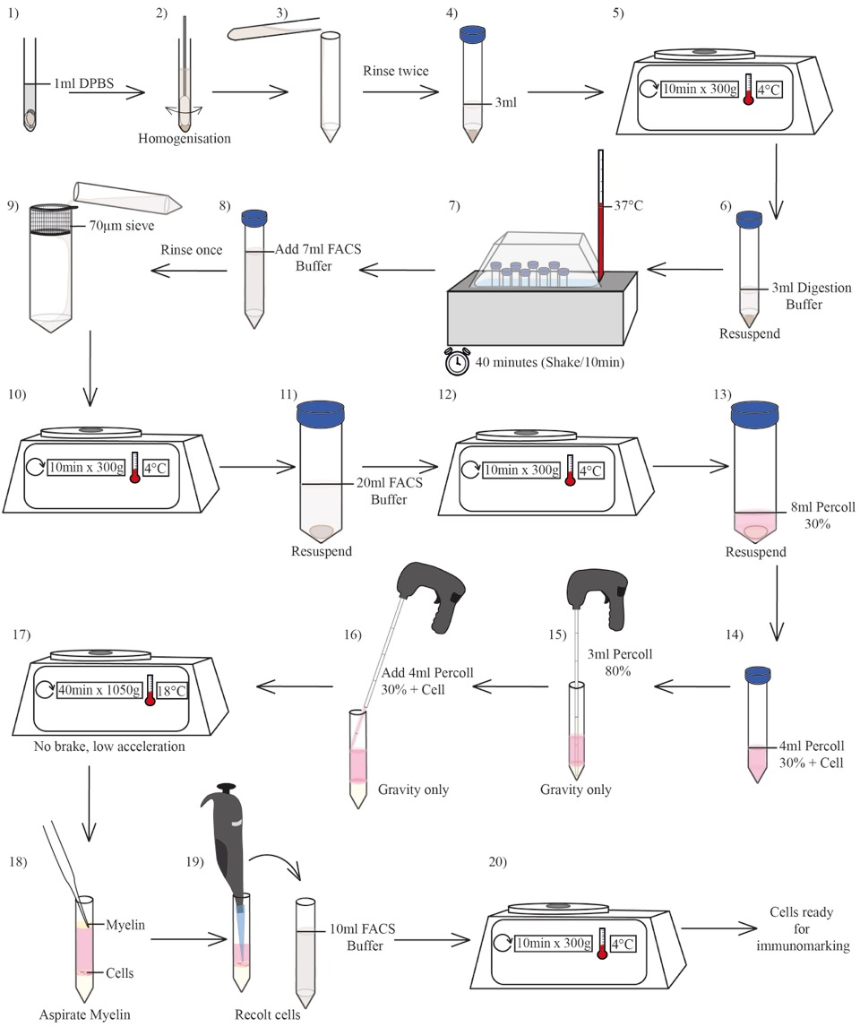
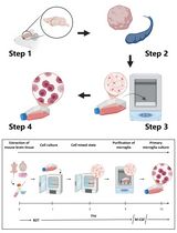
Figure 1. Schematic diagram of the gradient preparation
- Extract one brain and put the brain tissues into a tissue grinder containing 2 ml of ice-cold DPBS without Ca2+/Mg2+.
- Stage 3: Microglia cell markers staining and flow cytometry
- Transfer all cell suspensions (250 µl) into 96 v-shaped well plate.
- Centrifuge cells at 300 x g for 3 min at 4 °C and remove supernatants by inverting the plate.
- Resuspend cells into 200 µl of ice-cold FACS buffer containing purified rat anti-mouse CD16/CD32 (1:100) for Fc receptors blocking.
- Incubate on ice for 20 min.
- Centrifuge cells at 300 x g for 3 min at 4 °C and remove supernatants.
- Resuspend cells into 50 µl of ice-cold FACS buffer containing rat anti-mouse CD45 PEG/Cy5 (1:50), rat anti-mouse CD11b Alexa 700 (1:50) and Live/Dead Blue fluorescent reactive dye (1:50).
- Incubate on ice for 45 min.
- Add 200 µl of ice-cold FACS buffer.
Note: At this step, it is possible to fix cells for a subsequent analysis on the flow cytometer by adding 50 µl of 4% (w/v) paraformaldehyde (PFA) solution at pH = 7.4. Incubate at room temperature for 15 min and wash cells with ice-cold FACS buffer prior to their storage at 4 °C in the dark, for up to 4 days.
- Centrifuges cells at 300 x g for 3 min at 4 °C.
- Resuspend cells into 250 µl of ice-cold FACS buffer and transfer into 5 ml polystyrene tubes for flow cytometry.
- Analyze samples by using a two-laser, six-colors FACS Canto II flow cytometer and data acquisition with BD FACSDiva software (version 6.1.2).
- Transfer all cell suspensions (250 µl) into 96 v-shaped well plate.
Representative data
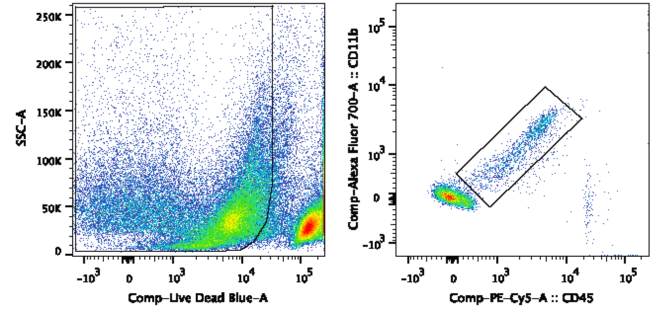
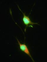
Figure 2. Example of results following an analysis of isolated microglial cells by flow cytometry. Dot plot showing a representative sample of isolated microglial cells analyzed by flow cytometry following a Live/Dead-Blue, CD45-PE-Cy5 and CD11b-AF700 immunofluorescent staining.
Recipes
- Digestion medium
TLCK 0.5 µg/ml
DNAse1 25 ng/ml
Liberase 8.125 pg/ml
HEPES 20 mM
In sterile water
- Ice-cold FACS buffer
5% FBS, EDTA (20 mM), HEPES (2 mM) dilute in PBS
- Percoll dilution medium
Red phenol HBSS with EDTA (2 mM) and HEPES (20 mM)
- PFA 4% (w/v) solution (pH = 7.4)
NaOH (50 mN) 4% (w/v) paraformaldehyde
50 mN NaOH
26.5 mM NaH2PO4
77 mM Na2HPO4
(77 mM)
Acknowledgments
This work was supported by grants from the Canadian Institutes for Health Research (CIHR) and the Multiple Sclerosis Scientific Research Foundation of Canada. This protocol was adapted from previous work of Martine Lessard and Antoine Lampron.
References
- Lampron, A., Larochelle, A., Laflamme, N., Prefontaine, P., Plante, M. M., Sanchez, M. G., Yong, V. W., Stys, P. K., Tremblay, M. E. and Rivest, S. (2015). Inefficient clearance of myelin debris by microglia impairs remyelinating processes. J Exp Med 212(4): 481-495.
- Video 1: www.youtube.com/watch?v=szvmT5iI0z0.
- Video 2: www.youtube.com/watch?v=3H8K7ooPWLs.
Article Information
Copyright
© 2016 The Authors; exclusive licensee Bio-protocol LLC.
How to cite
Theriault, P., Bordeleau, M. and Rivest, S. (2016). Isolation and Purification of Murine Microglial Cells for Flow Cytometry. Bio-protocol 6(1): e1703. DOI: 10.21769/BioProtoc.1703.
Category
Neuroscience > Cellular mechanisms > Cell isolation and culture
Cell Biology > Cell-based analysis > Flow cytometry
Cell Biology > Cell isolation and culture > Cell isolation
Do you have any questions about this protocol?
Post your question to gather feedback from the community. We will also invite the authors of this article to respond.