- Protocols

- Articles and Issues
- For Authors
- About
- Become a Reviewer

Histochemical Detection of Zn in Plant Tissues


(*contributed equally to this work) Published: Vol 5, Iss 10, May 20, 2015 DOI: 10.21769/BioProtoc.1470 Views: 11484
Reviewed by: Maria SinetovaAnonymous reviewer(s)
Original research article
The authors used this protocol in:
Jul 2014
Advertisement


Protocol Collections
Comprehensive collections of detailed, peer-reviewed protocols focusing on specific topics


Related protocols
Abstract
Accumulation of metals in plant tissues, and occasionally, different cells of the same tissue, may be highly non-uniform (Seregin and Kozhevnikova, 2008). Easy-to-use histochemical methods may greatly help to investigate the distribution and accumulation of metals within and among plant tissues, and also provide information on their subcellular localization (Seregin and Kozhevnikova, 2011). The histochemical techniques of zinc (Zn) visualization are based on the formation of the blue-colored complex of Zn with the metallochrome indicator Zincon (C20H15N4NaO6S), or the green-fluorescent complex with Zinpyr-1 (C46H36Cl2N6O5) (Seregin et al., 2011; Seregin and Kozhevnikova, 2011). A method for histochemical Zn detection in plant tissues using Zinpyr-1 was first proposed by Sinclair et al. (2007), and later modified by Seregin et al. (2011), and Seregin and Kozhevnikova (2011). Histochemical data supplement the results of quantitative analysis, thus allowing a detailed study of the distribution, accumulation, and translocation pathways of Zn within the plant, which are important topics in modern plant physiology. These histochemical techniques have been successfully applied in different plant species, for example Zea mays (Seregin et al., 2011), Noccaea caerulescens and Thlaspi arvense (Kozhevnikova et al., 2014a), Capsella bursa-pastoris and Lepidium ruderale (Kozhevnikova et al., 2014b), in which Zn was detected in different root and shoot tissues. Here, we present the full staining protocols for these methods, developed or modified in our lab (Seregin and Kozhevnikova, 2011; Kozhevnikova et al., 2014a; Kozhevnikova et al., 2014b).
Keywords: Zinc localizationMaterials and Reagents
- Staining with Zinpyr-1 (see Recipes)
- Zinpyr-1 or 4′, 5′-Bis[bis(2-pyridylmethyl)aminomethyl]-2′,7′-dichlorofluorescein (C46H36Cl2N6O5, Mr= 823.72) (Sigma-Aldrich, catalog number: 40667 or Millitech, catalog number: ZP1 )
- Dimethyl sulfoxide or DMSO (C2H6OS)
- Ethylenediaminetetraacetic acid (disodium salt) (Na2EDTA, C10H14O8N2Na2⋅2H2O, Mr = 372.24)
- Super demineralized water
- Zinpyr-1 or 4′, 5′-Bis[bis(2-pyridylmethyl)aminomethyl]-2′,7′-dichlorofluorescein (C46H36Cl2N6O5, Mr= 823.72) (Sigma-Aldrich, catalog number: 40667 or Millitech, catalog number: ZP1 )
- Staining with Zincon (see Recipes)
- Zincon sodium salt (C20H15N4NaO6S, Mr = 462.4) (Sigma-Aldrich, catalog number: 201332 )
- Borax (Na2B4O7⋅10 H2O)
- Sodium hydroxide (NaOH)
- Ethylenediaminetetraacetic acid, disodium salt (Na2EDTA, C10H14O8N2Na2⋅2H2O, Mr = 372.24)
- Super demineralized water
- Zincon sodium salt (C20H15N4NaO6S, Mr = 462.4) (Sigma-Aldrich, catalog number: 201332 )
Equipment
- Light microscope with a color digital camera attachment (staining with Zincon); confocal microscope or fluorescence microscope with appropriate filters and digital camera attachment (staining with Zinpyr-1; see below for spectral characteristics of the dye)
- Micro pipettes (100-1,000 µl) and pipette tips
- Vortex
- Precision balances
- Razor blades
- 50 ml, 100 ml and 1 L flasks
- 2 ml microtubes
- 5 ml or 15 ml centrifuge tubes
- Heat-resistant 20 ml flask
- Microscope slides and cover glasses
- Tweezers
- Dissecting needles
- Magnetic stirrer with heating
- Axio Imager Z2 microscope (ZEISS)
Procedure
- Preparation of plant materials
To get rid of the metal absorbed on the root surface, the roots should be incubated in Na2-EDTA (20 mM) for 10 min and then rinsed in demineralized water prior to the analysis. To prepare 20 mM Na2-EDTA solution, dissolve 7.44 g of Na2-EDTA in 1 L of demineralized water. - Staining procedure
- Make series of sections of the examined plant material on a glass slide using a safety razor blade. The optimal thickness is about 1-2 intact cells, but it depends on plant material. Leaf epidermis can be peeled with tweezers. It is important to use fresh living plant material. It is not useful to analyze the zinc distribution in fixed plant tissue owing to potential redistribution or wash-out of part of the cellular Zn during the procedure of chemical fixation. For example, large losses (up to 75-80%) of loosely bound Zn from roots to the fixative and dehydrating solutions during tissue preparation by conventional fixation for electron microscopy have been documented by Davies et al. (1991).
- Add 3-4 drops of analytical reagent.
- Cover the sections or peels with a cover glass. If needed, remove excessive reagent with filter paper. If during the examination the reagent dries out on the slide, it should be added under the cover glass.
- Make series of sections of the examined plant material on a glass slide using a safety razor blade. The optimal thickness is about 1-2 intact cells, but it depends on plant material. Leaf epidermis can be peeled with tweezers. It is important to use fresh living plant material. It is not useful to analyze the zinc distribution in fixed plant tissue owing to potential redistribution or wash-out of part of the cellular Zn during the procedure of chemical fixation. For example, large losses (up to 75-80%) of loosely bound Zn from roots to the fixative and dehydrating solutions during tissue preparation by conventional fixation for electron microscopy have been documented by Davies et al. (1991).
- Microscopy
Staining with Zinpyr-1
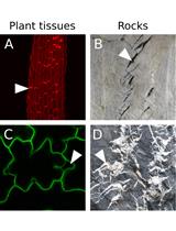
During the incubation keep the preparations in the dark. Preparations can be examined under a confocal scanning fluorescent light microscope, or fluorescent light microscope after 15-60 min (depending on the size and amount of plant material). The excitation and emission maxima of Zinpyr-1 are both within the visible spectrum: 490 and 525 nm, respectively. We used filter set 38 for the Axio Imager Z2 microscope, with excitation wavelength range 450-490 nm and emission wavelength range 500-550 nm. In order to avoid background fluorescence, it is important to remove the Zinpyr-1 solution from the slides after the treatment using filter paper and substitute it with superdemineralized water. The location of Zn in plant tissues is indicated by the green fluorescence of the Zn-Zinpyr-1 complex (Figure 1 A-C).
Staining with Zincon
Preparations can be examined under the light microscope after 5-15 min (depending on the size and amount of plant material). Preparations cannot be stored longer than a couple of hours. The location of Zn in plant tissues is indicated by the blue color of the Zn-Zincon complex (Figure 1 D-G).
Representative data

Figure 1. Zn localization in plant tissues using Zinpyr-1 (A-C) and Zincon (D-G). A. Root section, B. Root, C. Leaf petiole section of Capsella bursa-pastoris exposed to 20 µM Zn for 8 weeks; D. Root section, E-F. Leaf sections, G. Leaf epidermal peel of Noccaea caerulescens exposed to 800 µM Zn for 8 weeks. Green fluorescence indicates the location of the Zn-Zinpyr-1 complex, blue colour indicates the location of the Zn-Zincon complex. Designations: C-cortex; E-endodermis; Ep-epidermis; GC-stomata guard cells; M-mesophyll; P-pericycle; Ph-phloem; R-rhizodermis; RH-root hairs; SC-subsidiary cell; VB-vascular bundle; WSC-water-storage epidermal cell; X-xylem. Bar: 10 µm (A, D-G); 50 µm (B, C)

Notes
- To prove that green fluorescence observed after staining with Zinpyr-1 or blue coloring observed after staining with Zincon are Zn-dependent indeed, tissue sections can be incubated in a 1mM-solution of TPEN [N, N, N', N'-tetrakis (2-pyridylmethyl)ethane-1, 2-diamine] for 2 h at room temperature prior to staining with Zinpyr-1 or Zincon. TPEN is a chelator capable of penetrating cell membranes and showing high affinity and specificity toward Zn ions. Treatment of samples with TPEN should lead to almost complete absence of Zn-dependent green fluorescence after subsequent treatment with Zinpyr-1 or absence of blue staining after subsequent treatment with Zincon.
- The intensity of staining corresponds with the level of Zn accumulation in cells and tissues. Therefore, it is possible to semi-quantitatively estimate and compare the Zn contents within and among sections on a per unit area basis. The Zn contents of cells of different tissues can be compared only if the cell sizes in these tissues are similar. In particular, when Zn distribution in the growing parts of plants is analyzed, one should take into consideration that the reduced staining intensity of elongating cells, as compared with meristematic cells, may point to a decrease in the Zn content in elongating cells only on a per unit volume basis. The Zn content per cell may be the same or even higher as a result of ongoing Zn uptake during the elongation phase.
- In our hands, Zincon staining was visible at Zn concentrations of 10 μM or higher. The threshold Zn concentration of the fluorescence method (Zinpyr-1), as determined by analysis of the emission spectrum of Zinpyr-1 at various Zn concentrations, was 1 nM. One should bear in mind that the lack of coloration of certain tissues or organs only means that the Zn content in the tissues is below the detection limit of the histochemical method. However, plants can accumulate Zn at concentrations that are one or more orders of magnitude higher than in their nutrient or soil solution. Therefore, even when the Zn concentration in the medium is identical to or below the detection limit of these histochemical methods, the Zn content within the plants will most likely be sufficient for detection.
Recipes
- Staining with Zinpyr-1
- To prepare 1 mM Zinpyr-1 stock solution dissolve 1 mg of Zinpyr-1 in 500 µl of DMSO, stir it well using a vortex mixer, pipette the solution into a 2 ml microtube and add 714 µl of DMSO. Vortex it well again. Make 50 µl aliquots of Zinpyr-1 stock solution and store them in a freezer at -20 °C.
- Directly prior to the analysis, dilute an aliquot of Zinpyr-1 DMSO stock solution (1 mM) to a final concentration of 5 or 10 μM with superdemineralized water. To prepare 5 ml of 10 μM Zinpyr-1 working solution dissolve 50 µl of Zinpyr-1 DMSO stock solution in 4.95 ml of superdemineralized water. Alternatively, to prepare 5 ml of 5 μM Zinpyr-1 working solution dissolve 25 µl of Zinpyr-1 DMSO stock solution in 4.975 ml of superdemineralized water.
- To prepare 1 mM Zinpyr-1 stock solution dissolve 1 mg of Zinpyr-1 in 500 µl of DMSO, stir it well using a vortex mixer, pipette the solution into a 2 ml microtube and add 714 µl of DMSO. Vortex it well again. Make 50 µl aliquots of Zinpyr-1 stock solution and store them in a freezer at -20 °C.
- Staining with Zincon
- To prepare 50 ml of 1 M NaOH stock solution dissolve 2 g of NaOH in 50 ml of superdemineralized water.
- To prepare 10 ml of working solution dissolve 0.0065 g of Zincon and 0.1906 g of borax in 9 ml of superdemineralized water. Add 0.2 ml of 1 M NaOH and adjust the volume with superdemineralized water to 10 ml.
- Heat up the solution to 80-90 °C on a magnetic stirrer with heating and then cool down to room temperature. The solution may be stored for a week in darkness at room temperature.
- To prepare 50 ml of 1 M NaOH stock solution dissolve 2 g of NaOH in 50 ml of superdemineralized water.
Acknowledgments
This work was supported by the Russian Foundation for Basic Research (RFBR, # 11-04-00513, 15-04-02236). We thank Prof. V. B. Ivanov and Dr. A. Voronkov for fruitful discussions. The protocol for Zn staining with Zinpyr-1 was modified from the work of Dr. S. A. Sinclair and co-authors (2007).
References
- Davies, K. L., Davies, M. S. and Francis, D. (1991). Zinc-induced vacuolation in root meristematic cells of Festuca rubra L. Plant Cell Environ 14(4): 399-406.
- Kozhevnikova, A. D., Seregin, I. V., Erlikh, N. T., Shevyreva, T. A., Andreev, I. M., Verweij, R. and Schat, H. (2014a). Histidine-mediated xylem loading of zinc is a species-wide character in Noccaea caerulescens. New Phytol 203(2): 508-519.
- Kozhevnikova, A. D., Erlikh, N. T., Zhukovskaya, N. V., Obroucheva, N. V., Ivanov, V. B., Belinskaya, A. A., Khutoryanskaya, M. Y. and Seregin, I. V. (2014b). Nickel and zinc effects, accumulation and distribution in ruderal plants Lepidium ruderale and Capsella bursa-pastoris. Acta Physiologiae Plantarum 36(12): 3291-3305.
- Seregin, I. and Kozhevnikova, A. (2008). Roles of root and shoot tissues in transport and accumulation of cadmium, lead, nickel, and strontium. Russ J Plant Physiol 55(1): 1-22.
- Seregin, I. and Kozhevnikova, A. (2011). Histochemical methods for detection of heavy metals and strontium in the tissues of higher plants. Russ J Plant Physiol 58(4): 721-727.
- Seregin, I., Kozhevnikova, A., Gracheva, V., Bystrova, E. and Ivanov, V. (2011). Tissue zinc distribution in maize seedling roots and its action on growth. Russ J Plant Physiol 58(1): 109-117.
- Sinclair, S. A., Sherson, S. M., Jarvis, R., Camakaris, J. and Cobbett, C. S. (2007). The use of the zinc‐fluorophore, Zinpyr‐1, in the study of zinc homeostasis in Arabidopsis roots. New Phytol 174(1): 39-45.
Article Information
Copyright
© 2015 The Authors; exclusive licensee Bio-protocol LLC.
How to cite
Seregin, I., Kozhevnikova, A. and Schat, H. (2015). Histochemical Detection of Zn in Plant Tissues. Bio-protocol 5(10): e1470. DOI: 10.21769/BioProtoc.1470.
Category
Plant Science > Plant cell biology > Tissue analysis
Plant Science > Plant physiology > Ion analysis
Cell Biology > Cell staining > Other compound
Do you have any questions about this protocol?
Post your question to gather feedback from the community. We will also invite the authors of this article to respond.