- Protocols

- Articles and Issues
- For Authors
- About
- Become a Reviewer

Barrier Function Assay

Published: Vol 4, Iss 10, May 20, 2014 DOI: 10.21769/BioProtoc.1133 Views: 11135
Reviewed by: Lin FangFanglian HeAnonymous reviewer(s)
Original research article
The authors used this protocol in:
Feb 2013
Advertisement


Protocol Collections
Comprehensive collections of detailed, peer-reviewed protocols focusing on specific topics


Abstract
This protocol serves to determine the integrity of the barrier function of (murine) epidermis. Defects in the barrier function lead to dehydration and infection in neonatal mice/humans. One possible way to assess epidermal barrier integrity is by a dye penetration assay as described hereunder. This assay should be done on unfixed, untreated tissues (e.g. formaldehyde- or glutaraldehyde-fixed) or on whole mouse embryos (E18.5). This protocol was adapted from Hardman (1998).
Materials and Reagents
- E18.5 embryos from mice
- Chilled methanol in water (25%, 50%, 75% and 100% MetOH)
- 0.1% toluidine blue (in water) (Sigma-Aldrich)
- Chilled 1x PBS (pH 7.4) (Sigma-Aldrich, catalog number: P4417 ) (see Recipes)
Equipment
- Forceps
- Camera
Procedure
- Isolate uterus from pregnant females to obtain E18.5 embryos.
- Euthanize embryos by immersion in ice cold PBS for 30 min.
- At 18.5 days gestation, cull mother by cervical dislocation. Make a ventral incision and remove the uterine horn containing the embryos. To euthanize embryos, immerse entire uterus containing embryos in ice cold PBS for 30 min. Separate embryos and remove from uterus. Check that embryos have been successfully euthanized by pinching toe or tail with forceps. If embryo responds, place back into ice cold PBS and check every 5 min. Once embryos are unresponsive/culled, proceed to the next step.
- Finger/toe clip individual embryos for identification. This is especially important when crossing heterozygous mice. The digits can be used for genotyping the embryos.
- Passage embryos through chilled methanol gradient, taking 2 min per step. It is crucial to have all solutions at 4 degrees centigrade. Keep solutions on ice throughout protocol.
- 25% methanol in water
- 50% methanol in water
- 75% methanol in water
- 100% methanol
- 75% methanol in water
- 50% methanol in water
- 25% methanol in water
- 100% water or PBS
- 25% methanol in water
- Immerse embryos in 0.1% toluidine blue solution in water for 1-2 min on ice.
- Destain embryos in PBS (pH 7.4) until a dye pattern emerges (in case of barrier defect) or until dye is washed away (in case of intact barrier) on ice.
- Take pictures of individual embryos and if relevant genotype embryos. See also Figure 1 for some examples of intact barriers and barrier acquisition defects.
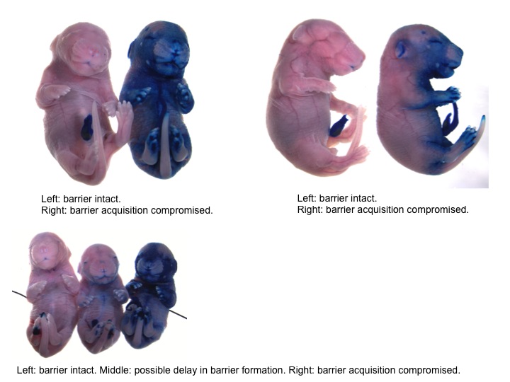
Figure 1. Examples of intact barriers and barrier acquisition defects
Recipes
- 1x PBS (pH 7.4)
1 tablet per 200 ml dH2O
Acknowledgments
This protocol was adapted from Hardman et al. (1998). Our work is supported by the Dutch Cancer Society (RUG 2012-5549), Stichting Kinder Oncologie Groningen (SKOG) and EMBO grants to FF.
References
- Hardman, M. J., Sisi, P., Banbury, D. N. and Byrne, C. (1998). Patterned acquisition of skin barrier function during development. Development 125(8): 1541-1552.
Article Information
Copyright
© 2014 The Authors; exclusive licensee Bio-protocol LLC.
How to cite
DiTommaso, T. and Foijer, F. (2014). Barrier Function Assay. Bio-protocol 4(10): e1133. DOI: 10.21769/BioProtoc.1133.
Category
Developmental Biology > Morphogenesis
Cell Biology > Tissue analysis > Tissue isolation
Cell Biology > Tissue analysis > Tissue staining
Do you have any questions about this protocol?
Post your question to gather feedback from the community. We will also invite the authors of this article to respond.