- Protocols

- Articles and Issues
- For Authors
- About
- Become a Reviewer

Purification and Fluorescent Labeling of Exosomes
Published: Vol 4, Iss 8, Apr 20, 2014 DOI: 10.21769/BioProtoc.1103 Views: 21068
Reviewed by: Anonymous reviewer(s)
Original research article
The authors used this protocol in:
Sep 2013
Advertisement


Protocol Collections
Comprehensive collections of detailed, peer-reviewed protocols focusing on specific topics


Related protocols
Abstract
Exosomes are small membrane vesicles of endocytic origin secreted into the extracellular environment from a variety of different cells, and are thought to play important roles in intercellular communications. Here, we provide a useful protocol to purify the exosomes released from cell lines using sucrose gradient centrifugation. In this protocol, we also applied a red-fluorescent lipophilic dye, DiI, which is incorporated in the outer membrane of exosomes. This fluorescently labeled exosomes allow us to visualize individual exosomes by a confocal laser scanning microscope.
Materials and Reagents
- Burkitt’s Lymphoma B cell lines (e.g. Mutu-, Mutu I, Mutu III cell lines)
- RPMI 1640 medium (Wako Chemicals USA, catalog number: 189-02025 )
- Sucrose (Sigma-Aldrich, catalog number: S7903 )
- Anti-CD63 monoclonal antibody (clone MEM-250) (Abnova, catalog number: MAB0931 )
- Bradford protein assay kit (Bio-Rad Laboratories, catalog number: 500-0006JA )
- 1, 1'-dioctadecyl-3, 3, 3', 3'-tetramethylindocarbocyanine perchlorate (DiI) (Life Technologies, catalog number: D3911 )
- Fetal Bovine Serum (FBS) (Sigma-Aldrich, catalog number: F9423 )
- Tris
- NaCl
- EDTA
- Exosome-depleted FBS (see Recipes)
- TNE buffer (see Recipes)
- 0.25-2.5 M sucrose gradient in TNE buffer (see Recipes)
Equipment
- 10 cm dish
- Centrifuge (Eppendorf, model: 5810R or that with equivalent equipment spec)
- Ultracentrifuge (Beckman Coulter, model: Optima L-80 XP or that with equivalent equipment spec)
- 37 °C, 5% CO2 cell culture incubator
- Autopipette
- 50 ml polypropylene concal plastic tubes (BD Biosciences, Falcon®, catalog number: 352070 or that with equivalent spec)
- SW28 rotor (Beckman Coulter, model: 342204 )
- SW40Ti rotor (Beckman Coulter, model: 331301 )
- Polyallomer centrifuge tubes 1 x 3½ in (25 x 89 mm) for SW28 rotor (Beckman Coulter, catalog number: 326823 )
- Polyallomer centrifuge tubes 9/16 x 3½ in (14 x 89 mm) for SW41Ti rotor (Beckman Coulter, catalog number: 331372 )
- Spectrometer
- Fluorescent or confocal laser scanning microscope
Procedure
- Purification of exosomes
- Burkitt’s Lymphoma cell lines are grown up from 1 x 10⁷ (in one 10 cm dish) to 2 x 10⁸ cells (in twenty 10 cm dishes) in 200 ml RPMI 1640 medium containing 10% exosome-depleted FBS in the 5% CO2 incubator at 37 °C.
- Culture medium containing exosomes are harvested and centrifuged in 50 ml conical tubes at 1,500 rpm for 10 min at room temperature to remove cells.
- The supernatant is centrifuged in 50 ml conical tubes at 3,500 rpm for 15 min at room temperature to remove cell debris.
- The supernatant is ultracentrifuged in polyallomer centrifuge tubes at 25,000 rpm for 1 h at 4 °C with an SW28 rotor.
- The pelleted exosomes are resuspended in 100 µl TNE buffer over night at 4 °C.
- The exosomes are fractionated by use of a 0.25-2.5 M sucrose gradient in TNE buffer in polyallomer centrifuge tubes at 25,000 rpm for 4 h at 4 °C with an SW40Ti rotor. After that, you will see a band derived from exosomes (if you collect 1 ml of each fraction from the top, exosome fraction usually locates around 6th fraction from the top).
- The band is collected (about 1 ml) carefully with an autopipette.
- Fractionated exosomes are ultracentrifuged at 25,000 rpm for 1 h at 4 °C with an SW40Ti rotor.
- The pelleted exosomes is resuspended in 100 ~ 200 µl TNE buffer over night at 4 °C.
- The total protein concentration in the fractions is determined by the Bradford protein assay.
- The fraction containing exosomes (4 µg, each) is confirmed by western blot analysis with anti-CD63 monoclonal antibody (1:1,000 dilution) (Figure 1).
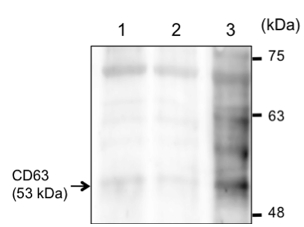
Figure 1. Purified exosomes derived from Burkitt’s lymphoma Mutu cell lines. Exosomes were purified from culture medium of Mutu- (1st lane), Mutu I (2nd lane), and Mutu III (3rd lane) cells. 4 µg of exosomes were analyzed by western blot with anti-CD63. The arrow indicates the bands that correspond to CD63.
- Burkitt’s Lymphoma cell lines are grown up from 1 x 10⁷ (in one 10 cm dish) to 2 x 10⁸ cells (in twenty 10 cm dishes) in 200 ml RPMI 1640 medium containing 10% exosome-depleted FBS in the 5% CO2 incubator at 37 °C.
- Fluorecent labeling of exosomes
- 1 ml of fractionated exosomes (100 ng/ml) is incubated with 6 µl of 10 µM stock solution of 1, 1'-Dioctadecyl-3, 3, 3', 3'-Tetramethylindocarbocyanine Perchlorate (DiI) in methanol for 1 h in the dark at room temperature with gentle agitation.
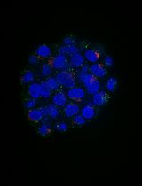
- Confirm the efficiency of labeling with small amount of exosomes under fluorescent or confocal laser scanning microscope.
- Aliquot, stored at -80 °C.
- 1 ml of fractionated exosomes (100 ng/ml) is incubated with 6 µl of 10 µM stock solution of 1, 1'-Dioctadecyl-3, 3, 3', 3'-Tetramethylindocarbocyanine Perchlorate (DiI) in methanol for 1 h in the dark at room temperature with gentle agitation.
Recipes
- Exosome-depleted FBS
Ultracentrifuge FBS at 25,000 rpm for 4 h at 4 °C
Collect supernatant and stored at 4 °C
- TNE buffer
10 mM Tris-HCl (pH 7.6)
100 mM NaCl
1 mM EDTA
Stored at room temperature
- 0.25-2.5 M sucrose gradient in TNE buffer
Prepare 0.25 M and 2.5 M sucrose in TNE buffer (Figure 2)
- Fill 2.5 M sucrose solution up to the half the height of polyallomer centrifuge tubes (approximately 6 ml)
- Add 0.25 M sucrose solution in layers up to the top of tubes (approximately 6 ml) and plug the tubes with rubber plugs
- Lay down the tubes with the top slightly higher than bottom (use thin tube floater as a pillow) for al leaset 1.5 h at room temperature
- Stand the tubes slowly and keep at 4 °C until just before use (storable up to 2 days)
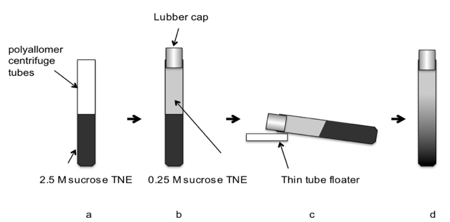
Figure 2. Preparation of 0.25-2.5 M sucrose gradient in TNE buffer. (a) Fill 2.5 M sucrose solution up to the half the height of polyallomer centrifuge tubes (approximately 6 ml). (b) Add 0.25 M sucrose solution in layers up to the top of tubes (approximately 6 ml) and plug the tubes with rubber plugs. (c) Lay down the tubes with the top slightly higher than bottom (use thin tube floater as a pillow) for at least 1.5 h at room temperature. (d) Stand the tubes slowly and keep at 4 °C until just before use (storable up to 2 days).
- Fill 2.5 M sucrose solution up to the half the height of polyallomer centrifuge tubes (approximately 6 ml)
Acknowledgments
This protocol has been adapted from a previously published paper (Nanbo et al., 2013). This work was supported in part by Grant for Funding from Basic Science research projects from The Sumitomo Foundation; Akiyama Life Science Foundation; Grant-in-Aid for Scientific Research; Shiseido Female Researcher Science Grant; The Sagawa Foundation for Promotion of Cancer Research.
References
- Nanbo, A., Kawanishi, E., Yoshida, R. and Yoshiyama, H. (2013). Exosomes derived from Epstein-Barr virus-infected cells are internalized via caveola-dependent endocytosis and promote phenotypic modulation in target cells. J Virol 87(18): 10334-10347.
- Nanbo, A., Imai, M., Watanabe, S., Noda, T., Takahashi, K., Neumann, G., Halfmann, P. and Kawaoka, Y. (2010). Ebolavirus is internalized into host cells via macropinocytosis in a viral glycoprotein-dependent manner. PLoS Pathog 6(9): e1001121.
Article Information
Copyright
© 2014 The Authors; exclusive licensee Bio-protocol LLC.
How to cite
Nanbo, A., Kawanishi, E., Yoshida, R. and Yoshiyama, H. (2014). Purification and Fluorescent Labeling of Exosomes. Bio-protocol 4(8): e1103. DOI: 10.21769/BioProtoc.1103.
Category
Cell Biology > Organelle isolation > Exosomes
Cell Biology > Cell imaging > Fluorescence
Do you have any questions about this protocol?
Post your question to gather feedback from the community. We will also invite the authors of this article to respond.