- Protocols

- Articles and Issues
- For Authors
- About
- Become a Reviewer

Neuron Culture from Mouse Superior Cervical Ganglion
Published: Vol 4, Iss 2, Jan 20, 2014 DOI: 10.21769/BioProtoc.1035 Views: 15572
Reviewed by: Xuecai Ge
Original research article
The authors used this protocol in:
Mar 2013
Advertisement


Protocol Collections
Comprehensive collections of detailed, peer-reviewed protocols focusing on specific topics


Abstract
The rodent superior cervical ganglion (SCG) is a useful and readily accessible source of neurons for studying the mechanisms of sympathetic nervous system (SNS) development and growth in vitro. The sympathetic nervous system (SNS) of early postnatal animals undergoes a great deal of remodeling and development; thus, neurons taken from mice at this age are primed to re-grow and establish synaptic connections after in situ removal. The stereotypic location and size of the SCG make it ideal for rapid isolation and dissociation. The protocol described here details the requirements for the dissection, culture and differentiation of SCG neurons. The protocol is suitable for culturing neurons from late embryonic gestation to approximately postnatal day 3. The culture technique discussed below utilizes glass coverslips for the microscopic examination of fixed cells.
Materials and Reagents
- Female mouse at desired gestational stage or early postnatal pups
- Sterile PBS (Corning Cellgro®, catalog number: 21-040-CV )
- L15 Leibovitz media (Corning Cellgro®, catalog number: 10-045-CV )
- Collagenase, Type 4 (1 mg) (Worthington Biochemical, catalog number: LS004182 )
- Collagenase enzyme solution (10 mg/ml in L15, filter-sterilized)
- Trypsin, 0.25% EDTA, Mg2+ Ca2+-free (Corning Cellgro®, catalog number: 25-053-Cl )
- Dulbecco’s Modified Eagle Medium (DMEM) (Corning Cellgro®, catalog number: 10-013-CV )
- Fetal bovine serum (FBS) (Equitech-Bio)
- Poly-D-lysine(PDL) (50 mg) (Sigma-Aldrich, catalog number: P0296 )
- Boric acid (Sigma-Aldrich, catalog number: B7660 )
- Sodium Tetraborate (Sigma-Aldrich, catalog number: B9876 )
- Laminin (1 mg) (BD Biosciences, catalog number: 354232 )
- 2.5 s Nerve Growth Factor (100 μg) (BD Biosciences, catalog number: 356004 )
- Concentrated nitric acid (Fisher Scientific, catalog number: A200-212 )
- Penicillin/streptomycin mix (Life Technologies, catalog number: 15140-122 )
- Sterile, deionized water
- Cytosine arabinoside (AraC) (Sigma-Aldrich, catalog number: C6645 )
- 0.1 M borate buffer (pH 8.5) (see Recipes)
- Regular plating medium (see Recipes)
Equipment
- Tissue culture incubator
- 35 mm or 6-well TC plates
- 35 mm tissue culture treated Petri dishes
- 100 mm Petri dishes
- 150 mm plastic Petri dish
- German glass coverslips, 25 mm (Electron Microscopy Sciences, catalog number: 72196-25 )
- Ceramic racks (Thomas Scientific, catalog number: 8542E40 )
- Basic gravity convection oven (VWR International, catalog number: 414005-108 )
- Silicon rubber dissection plates
- Scissors
- Fine tipped forceps
- 26 gauge needles
- Fire-polished, cotton-plugged, siliconized Pasteur pipets or Barrier tip 200
- Reduced bore siliconized Pasteur pipets
- Serological pipet
- Stereoscopic microscope
- Fume hood
- Water bath
- 37 °C incubator
Procedure
Note: All solutions and equipment coming into contact with cells must be sterile. Aseptic technique is critical throughout the procedure; thus, dissection taking place in a tissue culture hood modified for the use of a stereoscopic microscope may yield the best results.
- Preparation of glass coverslips
- Place coverslips in ceramic racks and rinse with water. Clean in concentrated nitric acid for 48 h in fume hood.
- Remove coverslips from acid and soak in MilliQ water for 1 h. Repeat three times.
- Cover racks in foil and place in hot air sterilization oven at 200 °C overnight.
- Place coverslips together in one 150 mm plastic Petri dish. Add ~50 ml of a filter-sterilized solution of 1 mg/ml poly-D-lysine (PDL) in borate buffer and leave overnight at RT.
- Rinse with sterile MilliQ water for 1 h. Repeat three additional times.
- Transfer individual coverslips to 35 mm or 6-well TC plates. Let dry completely.
Note: Dried PDL-coated slips can be stored for weeks at 4 °C in sterile conditions. - Apply ~400 μl laminin (10 μg/ml in sterile PBS) to coverslips intended for immediate use and incubate at 4 degrees until plating (≥ 4 h). Ensure that laminin doesn’t wick off the slips by centering them in the plates. 30 min before plating cells, place coverslips with laminin in tissue culture hood to bring to room temperature. Rinse slips 3 times with sterile PBS at room temperature. Do not allow coating to dry before plating neurons.
- Place coverslips in ceramic racks and rinse with water. Clean in concentrated nitric acid for 48 h in fume hood.
- Dissection
- Euthanize pregnant female and remove string of embryos or gather postnatal pups and euthanize in accordance with the requirements of your Institutional Animal Care and Use Committee. Place the pups or embryos in 100% ethanol to sterilize and then transfer to sterile PBS. In the case of embryos, placenta and other tissue can be removed in PBS. Allow pups or embryos to dry in a sterile 100 mm Petri dish.
- Decapitate pups/embryos into L15 remembering to clip the tails and reserve for later digestion if genotyping of animals is desired. The SCG are located in the bifurcation of the carotid arteries bilaterally coursing through neck region of the animal; decapitate animals as close to the “shoulder” area as possible to ensure full capture of the SCG (Figure 1A).
- Place the head with the cut surface up on a sterilized rubber dissection plate. Pin the head in place using two 26 gauge needles and flush with sterile PBS to clear the dissection field for viewing (Figure 1B).
- Under a stereoscopic microscope, locate the remnant of the common carotid artery and follow it deep into the head until the bifurcation is clearly seen. The SCG is stereotypically found at this junction between the internal and external carotid arteries and can be distinguished as a white to translucent, discrete spindle-shaped mass (Figure 1C-D). Often a large nerve can be seen branching from the superior aspect.
- Scoop beneath the SCG with fine forceps to gently remove the ganglion in one piece and place in a labeled plate with L15.
- Remove any excess tissue and debris from ganglia and place at 4 °C until dissociation.
Note: Intact SCG can be stored in sterile conditions for up to 4 days at 4 °C in L15 without appreciable loss in subsequent quality of culture.

Figure 1. Dissection of the superior cervical ganglion from a postnatal day 0 (P0) mouse pup. A. Whole P0 pup anesthetized and sterilized before dissection. Dotted line shows approximate level for decapitation. B. Inferior aspect of the isolated head. The cut trachea (arrowhead) is visible just superior to the bilateral zones (asterisk) where the carotid arteries will be located once surrounding tissue is removed. C. The trachea has been displaced upward and excess tissue removed from the dissection field, exposing the SCG (dotted outline). D. Image showing two dissected ganglia with small remnants of surrounding tissue attached. Scale bars = 1 mm (B, C, and D).
- Euthanize pregnant female and remove string of embryos or gather postnatal pups and euthanize in accordance with the requirements of your Institutional Animal Care and Use Committee. Place the pups or embryos in 100% ethanol to sterilize and then transfer to sterile PBS. In the case of embryos, placenta and other tissue can be removed in PBS. Allow pups or embryos to dry in a sterile 100 mm Petri dish.
- Dissociation and plating
- Place SCG into ~500 μl collagenase enzyme solution for 30 min in 37 °C water bath. Agitate every 5-10 min.
- Remove collagenase and replace with 500 μl pre-warmed 0.25% trypsin. Place in 37 °C water bath for 15-20 min, agitating every 5 min.
- Remove trypsin and rinse ganglia 3 times in L15.
- Replace L15 with approximately 500 μl pre-warmed regular plating medium. Triturate with reduced bore siliconized Pasteur pipets. 60-70 times should be sufficient if single cells are desired.
- Transfer 500 μl of the dissociated neuron suspension to uncoated 35 mm Petri dish, keeping drop in center of plate. This pre-plating step assists in the removal of glial cells before culturing the neurons. Glial cells will adhere to the uncoated dish while neurons will remain in suspension. Incubate 1 h in 37 °C incubator.
- Recover ~500 μl of the cell suspension with pipet tip. Dish can be gently tapped on the side to ensure good recovery of dissociated cells. Neurons can be spun down at 1,000 rpm for 10 min and re-suspended in pre-warmed medium as needed for high density (e.g., 2 SCG/70 μl for a 35 mm plate) or low density (e.g., 2/3-1 SCG/70 μl for a 35 mm plate) plating.
- After re-suspension, plate the cells on poly-D-lysine/laminin coated coverslips in 35 mm dishes or 6-well plates. Allow the neurons to adhere to the glass for 1.5-2 h in the incubator, then add 1.5-2 ml pre-warmed medium gently down the side of the dish with a serological pipet (dropwise). Incubate in 37 °C/5% CO2.
- If long-term culture is desired, new medium supplemented with 2 μM cytosine arabinoside (AraC) can be added at 3 days in vitro (DIV). Change the medium every 2-4 days thereafter to eliminate non-neuronal cells. Neurons can be cultured up to 28 DIV.

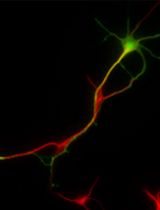
Figure 2. Image of cultured neurons of the superior cervical ganglion. Representative image showing neuronal class III β-tubulin (Tuj1) labeled SCG neurons at 48 h post-plating.
Recipes
- 0.1 M Borate buffer
Reagents Volume
Boric acid 1.24 g
Sodium tetraborate 1.90 g
dH2O 400 ml
pH 8.5, filter-sterilize
- Regular plating medium
Reagents Volume
FBS 5 ml
Penicillin/streptomycin mix 500 μl
DMEM 45 ml
Note: Nerve growth factor (NGF) is required for the growth and survival of sympathetic neurons in culture and should be added to plating medium. 100 ng/ml is suitable for signaling/long experiments; 10 ng/ml supports the survival of approximately 90% of neurons.
Acknowledgments
This protocol is adapted from Quach et al. (2013) and Eldredge et al. (2008).
References
- Eldredge, L. C., Gao, X. M., Quach, D. H., Li, L., Han, X., Lomasney, J. and Tourtellotte, W. G. (2008). Abnormal sympathetic nervous system development and physiological dysautonomia in Egr3-deficient mice. Development 135(17): 2949-2957.
- Quach, D. H., Oliveira-Fernandes, M., Gruner, K. A. and Tourtellotte, W. G. (2013). A sympathetic neuron autonomous role for Egr3-mediated gene regulation in dendrite morphogenesis and target tissue innervation. J Neurosci 33(10): 4570-4583.
- Xia, S., Lampe, P. A., Deshmukh, M., Yang, A., Brown, B. S., Rothman, S. M., Johnson, E. M., Jr. and Yu, S. P. (2002). Multiple channel interactions explain the protection of sympathetic neurons from apoptosis induced by nerve growth factor deprivation. J Neurosci 22(1): 114-122.
Article Information
Copyright
© 2014 The Authors; exclusive licensee Bio-protocol LLC.
How to cite
Readers should cite both the Bio-protocol article and the original research article where this protocol was used:
- Jackson, M. and Tourtellotte, W. (2014). Neuron Culture from Mouse Superior Cervical Ganglion. Bio-protocol 4(2): e1035. DOI: 10.21769/BioProtoc.1035.
-
Quach, D. H., Oliveira-Fernandes, M., Gruner, K. A. and Tourtellotte, W. G. (2013). A sympathetic neuron autonomous role for Egr3-mediated gene regulation in dendrite morphogenesis and target tissue innervation. J Neurosci 33(10): 4570-4583.
Category
Neuroscience > Development > Neuron
Neuroscience > Cellular mechanisms > Cell isolation and culture
Do you have any questions about this protocol?
Post your question to gather feedback from the community. We will also invite the authors of this article to respond.