

In vitro Tumorsphere Formation Assays
肿瘤干细胞球形成的体外试验
发布: 2013年02月05日第3卷第3期 DOI: 10.21769/BioProtoc.325 浏览次数: 56100
参见作者原研究论文
The authors used this protocol in:
May 2012


Abstract
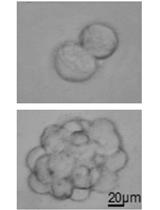
A tumorsphere is a solid, spherical formation developed from the proliferation of one cancer stem/progenitor cell. These tumorspheres (Figure 1a) are easily distinguishable from single or aggregated cells (Figure 1b) as the cells appear to become fused together and individual cells cannot be identified. Cells are grown in serum-free, non-adherent conditions in order to enrich the cancer stem/progenitor cell population as only cancer stem/progenitor cells can survive and proliferate in this environment. This assay can be used to estimate the percentage of cancer stem/progenitor cells present in a population of tumor cells. The size, which can vary from less than 50 micrometers to 250 micrometers, and number of tumorspheres formed can be used to characterize the cancer stem/progenitor cell population within a population of in vitro cultured cancer cells and within in vivo tumors (Lo et al., 2012; Liu et al., 2009). While several cell lines can be used for tumorsphere formation assay (e.g. primary mammary tumor cells from Her2/neu-transgenic mice, MCF7, BT474 and HCC1954), some cell lines may not form typical tumorsphere structures and may be difficult to count or classify definitively as tumorspheres.
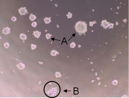
Figure 1. Shows tumorspheres formed byHCC1954 cells after 7 day incubation. The solid, circular formations represent tumorspheres (a). The individual oraggregated cells are not consideredtumorspheres (b).
Note: The tumorspheres should be solid, round structures, but the sizevaries greatly from less than 50 μm to around 250 μm. With aggregated cells, youcan still see individual cells attached to one another. With tumorspheres, thecells appear fused together and it is difficult to distinguish them asindividual cells.
Materials and Reagents
- 50x B27 Supplement (Life Technologies, InvitrogenTM, catalog number: 17504-044 )
- Basic Fibroblast Growth Factor (Sigma-Aldrich, catalog number: F0291 )
- Epidermal Growth Factor (Sigma-Aldrich, catalog number: E5036 )
- Insulin (Life Technologies, InvitrogenTM, catalog number: A11429IJ )
- Bovine Serum Albumin (Sigma-Aldrich, catalog number: A9576 )
- Dulbecco’s Modified Eagle Medium/F12 (Sigma-Aldrich, catalog number: D8437 )
- Sterile 1x Dulbecco’s Phosphate Buffered Saline (Sigma-Aldrich, catalog number: D8537 )
- Trypan Blue (Life Technologies, InvitrogenTM, catalog number: 15250-061 )
- Tumorsphere medium (500 ml) (see Recipes)
Equipment
- Incubator with CO2 input
- Counting Chamber/Hemocytometer (Hausser Scientific, catalog number: 3200 )
- 96-well Ultra-low Attachment Plates (Corning Incorporated, catalog number: 3474 )
Procedure
文章信息
版权信息
© 2013 The Authors; exclusive licensee Bio-protocol LLC.
如何引用
Johnson, S., Chen, H. and Lo, P. (2013). In vitro Tumorsphere Formation Assays. Bio-protocol 3(3): e325. DOI: 10.21769/BioProtoc.325.
分类
癌症生物学 > 癌症干细胞 > 细胞生物学试验 > 自我更新
癌症生物学 > 增殖信号转导 > 肿瘤微环境 > 增殖分析
细胞生物学 > 细胞分离和培养 > 细胞分离
您对这篇实验方法有问题吗?
在此处发布您的问题,我们将邀请本文作者来回答。同时,我们会将您的问题发布到Bio-protocol Exchange,以便寻求社区成员的帮助。