

Isolation and Quantification of Plant Extracellular Vesicles
植物细胞外囊泡的分离和定量

发布: 2017年09月05日第7卷第17期 DOI: 10.21769/BioProtoc.2533 浏览次数: 22206
评审: Marisa RosaSibongile MafuAnonymous reviewer(s)
参见作者原研究论文
The authors used this protocol in:
Jan 2017


Abstract
Extracellular vesicles (EVs) play an important role in intercellular communication by transporting proteins and RNA. While plant cells secrete EVs, they have only recently been isolated and questions regarding their biogenesis, release, uptake and function remain unanswered. Here, we present a detailed protocol for isolating EVs from the apoplastic wash of Arabidopsis thaliana leaves. The isolated EVs can be quantified using a fluorometric dye to assess total membrane content.
Keywords: Arabidopsis thaliana (拟南芥)Background
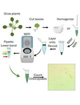
Extracellular vesicles (EVs) are membrane-bound structures that mediate the cell-to-cell transfer of proteins, lipids and genetic material. Interest in mammalian EVs has grown over the years due to their ability to transfer RNA and modulate immune responses. Mammalian EVs are routinely isolated for study from the medium of cultured cells, as well as a growing list of biological fluids (Colombo et al., 2014). Plant EVs are also thought to have a role in the immune response but are comparatively understudied (An et al., 2007; Davis et al., 2016). This is due, in large part, to the absence of a method of isolation.
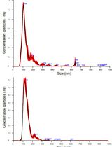
While plant EVs have been observed since 1967 using transmission electron microscopy, methods for their isolation were not developed until 2009 (Halperin and Jensen, 1967). Regente et al. (2009) isolated small (50-200 nm in diameter) vesicle-like structures from water-imbibed sunflower (Helianthus annuus) seeds. We modified the methods presented in Regente et al. (2009) to isolate vesicles from the apoplastic wash of Arabidopsis thaliana rosettes. To determine which conditions induce or impair EV secretion, we also designed a method for staining the EV pellet with 3,3’-dihexyloxacarbocyanine iodide (DIOC6(3)), a fluorescent lipophilic dye. In the absence of sophisticated forms of nanoparticle tracking, this relatively simple approach quantifies the total membrane content and can be used to indirectly measure the concentration of EVs (Rutter and Innes, 2017). For more precise measurements, and to assess the size distributions of EVs, nanoparticle tracking can be used. Our protocols enable the study of plant EV content and composition, as well as the pathways and conditions that mediate EV biogenesis and release.
Materials and Reagents
- MicroporeTM surgical tape (3M, catalog number: 1530-1 )
- Clear plastic domes (Hummert International, catalog number: 11-3348 )
- 30 ml needle-less syringes (BD, catalog number: 309650 )
- 50 ml conical tubes (VWR, catalog number: 89039-656 )
- Pipette tips
- Paper towels
- 250 ml plastic bottles (w/out cap) (Thermo Fisher Scientific, Thermo ScientificTM, catalog number: 3120-0250 )
- 15 ml conical tube (VWR, catalog number: 89039-668 )
- 1.5 ml microcentrifuge tubes (VWR, catalog number: 20170-038 )
- 10 ml syringe
- 5 ml needle-less syringes (BD, catalog number: 309646 )
- Acrodisc 0.45 μm syringe filter (Pall, catalog number: 4454 )
Note: An Acrodisc 0.22 μm filter (Pall, catalog number: 4192 ) can also be used if one is concerned about bacterial contamination. - Pryme PCR 8 strip 0.2 ml tubes (MIDSCI, catalog number: AVSST )
- Kim-wipes (KCWW, Kimberly-Clark, catalog number: 34120 )
- COSTAR EIA/RIA Plate, 96 well, half area, no lid, flat bottom, non-treated, black polystyrene plate (Corning, Costar®, catalog number: 3694 )
- 5.8 ml transfer pipets (Thermo Fisher Scientific, catalog number: 222-1S )
- Petri dishes, 100 x 15 mm (VWR, catalog number: 25384-302 )
- Arabidopsis seeds
- Bleach (Austin’s, catalog number: 90000360 )
- PRO-MIX PGX Biofungicide potting mix (Premier Tech Horticulture, catalog number: 10382RG )
- OptiprepTM density gradient medium (Sigma-Aldrich, catalog number: D1556 )
- Murashige Skoog basal salt mixture (Sigma-Aldrich, catalog number: M5524 )
- Sodium hydroxide (NaOH) pellets (Avantor Performance Materials, MACRON, catalog number: 7708-10 )
- Potassium hydroxide (KOH) pellets (Fisher Scientific, catalog number: P250-500 )
- Agar (Sigma-Aldrich, catalog number: 05040 )
- MES hydrate (Sigma-Aldrich, catalog number: M8250 )
- Calcium chloride (CaCl2) (Sigma-Aldrich, catalog number: C1016 )
- Sodium chloride (NaCl) (Sigma-Aldrich, catalog number: S7653 )
- Trizma® base (Sigma-Aldrich, catalog number: T1503 )
- Hydrochloric acid (HCl) (EMD Millipore, catalog number: HX0603-3 )
- 3,3’-Dihexyloxacarbocyanine iodide (DiOC6(3)) (Thermo Fisher Scientific, InvitrogenTM, catalog number: D273 )
- Plant protease inhibitor cocktail (Sigma-Aldrich, catalog number: P9599 )
- 2,2’-Dipyridyl disulfide (DPDS) (Sigma-Aldrich, catalog number: 149225 )
- 0.5x Murashige and Skoog agar (see Recipes)
- Vesicle isolation buffer (VIB) (see Recipes)
- 20 mM Tris-HCl, pH 7.5 (see Recipes)
- DiOC6 staining solution (see Recipes)
- Vesicle resuspension buffer (see Recipes)
Equipment
- Metal forceps
- Scissors
- Stainless steel tweezers, type 3 (Techni-Tool, catalog number: 758TW474 )
- Scale
- 1 L plastic beakers
- Titanium French press coffee maker, 24 fl oz (Snow Peak, catalog number: CS-111 )
- Vacuum chamber, 0.20 Cu. Ft. (SP Scienceware - Bel-Art Products - H-B Instrument, catalog number: F42031-0000 )
- Vacuum pump, 3.5 CFM (Ideal Vacuum Products, catalog number: P101532 )
- JA-14 fixed-angle rotor (Beckman Coulter, model: JA-14, catalog number: 339247 )
- Pipettes
- MJ research PTC-200 thermal cycler (MJ Research, catalog number: 8252-30-0001 )
- Fluorometric microplate reader
Note: We use an AppliskanTM fluorometric plate reader (Thermo Fisher Scientific, Thermo ScientificTM, catalog number: 5230000 ) to measure DiOC6 fluorescence, but any fluorometer capable of detecting multiple wavelengths could work. - SW 41 Ti rotor, swinging bucket, titanium, 6 x 13.2 ml, 41,000 rpm, 288,000 x g (Beckman Coulter, model: SW 41 Ti, catalog number: 331362 )
- Thickwall polycarbonate centrifuge tubes (3.5 ml, 13 x 51 mm) (Beckman Coulter, catalog number: 349622 )
- Thinwall, Ultra-ClearTM 13.2 ml, 14 x 89 mm ultracentrifuge tubes (Beckman Coulter, catalog number: 344059 )
- TLA100.3 fixed-angle rotor (Beckman Coulter, model: TLA-100.3, catalog number: 349481 )
- Avanti J-26S XP centrifuge (Beckman Coulter, model: Avanti J-26S XPI , catalog number: B22989)
- Fluorescent light microscope
- Growth chamber
- Ice bucket (schuett-biotech, Spongex, catalog number: 3.680 052 )
- Optima TLX ultracentrifuge (Beckman Coulter, model: OptimaTM TLX , catalog number: 361545)
- Nanoparticle trackers (Particle Metrix, model: ZetaView® , or Malvern Instruments, model: Nanosight NS300 )
Procedure
文章信息
版权信息
© 2017 The Authors; exclusive licensee Bio-protocol LLC.
如何引用
Readers should cite both the Bio-protocol article and the original research article where this protocol was used:
- Rutter, B. D., Rutter, K. L. and Innes, R. W. (2017). Isolation and Quantification of Plant Extracellular Vesicles. Bio-protocol 7(17): e2533. DOI: 10.21769/BioProtoc.2533.
- Rutter, B. D. and Innes, R. W. (2017). Extracellular vesicles isolated from the leaf apoplast carry stress-response proteins. Plant Physiol 173(1): 728-741.
分类
植物科学 > 植物细胞生物学 > 细胞器分离
植物科学 > 植物细胞生物学 > 细胞间通讯
细胞生物学 > 细胞器分离 > 胞外囊泡
您对这篇实验方法有问题吗?
在此处发布您的问题,我们将邀请本文作者来回答。同时,我们会将您的问题发布到Bio-protocol Exchange,以便寻求社区成员的帮助。